


Complications after hip disarticulation and hemipelvectomy
- Institute for Orthopedic Surgery “Banjica”, Belgrade, Serbia
ABSTRACT
Introduction: Hip disarticulation and hemipelvectomy are surgical procedures with a high rate of local complications: swollen scrotum/labia, superficial necrosis, infection, and wound dehiscence.
Aim: We present local complications in our series.
Material and methods: 17 patients were included in the study, there were 16 cases of malignant tumors and one case of osteomyelitis. Twelve of the tumors were soft tissue tumors, and four were malignant bone tumors. The descriptive statistical methods and methods for hypothesis testing used were the following: mean value, standard deviation and absolute numbers as structure indicators, the t-test, and Fisher’s exact test.
Results: Local complications were observed in ten patients. Scrotum/labia edema was observed in five, superficial necrosis in four, wound infection in six and wound dehiscence in seven cases. A single complication was observed in three patients; two and three complications were observed in three cases, each; and four complications were observed in one patient. No significant statistical difference was found between male and female patients (p=1.000). Patients with complications were significantly older (p=0.008). Correlation was found only between wound infection and dehiscence (p=0.001). Staphylococcus aureus was found in three cases, Enterococcus faecalis in one, Pseudomonas aeruginosa in three, Proteus mirabilis in two and Acinetobacter spp. in one. Two patients had a single agent infection, the remaining four had polymicrobial infection. The complication rate was 58.8%, with wound infection being the most common one. Age was found as a possible predictor. This might be in relation to the condition of the blood vessels, but also with the general condition of the patient.
Conclusion: Complications in hip disarticulation/hemipelvectomy vary from insignificant to life threatening, prolonging recovery and rehabilitation.
INTRODUCTION
Hip disarticulation and hemipelvectomy are surgical techniques of leg amputation, through the hip joint or through the pelvic bone, which entail removing the acetabulum along with the rest of the leg. Both are some of the most radical surgical procedures, not only in orthopedic, but in surgery in general [1].
The most common indications for these procedures are unresectable musculoskeletal malignant tumors, limb ischemia, and severe trauma [2]. Disarticulation of the hip due to an infected lower extremity in this day and age is not performed often, but in cases of gangrene, necrotizing fasciitis, infection following hip arthroplasty, proximal femur osteomyelitis, infected vascular grafts, and severe decubitus ulcers in paraplegics, it is still the procedure of choice [3]. In most of these cases there is a disease involving both bone and soft tissue in the thigh, especially the neurovascular bundle or the sciatic nerve, or both. Hindquarter amputation is indicated in cases where a tumor or disease process involve the hip joint or pelvis. [4].
As it is so radical, this procedure is followed by a perioperative mortality rate ranging from 0% up to as much as 37% [5]. These controversial results regarding mortality vary depending on the indication, clinical status of the patient, and the degree of urgency of the surgery. Elective surgery is followed by the best results, particularly in tumor cases. Conversely, the worst results are noted in severe polytrauma patients, associated with hemodynamic instability [6].
A high rate of complications of around 65% has been noted [5],[6]. Different local complications have been described: swollen scrotum or labia, superficial necrosis, infection, and wound dehiscence [7]. In many described cases more than one complication was present.
The aim of this retrospective study is to present local complications in our series.
MATERIALS AND METHODS
The study included 17 patients who had undergone hip disarticulation or hemipelvectomy during a twoyear period. There were 10 male and 7 female patients, ranging from 19 to 78 years of age (mean 56). All, but one, were malignant tumor patients and one was a case of severe chronic femur osteomyelitis. There were 12 cases of soft tissue tumors (all high-grade malignant) and four cases of malignant bone tumors.
The descriptive statistical methods used were measures of central tendency (mean value), variability measures (standard deviation) and absolute numbers (structure indicators). The methods used for hypothesis testing were the t-test and Fisher’s exact test (with Holm adjustments for multiple comparisons).
RESULTS
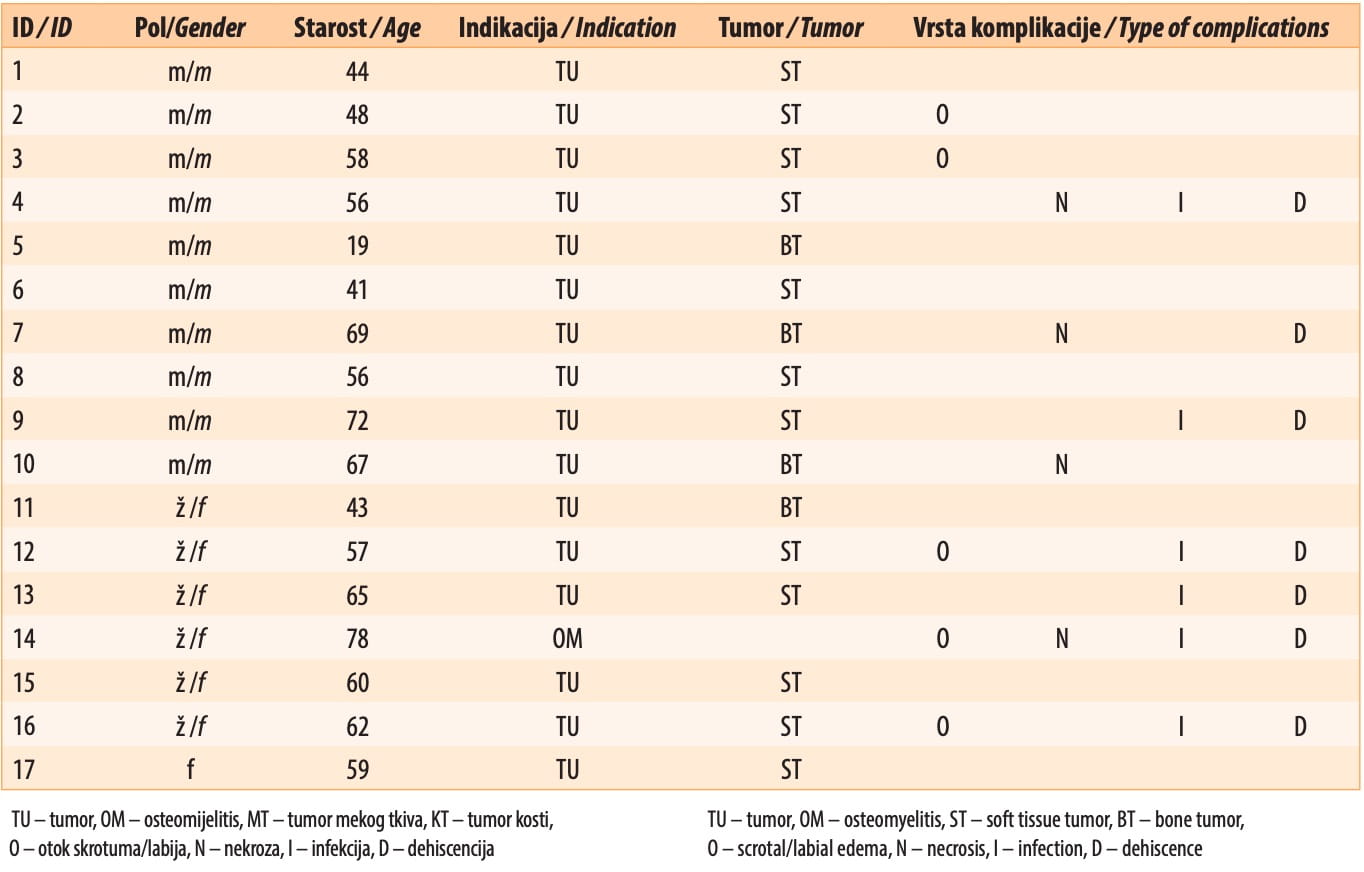
Local complications were observed in 10 patients. Scrotal or labial edema was observed in 5, superficial necrosis in four, wound infection in 6 and wound dehiscence in 7 cases. There were two and three complications, in three cases each, and four complications in one patient.
Six out of 10 male patients and four out of 7 female patients had complications, with no significant statistical difference (p=1.000) with respect to gender. The mean age in patients with complications was 63.2 ± 8.8 years, and in patients without complications it was 46.0 ± 14.3. Patients with complications were significantly older (p=0.008).
No correlation was found between complications except between wound infection and dehiscence, where correlation was highly statistically significant (p=0.001).
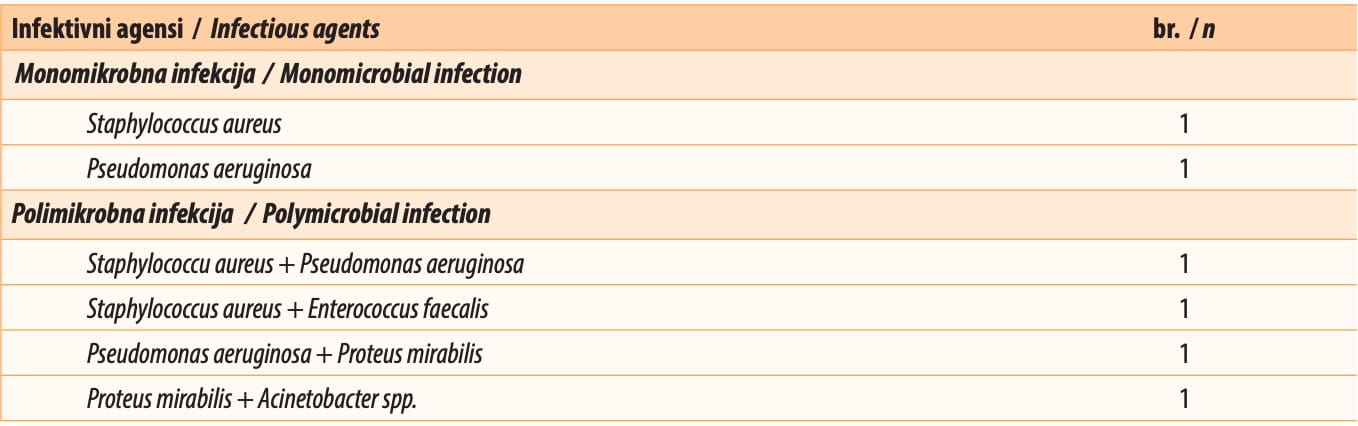
The infectious agents were as follows: Staphylococcus aureus in three cases, Enterococcus faecalis in one, Pseudomonas aeruginosa in three, Proteus mirabilis in two and Acinetobacter spp. in one case. Two patients had a single agent infection, the remaining four had polymicrobial infection.
DISCUSSION
Not many papers deal with this type of surgery, especially when it comes to the details of its complications. However, almost all of them have reported a high incidence of complications. In their series involving 53 patients, Endean et al. [5] found a 60% complication rate, most commonly associated with previous site infection (osteomyelitis, septic arthritis). The most common types of complication were necrosis and wound infection.
In their study, Daigeler et al. [8] reviewed only oncological patients, and found a complication rate of 44%. They noted flap necrosis as the most common (20%), followed by wound infection (16%), hematoma, seroma and wound dehiscence (4%). They also found one hip hernia after hip disarticulation. In other studies, obturator hernias were found as complications. They were caused by iatrogenic anatomic alteration leading to possible life-threatening hernia incarceration, which makes it essential to make an accurate diagnosis and repair the defect as an emergency [9].
The most comprehensive paper on this subject is the one by Senchenkov et al. [10]. Their study included 160 patients. Wound infection was the leading cause of morbidity, in 39% of patients, followed by 26% of cases with dehiscence. The authors investigated different factors which could influence this – age, sex, smoking, diabetes, obesity, preoperative radiation therapy, chemotherapy, ASA physical status classification, the type and extent of hemipelvectomy, the flaps, the operation duration, and blood transfusion. Only the operation duration and the extent of hemipelvectomy were found to be statistically significant predictors of wound infection and healing problems. Other studies [11],[12],[13] indicate that the surgical technique is of great importance for the outcome of this procedure. The separation of the m. gluteus maximus from the fasciocutaneous flap and extensive blood vessel ligation was significantly correlated with loss of the flap. Flap size and thorough postoperative wound care are of high importance for proper hip disarticulation [14]. Certain papers note patient age of over 65 years and a lower economic status as possible predictors for the development of complications [15].
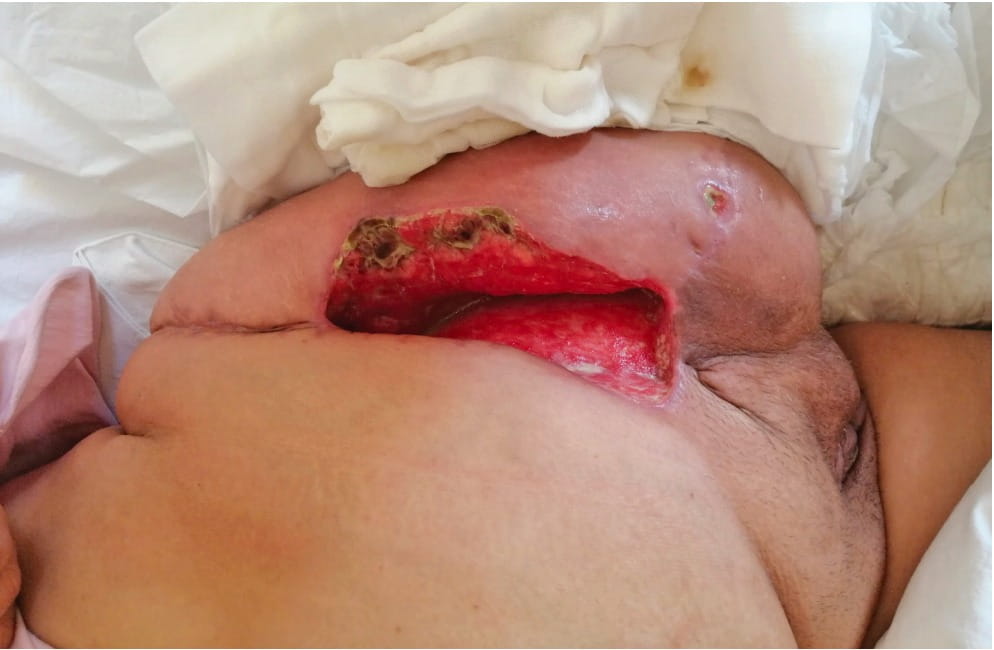
Similar results were found in our study. The complication rate was 58.8%, with wound dehiscence as the most common one. We recorded patients with superficial necrosis but without dehiscence or infection and those with dehiscence without infection, which is why we decided to separate these complications. Later, the two most common complications were found together, with infection preceding dehiscence. No correlation was found between other complications, so we may say that if scrotal/labial edema or necrosis is present this is not necessarily a sign of infection or impending dehiscence. On the other hand, if an infection is present, it is certain that dehiscence will develop in a matter of days.
Age was found as a possible predictor. This might be in relation to the condition of the blood vessels, but also in relation to the general status of the patient. Younger patients tend to recover faster, ambulating with crutches and using the toilet. Elderly patients stay in bed longer, not using the toilet, so local hygiene is poorer than in their younger counterparts, which is possibly a factor in infection development.
In our study, we found two monomicrobial and four polymicrobial infections. Different results have been published on this matter, some stating monomicrobial infection as more common [16]. Staphylococcus aureus was found as the most common, followed by Pseudomonas aeruginosa and Proteus mirabilis, but other microorganisms were also found. Further investigation should be undertaken with respect to this matter exploring whether one or more agents or a particular type of agent may be correlated with the severity of local complications. In the treatment of complications, it is important to emphasize the importance of the following: targeted antibiotic therapy based on microbiology examination results, wound management aimed at preventing contamination with intrahospital germs, and the cooperation between the orthopedic surgeon the microbiologist, the anesthesiologist, the nutrition and rehabilitation specialist and the nursing staff [17].
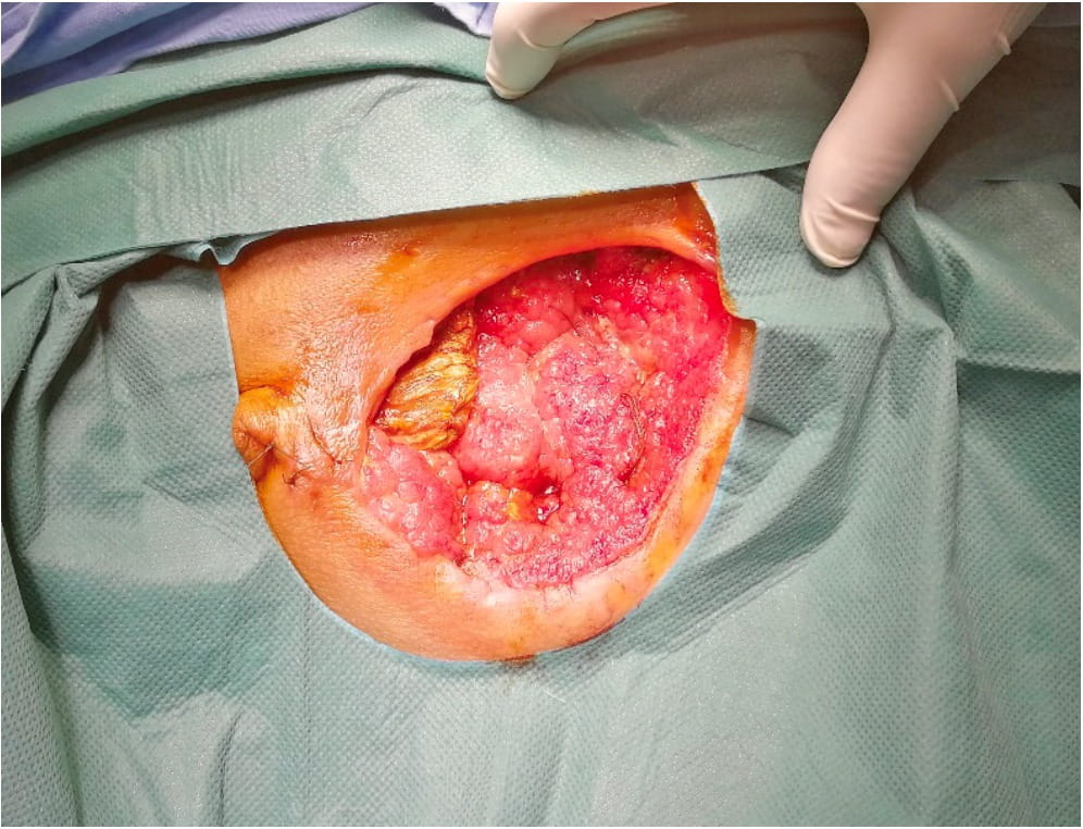
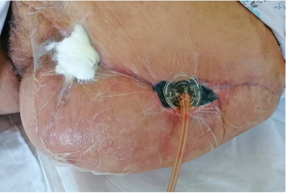
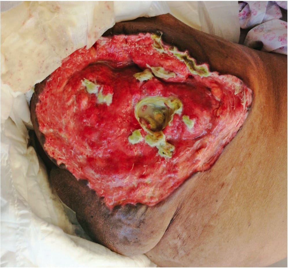
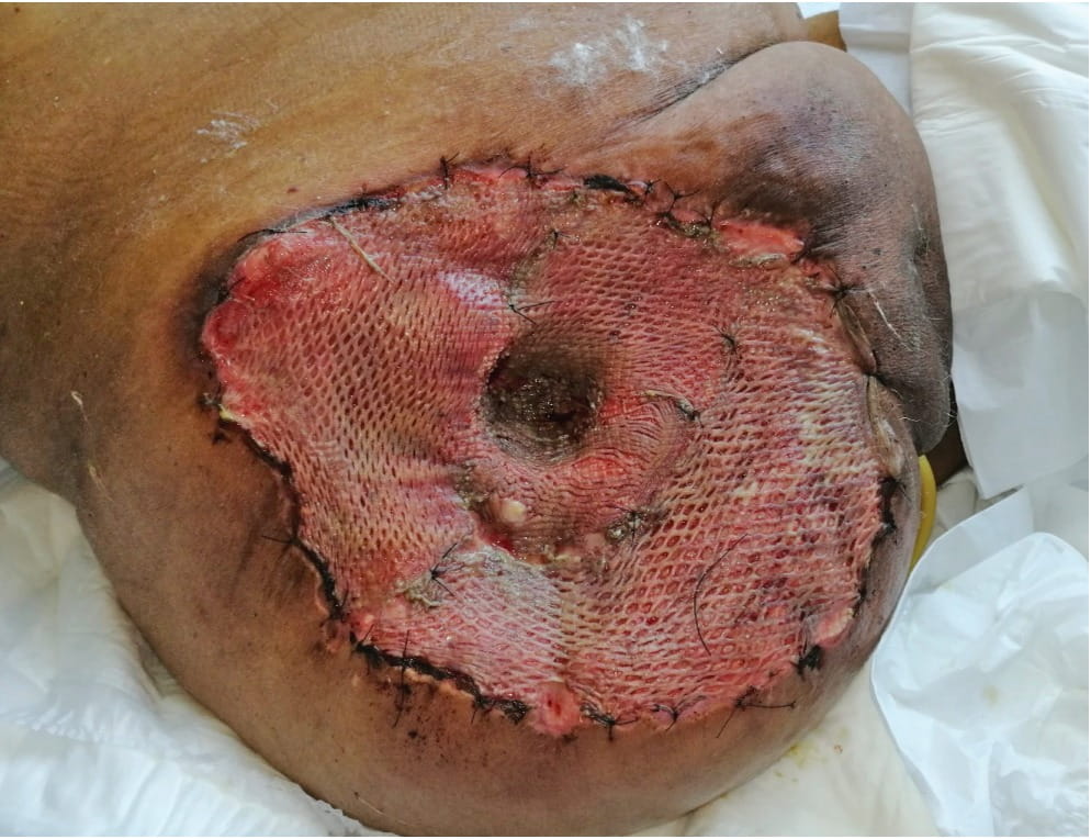
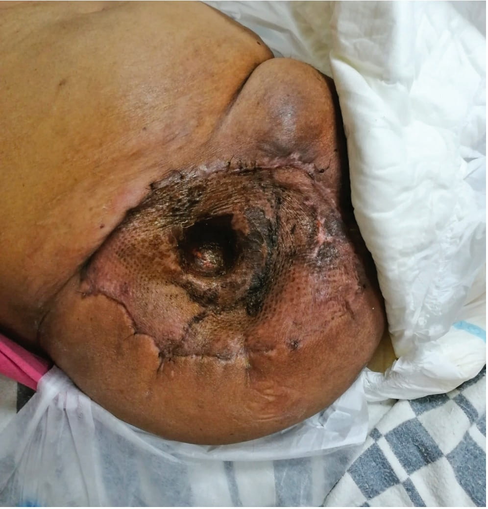
In our series, all of the patients with wound dehiscence were treated surgically, some multiple times. In some cases, vacuum drainage was used before final surgical wound closure. Parenteral antibiotics were used in all cases. Scrotal/labial edema was an independent complication which, although unpleasant and sometimes painful, was not connected with other complications. It was easily treated with ice packs and elevation. The only case of superficial necrosis without infection and dehiscence was treated with prolonged care and bandaging.
The limitations of the present study were the small number of individuals in the sample and the fact that it was a retrospective observational study. In future, other factors, such as preoperative radiation therapy and chemotherapy, should be taken into account.
CONCLUSION
Complications in hip disarticulation/hemipelvectomy vary from insignificant to life threatening, prolonging recovery and rehabilitation. It is hard to predict which patients are likely to have severe complications, since these depend on multiple factors – age, general physical condition, diagnosis, and surgical technique. At present, even with the risk of complications, these surgical procedures remain significant, especially for oncological patients, as for some of them this is the last resort.
Informations

-
Keywords:
-
Received:
-
Revised:
-
Accepted:
-
Online first:
-
DOI:
-
Cite this article:

Stanislav Rajković
Institute for Orthopedic Surgery “Banjica”
28 Mihaila Avramovica Street, 11000 Belgrade, Serbia
E-mail:
1. Dillingham TR, Pezzin LE, MacKenzie EJ. Limb amputation and limb deficiency: epidemiology and recent trends in the United States. South Med J. 2002;95(8):875–83.2[CROSSREF]
2. Loon HE. The past and present significance of hip disarticulation. Artif Limbs. 1957; 4:4–21.
3. Charalampos G, Zalavras MD, Nick Rigopoulos MD, Elke Ahlmann MD, Michael J. Patzakis MD. Hip Disarticulation for Severe Lower Extremity Infections. Clin Orthop Relat Res. 2009; 467:1721–1726.[CROSSREF]
4. Jain R, Grimer RJ, Carter SR, Tillman RM, Abudu AA. Outcome after disarticulation of the hip for sarcomas. EJSO. 2005; 31:1025–1028.[CROSSREF]
5. Endean ED, Schwarcz TH, Barker DE, Munfakh NA, Wilson-Neely R, Hyde GL. Hip disarticulation: factors affecting outcome. J Vasc Surg. 1991; 14:398–404.[CROSSREF]
6. Unruh T, Fisher DF Jr, Unruh TA, Gottschalk F, Fry RE, Clagett GP. Hip disarticulation. An 11-year experience. ArchSurg. 1990; 125(6):791–3.[CROSSREF]
7. László G, Kullmann L. Hip disarticulation in peripheral vascular disease. Arch Orthop Trauma Surg. 1987; 106(2):126–8.[CROSSREF]
8. Daigeler A, Lehnhardt M, Khadra A, Hauser J, Steinstraesser L, Langer S. Proximal major limb amputations – a retrospective analysis of 45 oncological cases. World J Surg Oncol. 2009; 7:15.[CROSSREF]
9. Rodolfo JO, Alexander HM. Emergency incarcerated obturator hernia repair with biologic mesh in a male patient after ipsilateral hip disarticulation: A case report. Int J Surg Case Rep. 2017; 37:41-44.[CROSSREF]
10. Senchenkov A, Moran SL, Petty PM, Knoetgen J 3rd, Clay RP, Bite U, et al. Predictors of complications and outcomes of external hemipelvectomy wounds: account of 160 consecutive cases. Ann Surg Oncol. 2008; 15:355-363.[CROSSREF]
11. Karakousis CP, Emrich LJ, Driscoll DL. Variants of hemipelvectomy and their complications. Am J Surg. 1989; 158:404–8.[CROSSREF]
12. Apffelstaedt JP, Zhang PJ, Driscoll DL, Karakousis CP. Various types of hemipelvectomy for soft tissue sarcomas: complications, survival and prognostic factors. Surg Oncol. 1995; 4:217–22.
13. Apffelstaedt JP, Driscoll DL, Spellman JE. Complications and outcome of external hemipelvectomy in the management of pelvic tumors. Ann Surg Oncol. 1996; 3:304–9.[CROSSREF]
14. Adam JS, Darrin JT, Katherine AB, Kristine TH, David AE, Elizabeth BH. Hip Disarticulation for Periprosthetic Joint Infection: Frequency, Outcome, and Risk Factors. J Arthroplasty. 2020 Jun 15; S0883-5403(20)30661-6.[CROSSREF]
15. Colosimo C, Fredericks C, James RY, John CK, Faran B, Stathis P. Damage control hip disarticulation: two-stage operation with index creation of a large medial flap for the septic hip. Trauma Surg Acute Care Open. 2020 Sep 1; 5(1):e000502.[CROSSREF]
16. Moura DL, Garruço A. Hip disarticulation - case series analysis and literature review. Rev Bras Ortop. 2017; 3:52(2):154-158.[CROSSREF]
17. Patera M, Mizera R. Severe Lower Extremity Infections Treated with Hip Disarticulation - Case Series. Acta Chir Orthop Traumatol Cech. 2017; 84(5):396-400.[GoogleScholar]
- Table 1. Local complications after hip disarticulation and hemipelvectomy
- Table 2. Infectious agents
- Figure 1. Wound dehiscence after superfitial necrosis. Some fields of necrosis still visible
- Figure 2. Surgical debridement of necrosis and fibrotic tissue
- Figure 3. Initial wound closure by vaccum suction
- Figure 4. Total wound dehiscence with infection
- Figure 5. Wound closue using free skin graft after Tiersche
- Figure 6. Final result after six weeks