


Laparoscopic management of a giant simple hepatic cyst
- Neurosurgery Department, “Zemun” Medical Center, Belgrade, Serbia
- Surgery Department, “Zemun” Medical Center, Belgrade, Serbia
- Pathology Department, “Zemun” Medical Center, Belgrade, Serbia
- Faculty of Dental Medicine, Belgrade, Serbia
ABSTRACT
Introduction: Congenital simple hepatic cysts are extremely rare and are usually asymptomatic. The best therapeutic approach to treating simple liver cysts remains the subject of scientific debate.
Case report: We present the case of a giant liver cyst removed with a minimally invasive, laparoscopic approach in a 68-year-old woman, who presented with rapid weight loss.
Conclusion: Due to the lack of randomized clinical trials comparing minimally invasive (laparoscopic) and open surgery approaches in liver cyst treatment, we recommend a personalized medical approach, applied after analyzing the characteristics of both the cyst and the patient.
INTRODUCTION
A hepatic cyst is typically a solitary non-parasitic lesion of the liver, also known as a simple hepatic cyst. The origin of simple hepatic cysts is still unknown, but it appears that they are congenital. These cysts are exceedingly uncommon [1],[2]. Also, they are usually without clinical symptoms. Extremely large, giant cysts can become symptomatic due to rupture, mass effect, hemorrhage, and/or infection. They can also produce liver tissue atrophy, sometimes with compensatory hypertrophy of the unaffected liver part [3]. The therapeutic approach for simple liver cysts is still the subject of scientific debate [4]. Therapy may include percutaneous needle aspiration, injection of different sclerosing agents, laparoscopic or open surgery fenestration, surgical cystectomy or marsupialization [1].
We present the case of a giant liver cyst removed with a minimally invasive, laparoscopic approach in a 68-yearold woman, who presented with rapid weight loss.
CASE REPORT
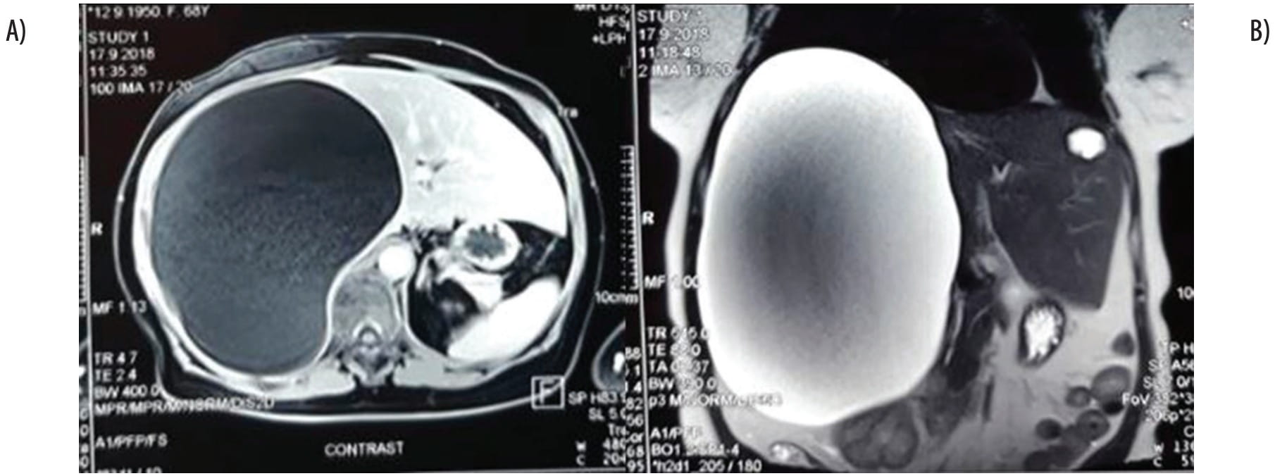
A 68-year-old female patient presented to our emergency department with abdominal discomfort. The patient reported significant weight loss of 17 kg in three months. On physical examination, the abdomen was slightly distended, and the anterior abdominal wall was above the level of the chest. Indirect finger percussion of the liver span produced a dull sound. There was minimal abdominal palpation tenderness, but no muscular defense. Also, there were no signs of jaundice, ascites, or edema of the legs. With the exception of a minimally elevated aspartate aminotransferase (AST) level, laboratory findings were within the physiological range. Tumor marker levels were also within normal ranges. The echinococcus antibody detection finding was negative. Abdominal ultrasound was performed and showed a giant, anechoic, oval-shaped, sharply edged cyst, which occupied the entire right liver lobe. The dimensions of the cyst could not be determined. Abdominal magnetic resonance imaging (MRI) was performed and revealed a 200 mm x 260 mm x 260 mm cystic mass in the right liver lobe, which extended distally into the abdomen and compressed the inferior vena cava, without signs of thrombosis or infiltration. The T1-weighted imaging MRI sequence showed that the cyst had very low homogeneous signal intensity, while the T2-weighted imaging MRI sequence showed increased signal intensity, without enhancement of the signal intensity after contrast administration. The cyst also compressed the duodenum and the pancreas head, as well as the right kidney, caudally (Figure 1).
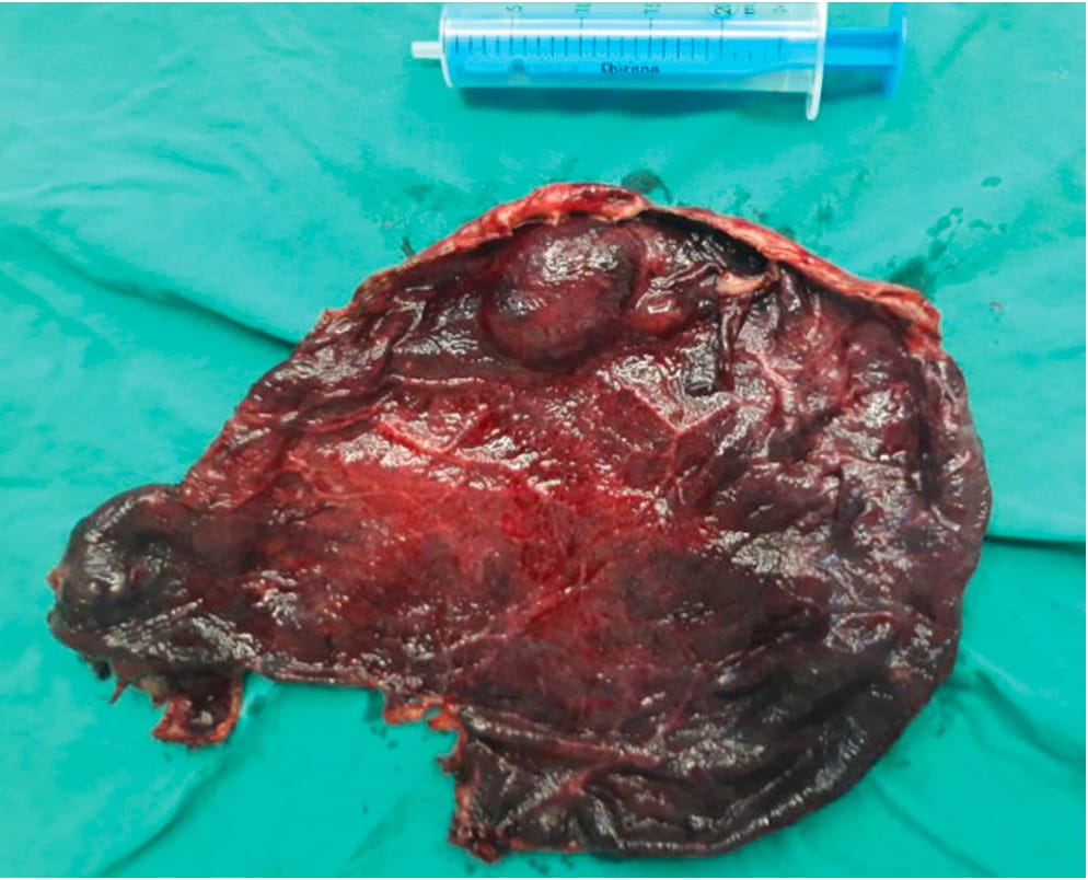
The patient was operated on, electively. The laparoscopic approach was used with three trocars. After creating pneumoperitoneum, via the umbilical port, visualization of the cyst was performed. The next step performed was percutaneous aspiration of the cyst, and approximately 6 liters of serous fluid was collected. Cystectomy was then performed with harmonic surgical scissors (Video1); during the cystectomy, a partial excision of the cyst wall was performed and it was extracted through the umbilical port (Video 2) (Figure 2). However, the intrahepatic part of the cyst wall adherent to the hepatobiliary vascular elements was left intact.
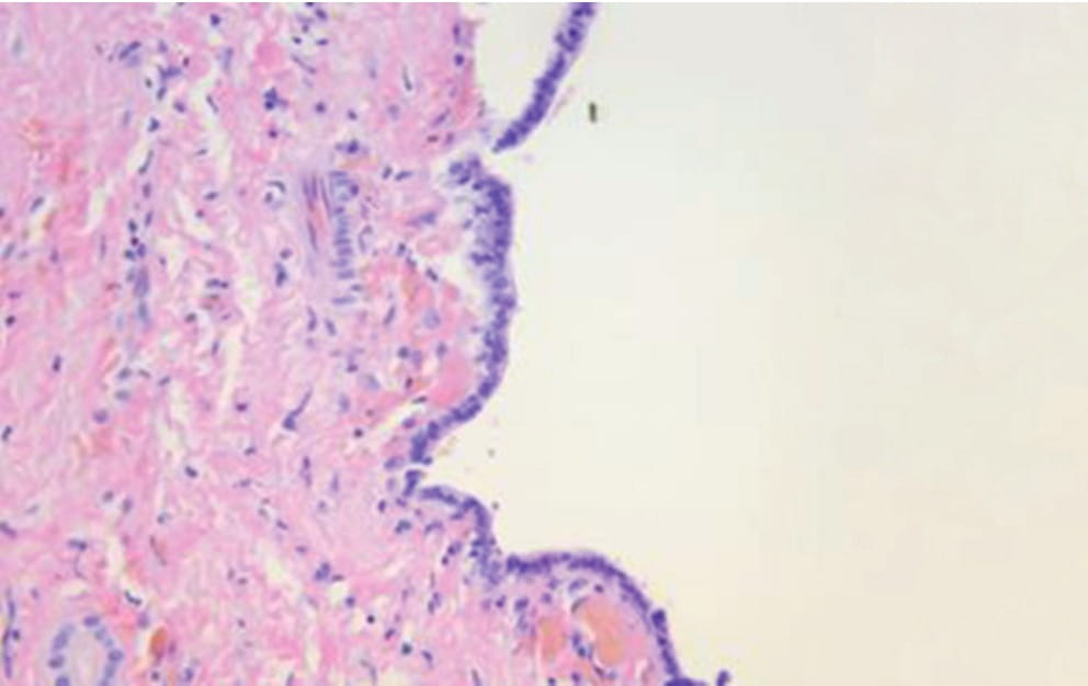
All port sites were closed manually. The postoperative period was uneventful, and the patient was discharged on the third postoperative day. The pathohistological finding showed a simple congenital cyst originating from the bile duct (Figure 3). At the first check-up, one month after surgery, the patient was feeling well, she had no complaints, and she regained about three kilograms. After this point the patient was lost to follow-up.
DISCUSSION
The prevalence of liver cysts is about 2.5%, based on ultrasound examinations. However, the precise frequency of hepatic cysts is not known since most of them are asymptomatic. Hepatic cysts are classified into four main groups, based on the origin of formation: congenital, neoplastic, inflammatory, and traumatic. Simple congenital cysts are mainly solitary, and are believed to arise from the abnormal development of intrahepatic bile ducts [5],[6]. In the presented case, the cyst was solitary, unilocular, and, according to pathohistological findings, it was congenital in origin.
Simple cysts vary in diameter from a few millimeters to more than 20 centimeters. The largest cyst reported in literature contained 17 liters of serous fluid [7]. In the presented case, the cyst was massive, and contained approximately 6 liters of serous fluid.
The majority of simple congenital cysts are asymptomatic, but they can become symptomatic and complicated due to rupture, intracystic hemorrhage or secondary bacterial infection. Sometimes, compression of adjacent structures can cause characteristic symptoms, such as edema of the lower extremities (due to inferior vena cava compression) portal hypertension and its complications (due to the compression of the portal veins), or cholestasis (due to biliary tree compression) [5],[8],[9]. Sometimes, massive cysts can cause abdominal pain, mainly in the right upper abdominal quadrant, due to the stretching of the liver capsule. Rarely, a palpable mass or an abdominal lump can be detected. In the case presented here, the patient experienced abdominal discomfort and reported significant weight loss in the three months prior to admission. The abdomen was slightly distended and there was minimal abdominal palpation tenderness.
Radiological imaging is useful for detection and determination of the characteristics of hepatic cysts. Computed tomography (CT) scans of liver cysts show water attenuation, typically homogeneous hypo-attenuation of about 0-10 Hounsfield units. A typical MRI finding of this type of cyst demonstrates the same signal intensity as water; the cyst wall is usually imperceptible; its signal does not increase in intensity after intravenous contrast material administration. The same applies to a CT scan of such a cyst. Thus, the T1-weighted imaging sequence demonstrates uniform, very low signal intensity, while the T2-weighted imaging sequence shows an increased signal intensity, greater than other T2 hyperintense liver lesions, such as hemangioma. In case of intracystic hemorrhage, which is an uncommon complication, the signal intensity is high and heterogeneous, with a fluid-fluid level on both T1- and T2-weighted images, when mixed-blood products are present. Also, when the cyst content is rich in protein, an elevated T1-weighted imaging sequence signal can be detected. The use of imaging subtraction can be helpful when assessing the presence of signal enhancement. Although these findings are generally accurate, a surgeon should always bear in mind the possibility of the presence of a neoplastic cyst before surgery [5],[10]. Understanding the typical CT and MR imaging appearance of cystic focal liver lesions allows a more definitive diagnosis and shortens the diagnostic work-up time. Also, the majority of authors suggest performing ultrasound imaging along with CT and MRI, for the purpose of collecting as much information as possible, and combining these findings with clinical information, in order to establish an accurate diagnosis, since it is extremely important to differentiate between benign cysts and potentially harmful cysts, such as cystadenoma, cystadenocarcinoma, and echinococcosis, which require specific treatment and therapy [11]. In the case presented here, the MRI showed a typical finding of a large simple hepatic cyst, which required surgical removal.
Liver cysts can be treated with nonsurgical methods or surgically. Percutaneous aspiration is considered inadequate, since the recurrence rate is high, and there is risk of infection [12]. Saini et al. conducted a study with 13 patients with liver cysts, which had been treated with percutaneous needle aspiration. All patients had a cyst recurrence within the space of two years [13]. Several other methods were described in literature with different outcome success: percutaneous aspiration with sclerosis, transdermal transhepatic ethanol injection therapy, aspiration with injection of alcohol, formalin, minocycline, monoethanolamine, doxycycline or oleate. However, these procedures may cause irreversible sclerosing cholangitis, because of the possible communication with the biliary system [5],[14]. In the past 20 years, open surgical procedures have mainly been replaced with minimally invasive surgery. There are many studies and case report series which describe large and massive hepatic cysts being operated on with the laparoscopic approach, with the recurrence rate varying from 10% to 25% [5],[15],[16]. The risk of conversion to open surgery is about 8% [5]. A majority of simple liver cysts can be managed with the laparoscopic approach, but there are patients who require the open surgical approach. A study by Gall et al. conducted on 102 patients with giant liver cysts, who were operated on with different techniques, showed that there was no difference in the quality of life with regards to the type of surgery (minimally invasive vs. open surgery) [6]. Our patient was operated on laparoscopically and demonstrated no signs of recurrence on first follow-up. Many authors conclude that open surgery is the treatment of choice, especially if there is a suspicion of malignancy, or if the cyst is located posteriorly or in the proximity of large hepatic blood vessels. However, since there are no randomized clinical trials, or trials with a large number of patients comparing minimally invasive and open surgery approaches, we recommend a personalized medical approach upon analyzing the characteristics of both the cyst and the patient.
Bearing in mind the words of the famous physician, Sir William Osler, who sad that the whole art of medicine is in observation, and in the light of the presented case, we advocate a personalized approach with every patient, combining crucial clinical information with radiological findings in order to establish an accurate diagnosis.
Informations

-
Keywords:
-
Received:
-
Revised:
-
Accepted:
-
Online first:
-
DOI:
-
Cite this article:

Vuk Aleksić
Neurosurgery Department, “Zemun” Medical Center
9 Vukova Street, 11000 Belgrade, Serbia
E-mail:
1. Asuquo M, Nwagbara V, Agbor C, Otobo F, Omotoso A. Giant simple hepatic cyst: a case report and review of relevant literature. African Health Sciences. 2015; 15(1):293-8.[CROSSREF]
2. Ismali KA, Mousa GI, El Khadrawy OH, Mohamed HA. Symptomatic non-parasitic benign hepatic cyst: Evaluation of management by deroofing in ten consecutive cases. Ann Ped Surg. 2010;6(2):83–9.[GoogleScholar]
3. Yawai H, Tada N. A simple hepatic cyst with elevated serum and cyst fluid CA19-9 levels: a case report. J Med Case Report. 2008;2:329.[CROSSREF]
4. Mazza OM, Fernandez DL, Pekoli J, Pfaffen G, Sanchez CR, Molmenti EP, et al. Management of non-parasitic hepatic cysts. 2009. J Am Coll Surg. 2009;209(6):733–9.[CROSSREF]
5. Ozbalci GS, Tanrikulu Y, Erel S, Kismet K, Akkus MA. Giant Simple Hepatic Cyst (A Case Report) and Review of the Literature. Eur J Surg Sci. 2010;1(2):53-7.
6. Gall TM, Oniscu GC, Madhavan K, Parks RW, Garden OJ. Surgical management and long term follow-up of non-parasitic hepatic cysts. HPB (Oxford). 2009;11: 235-41.[CROSSREF]
7. Burch JC, Jones HE. Large non-parasitic cyst of the liver simulating an ovarian cyst. Am J Obstet Gynecol. 1952;63:441.[CROSSREF]
8. Nanivadekar SA, Bichile LS, Pai RR. Giant soli9. Haap M, Seeger A, Haas CS. Giant solitary hepatic cyst. Liver Int. 2008;28: 840.[CROSSREF]
9. Haap M, Seeger A, Haas CS. Giant solitary hepatic cyst. Liver Int. 2008;28: 840.[CROSSREF]
10. Horton KM, Bluemke DA, Hruban RH, Soyer P, Fishman EK. CT and MR imaging of benign hepatic and biliary tumors. Radiographics. 1999;19:431-51.[CROSSREF]
11. Lantinga MA, Gevers TJ, Drenth JP. Evaluation of hepatic cystic lesions. World J Gastroenterol. 2013;19(23):3543-54.[CROSSREF]
12. Tucker ON, Smith J, Fenlon HM, McEntee GP. Giant solitary non-parasitic cyst of the liver. Ir J Med Sci. 2005;174: 60-2.[CROSSREF]
13. Saini S, Mueller PR, Ferrucci JT Jr, Simeone JF, Wittenberg J, Butch RJ. Percutaneous aspiration of hepatic cysts does not provide definitive therapy. AJR Am J Roentgenol. 1983;141: 559-60.[CROSSREF]
14. Tocchi A, Mazzoni G, Costa G, Cassini D, Bettelli E, Agostini N, et al. Symptomatic nonparasitic hepatic cysts: options for and results of surgical management. Arch Surg. 2002;137: 154-8.[CROSSREF]
15. Descottes B, Lachachi F, Durand-Fontanier S, Sodji M, Pech de Laclause B, Valleix D. Laparoscopic treatment of solid and cystic tumors of the liver. Study of 33 cases. Ann Chir. 2000;125: 941-7.[CROSSREF]
16. Katkhouda N, Hurwitz M, Gugenheim J, Mavor E, Mason RJ, Waldrep DJ, et al. Laparoscopic management of benign solid and cystic lesions of the liver. Ann Surg. 1999;229: 460-6.[CROSSREF]
- Figure 1. Abdominal MRI showing the large liver cyst. (A) T1-weighted imaging MRI sequence, transverse plane. (B) T2-weighted imaging MRI sequence, coronal plane.
- Figure 2. Cyst after extraction next to a 25 ml syringe. (The cyst was previously aspirated.)
- Figure 3. Pathohistological finding showing the simple congenital cyst formed from the bile duct (hematoxylin-eosin, magnification x400).