


Esophageal, gastric, colorectal, pancreatic, hepatocellular carcinomas and cholangiocarcinomas in Northern Macedonia: a series of patients treated at the University clinic, between 2015 and 2019
- University Clinic for Gastroenterohepatology, Skopje, North Macedonia
- Ministry of Health of the Republic of North Macedonia, Skopje, North Macedonia
- Directorate for e-Health, Ministry of Health of the Republic of North Macedonia, Skopje, North Macedonia
ABSTRACT
Introduction: The global burden of gastrointestinal cancer (GIC) is growing. Stomach, colon and liver are among the five most common sites for GIC in men and women worldwide. The incidence of GIC shows significant variation in Europe and North America.
Aim: The aim of this paper is to describe hospital morbidity from GI cancer at the University Clinic in Northern Macedonia.
Materials and methods: A retrospective longitudinal analysis included a series of cases with GIC, at the University Clinic of Gastroenterohepatology (UCG) in Skopje, in the period 2015-2019. Descriptive statistical methods were used to describe hospital morbidity from GIC, and its distribution by age, sex, and cancer site.
Results: In a five-year period, a total of 2,831 patients with GIC were treated at the UCG, of which 1,484 patients had colorectal cancer, 763 patients had gastric cancer and 88 patients had esophageal cancer. Although liver cancers were less common, as many as one eighth of such patients (355 or 13%) had nonspecific liver malignancy. Most patients were in the 60 – 69 age group, with the exception of esophageal cancer. An increase in the incidence of pancreatic cancer was observed, almost equal, when considering the distribution by sex, and mainly in the age groups 60 –69 and 70–79 years.
Conclusion: Hospital morbidity due to GIC in North Macedonia shows an increasing trend, so it is important to determine how much screening has contributed to the early detection of these cancers and to ensure access to and availability of therapy for hepatitis B and C.
INTRODUCTION
The global burden of gastrointestinal cancers (GIC) is growing. The stomach, colon, and liver are among the five most common sites for GIC, in men and women worldwide [1],[2],[3]. The incidence of GIC shows significant geographic variation, with colorectal cancer incidence being higher in Western Europe and North America, and gastric and liver cancer incidence being higher in Asia and Africa. Based on the Global Cancer Observatory (GLOBOCAN) [4], the incidence and mortality rates in North Macedonia are similar to those of Western European countries. In 2020, the incidence of colorectal cancer in North Macedonia was 10.9 and 14.4 per 100,000 population, for men and women, as compared to 9.9% and 10%, respectively, in Western Europe [4].
According to the E-health Directorate (internal data), in 2018, the highest incidence in North Macedonia was observed for GIC. These data suggest that more than one third of all colorectal and gastric carcinoma cases are new cases, as opposed to liver and pancreatic cases where two thirds of all cases are new cases. For example, 74% (304 of a total of 402 cases) of liver and bile duct carcinomas (C22 of the International Classification of Diseases version X (ICD-X)), and 70% (338 of a total of 486 cases) of pancreatic carcinoma (ICD-X: C25), as opposed to 35% (747 of a total of 2,121 cases) of colon carcinoma (ICD-X: C18), 33% (486 of a total of 1,486 cases) of rectal carcinoma (ICD-X: C20), and 36% (403 of a total of 1,129 cases) of carcinoma at the recto-sigmoid junction (ICD-X: C19), and 49% (516 of a total of 1,045 cases) of gastric carcinoma (ICD-X: C16). Similarly to these data, the worldwide incidence of pancreatic and liver cancer has an aggressive trend, despite being ranked, according to prevalence data, below colon, rectal and gastric carcinoma.
The discovery of the group 1 carcinogen gastric pathogen [5], Helicobacter pylori (H. pylori), has considerably altered the treatment and concept of the disease towards an infectious disease, which is predictable and preventable [6],[7],[8].
Based on GLOBOCAN 2018 estimates, pancreatic cancer has ranked the 11th most common cancer in the world, making up 4.5% of all deaths caused by cancer in 2018, while it is the third most common cancer in the United States [9]. To date, it is known that incidence and mortality of pancreatic cancer correlate with increasing age and the male sex, worldwide [10],[11].
Among the various types of primary liver cancers, the fifth most common cancer globally [12],[13], hepatocellular carcinoma (HCC) and intrahepatic cholangiocarcinoma (ICC) are the most common, accounting for roughly 70% and 15% of cases, respectively [14],[15],[16].
Given the significant variation of incidence of GIC in Europe and North America, this paper discusses the signs of convergence of hospital morbidity in North Macedonia towards the global morbidity. The aim of this paper is to describe hospital morbidity from GI cancer at the University clinic in Northern Macedonia.
MATERIALS AND METHODS
A retrospective longitudinal analysis included a series of GIC cases at the University Clinic of Gastroenterohepatology (UCG) in Skopje, during the period between 2015 and 2019.
We applied descriptive statistical methods to present hospital morbidity, by GIC distribution, by age, by sex, and by cancer site, for a five-year period, from January 1, 2015 to December 31, 2019.
The original dataset was cleaned from duplicates and filtered by unique patients. Data was analyzed in MS Excel software for macOS (Microsoft Corporation, Redmond, Washington, US).
RESULTS
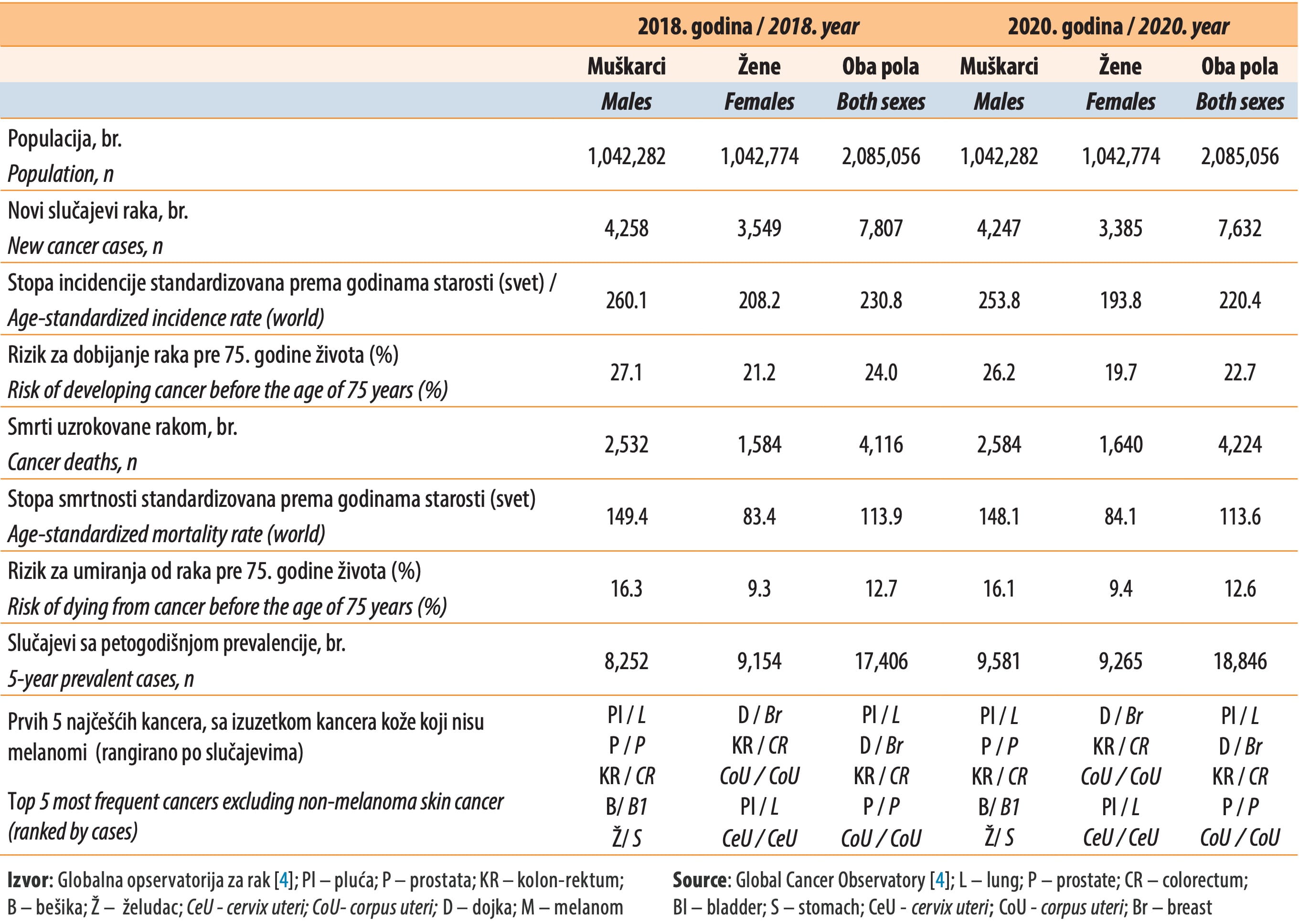
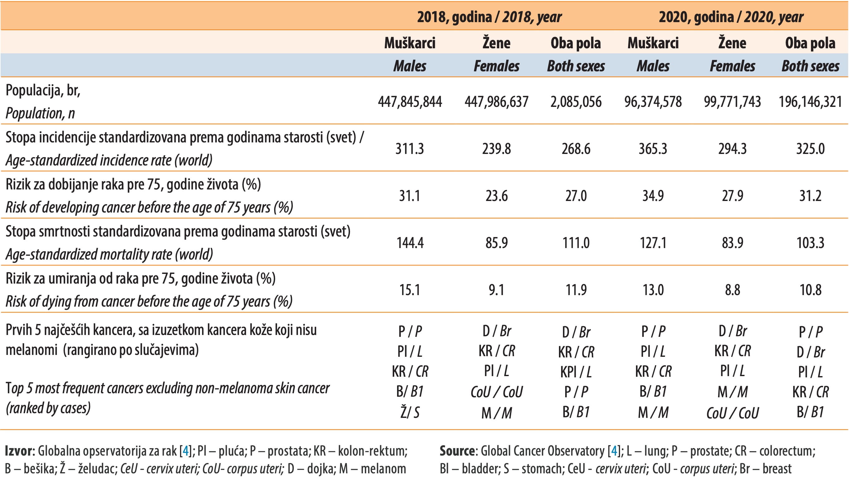
The latest GLOBOCAN data [4] shows that the overall age-standardized incidence rate and the risk of developing cancer have decreased between 2018 and 2020, along with the mortality indicators in North Macedonia (Table 1a). While the incidence rate is lower, the mortality rate is higher than in the countries of the WHO European Region and Western Europe (Table 1b).
During a 5-year period, a total of 2,831 patients with GIC were treated at the UCG, of which 1,484 (53%) patients had colorectal cancer, 763 (27%) patients had gastric cancer and 88 (3%) patients had esophageal cancer. Although liver cancers were less common, as many as over one eighth of such patients (355 or 13%) had nonspecific liver malignancy. Esophageal cancer was diagnosed in 88 patients, of which 73 male and 15 female, in a range of age groups. The following results present series of patients at the UCG, by carcinoma site, age, and sex, over the observed period (Table 2).
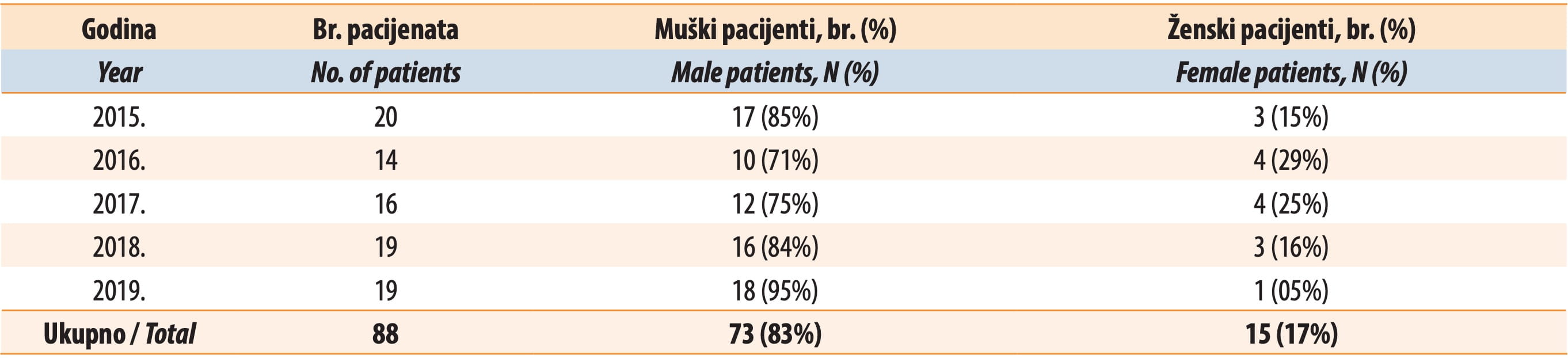
It is evident that esophageal carcinoma was far more dominant in men (85% of total cases, range: 71 – 95%) compared to women (15% of total cases, range: 5 – 29%). Over the observed period, the number of cases with esophageal carcinoma had a stable trend with decreases in years 2016 and 2017.
During the analyzed period, at the UCG, gastric cancer was detected in 763 patients, of which 532 male and 231 female, with a higher distribution in the 60 – 69 age group. Data of the existence or eradication of Helicobacter pylori, or other risk factors, were not available (Table 3).
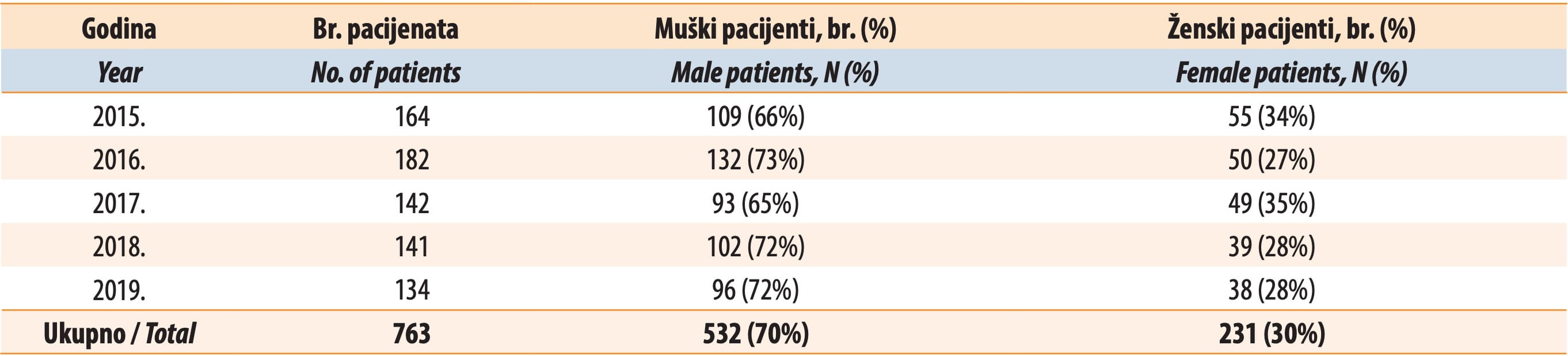
The data on gastric cancer reveal that, for this type of cancer, as well, the number of cases in male patients (70% of total cases, range: 65 – 73%) was much higher than in female patients (30% of total cases, range: 27 – 35%). Over the observed period, the number of gastric cancer cases had a stable trend, with an increase in 2018.
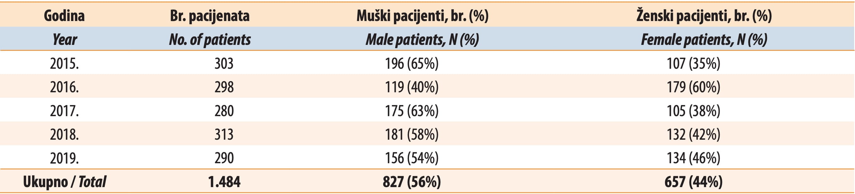
During the analyzed period, at the UCG, colorectal carcinoma was detected in 1,484 patients, of which 827 male and 657 female, mainly in the 60 – 69 age group (Table 4). The number of cases with colorectal carcinoma had a variable trend over the observed period.
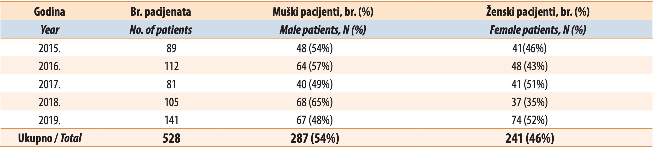
There was an observed increasing trend of the number of cases with pancreatic carcinoma at the UCG, in the analyzed period (Table 5). A total of 528 patients were diagnosed with pancreatic cancer, of whom 287 male and 241 female, mostly in the 60 – 69 and 70 – 79 age groups. Only two of them were diagnosed with pan-NET (one male and one female). Data on their habits or other risk factors were not available (Table 5).
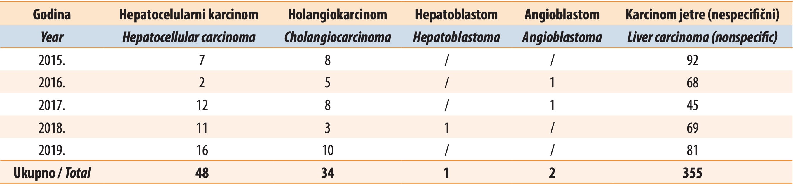
The analyzed data from the UCG showed that hepatocellular carcinoma was diagnosed in 48 patients, cholangiocarcinoma in 34 patients, and carcinoma of extrahepatic bile ducts in 59 patents. Nonspecific liver malignancy was diagnosed in 355 patients (Table 6). There was a significant increasing trend of the number of liver site carcinoma cases at the UCG, in the analyzed period (Table 6).
DISCUSSION
In the analyzed period, among a series of patients with GIC, treated at the UCG, a considerably increasing trend of the number of carcinoma cases at the site of the liver and the pancreas was observed; the number of cases with gastric and esophageal carcinoma had a stable trend, while the number of cases with colorectal carcinoma had a variable trend. These trends converge towards the trends of GIC global incidence data. The incidence and mortality of hepatocellular carcinoma have been increasing in North America and Europe [17], and, for pancreatic cancers, both incidence and mortality rates in high-income economies have been either stable or slightly increasing [18]. Gastric cancers have shown a uniform decline in incidence over the decades, which has been attributed to better food preservation practices [18].
With regard to the histological type, globally, squamous cell carcinoma remains the most common type of esophageal carcinoma, although in Western countries, it is adenocarcinoma. It is believed that the rise of the number of adenocarcinoma subtype cases corresponds to a rise in the incidence of obesity, gastro-esophageal reflux disease, and Barrett’s esophagus, and depends on the genomic instability, race, and gender of the patient [19].
The development of diagnostic procedures and antibiotics for H. pylori have improved the treatment of peptic ulcers [20],[21],[22] and patients with increased risk of gastric cancer [23],[24],[25],[26],[27]. Even though eradication of H. pylori can reduce the number of gastric cancer cases, the number of cases treated with eradication therapy is increasing [28],[29],[30],[31],[32]. A review by Satoki Shichijo and Yoshihiro Hirata confirmed the need for the establishment of a surveillance program of patients after a successful H. Pylori eradication, according to risk stratification i.e., the characteristics and predictors of gastric cancer [33].
Colorectal cancer is the most common type of GIC in Europe, with 342,137 new cases (14.3% of all cancers) [26],[34],[35],[36],[37],[38],[39],[40],[41]. The same incidence is in North Macedonia, as evident in data from the Directorate for E-health and confirmed with the analysis of the UCG data. Literature suggests that diet and obesity are among the main risk factors for colorectal cancer [42]. In North Macedonia, unhealthy dietary habits are very prevalent, with average daily intake of fats of 34.1% (compared with < 30% recommended intake), exceptionally high sodium intake of 7,883 mg (compared with the recommended value of 500 – 2500 mg), and high salt intake, which are all the result of a high consumption of processed foods [43].
Despite advancement in the knowledge of potential risk factors that cause pancreatic cancer, as well as newly available tools for early diagnosis, its incidence is estimated to increase. Although the cause of pancreatic cancer is complex and multifactorial, cigarette smoking [10] and family history are dominant [11]. Pancreatic cancer is mainly divided into two types: pancreatic adenocarcinoma, which is the most common (85% of cases) arising in the exocrine glands of the pancreas, and pancreatic neuroendocrine tumor (Pan-NET), which is less common (less than 5%) and occurs in the endocrine tissue of the pancreas [44].
Pancreatic adenocarcinoma has a very poor prognosis, typically after diagnosis, only 24% of people survive 1 year, and 9% live for 5 years [44].
Hepatocellular carcinoma represents about 90% of primary liver cancers and constitutes a major global health problem. There are a number of major infectious, lifestyle, metabolic, and hereditary risk factors for both HCC and ICC. Some of these risk factors are either potentially preventable (e.g., alcohol and tobacco use) or are currently treatable (e.g., hepatitis infection). In most cases, the molecular pathway or mechanism by which these etiological factors cause primary liver cancer, has not been well delineated [45].
Cholangiocarcinoma is the second most common liver cancer with a clinically silent development and an increasing global incidence [46]. Our results show that North Macedonia has a similar incidence and an increasing trend. Due to the absence of early markers for its diagnosis, most cholangiocarcinoma patients are identified at an advanced stage and die of metastases [46].
A very large number of non-specific liver carcinoma cases was observed in our sample. In most of these cases histopathological examination was performed, but the results were insufficient to give a definitive and more differentiating diagnosis. This can potentially impede choosing the appropriate treatment and ensuring a better patient outcome [47].
For a precise analysis of gastrointestinal carcinomas, more detailed data for the entire country are needed, including risk factors, localization, and staging. In addition, a large number of non-specific liver carcinoma cases was observed, and, while some level of diagnostics was performed, it is evident that there is a need for more diagnostic resources and tools, in order to enable a precise determination of the diagnosis. This will potentially assist in determining the most appropriate therapy and treatment and enable better quality of care and patient outcome.
Prevention, however, remains one of the most cost-effective interventions. To decrease the incidence of GI cancers, it is important to have well-organized national screening programs for prevention and early detection, especially of colorectal cancer. In addition, access to therapy for hepatitis B and C is important in the prevention of liver carcinoma.
Last but not the least, prevention of GI cancers in the long term largely depends on having and implementing policies for the prevention of major risk factors, such as obesity, smoking, unhealthy diet and physical inactivity. This should include, among other things, promoting healthy lifestyles and reducing access to unhealthy choices.
CONCLUSION
The UCG data show an increasing trend of the number of GIC cases, especially in men, with a predominance of colorectal, pancreatic and liver cancer. The establishing of a surveillance program of risk stratified patients with GIC, after treatment, could help in understanding the ways to reverse the progress of disease. The UCG data show a convergence trend toward global estimates of GIC incidence and prevalence. In future, it is important to determine how much screening has contributed to the early detection of these cancers and to ensure access to and availability of therapy for hepatitis B and C.
Informations

-
Keywords:
-
Received:
-
Revised:
-
Accepted:
-
Online first:
-
DOI:
-
Cite this article:

Kalina Grivčeva Stardelova
University Clinic of Gastroenterohepatology, Clinical Center, North Macedonia
E-mail:
1. DeSantis CE, Lin CC, Mariotto AB, Siegel RL, Stein KD, Kramer JL, et al. Cancer treatment and survivorship statistics, 2014. CA Cancer J Clin. 2014 JulAug;64(4):252-71.[CROSSREF]
2. Rustgi AK, El-serag HB. Esophageal carcinoma. N Engl J Med 2014; 371:2499- 509.[CROSSREF]
3. Arnold M, Soerjomataram I, Ferlay J, Forman D. Global incidence of oesophageal cancer by histological subtype in 2012. Gut. 2015 Mar;64(3):381-7.[CROSSREF]
4. Ferlay J, Lam F, Colombet M, Mery L, Pineros M, Znaor A, et al. Global cancer observatory: cancer today. Lyon, France: International Agency for Research on Cancer, dostupno na: https://gco.iarc. Fr/today, pristupljeno: 20. januar 2021.
5. Shichijo S, Hirata Y. Characteristics and predictors of gastric cancer after H. pylori eradication; World J Gastroenterol 2018; 24(20): 2163-72.[CROSSREF]
6. Hsu PI, Lai KH, Hsu PN, Lo GH, Yu HC, Chen WC, et al. Helicobacter pylori infection and the risk of gastric malignancy. Am J Gastroenterol 2007; 102:725-30.[CROSSREF]
7. Shichijo S, Hirata Y, Niikura R, Hayakawa Y, Yamada A, Koike K. Association between gastric cancer and the Kyoto classification of gastritis. J Gastroenterol Hepatol 2017; 32: 1581-6.[CROSSREF]
8. Handa Y, Saitoh T, Kawaguchi M, Misaka R, Ohno H, Tsai CR, et al. Association of Helicobacter pylori and diffuse type gastric cancer. J Gastroenterol 1996; 31 Suppl 9: 29-32.
9. Klein AP, Brune KA, Petersen GM, Goggins M, Tersmette AC, Offerhaus GJ, et al. Prospective risk of pancreatic cancer in familial pancreatic cancer kindreds. Cancer Res. 2004; 64(7):2634-8.[CROSSREF]
10. Hidalgo M, Cascinu S, Kleeff J, Labianca R, Lohr JM, Neoptolemos J, et al. Addressing the challenges of pancreatic cancer: future directions for improving outcomes. Pancreatology. 2015; 15(1):8-18.[CROSSREF]
11. Wild C. World cancer report 2014. Wild CP, Stewart BW, editors. Geneva, Switzerland: World Health Organization; 2014.
12. Starley BQ, Calcagno CJ, Harrison SA. Nonalcoholic fatty liver disease and hepatocellular carcinoma: a weighty connection. Hepatology. 2010; 51(5):1820-32.[CROSSREF]
13. Vernon G, Baranova A, Younossi ZM. Systematic review: the epidemiology and natural history of non-alcoholic fatty liver disease and non-alcoholic steatohepatitis in adults. Aliment Pharmacol Ther. 2011; 34(3):274-85.[CROSSREF]
14. Noureddin M, Rinella ME. Nonalcoholic Fatty liver disease, diabetes, obesity, and hepatocellular carcinoma. Clin Liver Dis 2015; 19: 361-79.[CROSSREF]
15. Satapathy SK, Sanyal AJ. Epidemiology and Natural History of Nonalcoholic Fatty Liver Disease. Semin Liver Dis 2015; 35: 221-35.[CROSSREF]
16. El-Serag HB. Hepatocellular carcinoma. N Engl J Med 2011; 365: 1118-1127.[CROSSREF]
17. Kulik L, El-Serag HB. Epidemiology and management of hepatocellular carcinoma. Gastroenterology. 2019 Jan 1;156(2):477-91.[CROSSREF]
18. Arnold M, Abnet CC, Neale RE, Vignat J, Giovannucci EL, McGlynn KA, et al. Global burden of 5 major types of gastrointestinal cancer. Gastroenterology. 2020 Jul 1;159(1):335-49.[CROSSREF]
19. Lepage C, Drouillard A, Jouve JL, Faivre J. Epidemiology and risk factors for oesophageal adenocarcinoma. Dig Liver Dis. 2013 Aug;45(8):625-9.[CROSSREF]
20. Wang C, Yuan Y, Hunt RH. The association between Helicobacter pylori infection and early gastric cancer: a meta-analysis. Am J Gastroenterol 2007; 102: 1789-98.[CROSSREF]
21. Fukase K, Kato M, Kikuchi S, Inoue K, Uemura N, Okamoto S, et al. Japan Gast StudyGroup. Effect of eradication of Helicobacter pylori on incidence of metachronous gastric carcinoma after endoscopic resection of early gastric cancer: an open-label, randomised controlled trial. Lancet 2008; 372: 392-7.[CROSSREF]
22. Hanaoka N, Uedo N, Shiotani A, Inoue T, Takeuchi Y, Higashino K, et al. Autofluorescence imaging for predicting development of metachronous gastric cancer after Helicobacter pylori eradication. J Gastroenterol Hepatol 2010; 25: 1844-9.[CROSSREF]
23. Mori G, Nakajima T, Asada K, Shimazu T, Yamamichi N, MaekitaT, et al. Incidence of and risk factors for metachronous gastric cancer after endoscopic resection and successful Helicobacter pylori eradication: results of a large-scale, multicenter cohort study in Japan. Gastric Cancer 2016; 19: 911-8.[CROSSREF]
24. Sugimoto T, Yamaji Y, Sakitani K, Isomura Y, Yoshida S, Yamada A, et al. Neutrophil infiltration and the distribution of intestinal metaplasia is associated with metachronous gastric cancer following endoscopic submucosal dissection. Can J Gastroenterol Hepatol 2015; 29: 321-5.[CROSSREF]
25. Maeda M, Nakajima T, Oda I, Shimazu T, Yamamichi N, Maekita T, et al. High impact of methylation accumulation on metachronous gastric cancer: 5-year follow-up of a multicentre prospective cohort study. Gut 2017; 66: 1721-3.[CROSSREF]
26. Shiotani A, Haruma K, Graham DY. Metachronous gastric cancer after successful Helicobacter pylori eradication. World J Gastroenterol 2014; 20: 11552-9.[CROSSREF]
27. Kobayashi M, Sato Y, Terai S. Endoscopic surveillance of gastric cancers after Helicobacter pylori eradication. World J Gastroenterol 2015; 21:10553-62.[CROSSREF]
28. Ohba R, Iijima K. Pathogenesis and risk factors for gastric cancer after Helicobacter pylori eradication. World J Gastrointest Oncol 2016; 8: 663-72.[CROSSREF]
29. Kamada T, Hata J, Sugiu K, Kusunoki H, Ito M, Tanaka S, et al. Clinical features of gastric cancer discovered after successful eradication of Helicobacter pylori: results from a 9-year prospective follow-up study in Japan. Aliment PharmacolTher2005; 21: 1121-6.[CROSSREF]
30. Maehata Y, Nakamura S, Esaki M, Ikeda F, Moriyama T, Hida R, et al. Characteristics of Primary and Metachronous Gastric Cancers Discovered after Helicobacter pylori Eradication: A Multicenter Propensity Score-Matched Study. Gut Liver 2017; 11: 628-34.[CROSSREF]
31. Nishizawa T, Suzuki H, Arano T, Yoshida S, Yamashita H, Hata K, et al. Characteristics of gastric cancer detected within 1 year after successful eradication of Helicobacter pylori. J Clin BiochemNutr2016; 59: 226-30.[CROSSREF]
32. Matsuo T, Ito M, Tatsugami M, Boda T, Takata S, Tanaka S, et al. Gastric cancer development after Helicobacter pylori eradication therapy: a new form of gastric neoplasia. Digestion 2012; 85: 61-7.[CROSSREF]
33. Yamamoto K, Kato M, Takahashi M, Haneda M, Shinada K, Nishida U, et al. Clinicopathological analysis of early-stage gastric cancers detected after successful eradication of Helicobacter pylori. Helicobacter 2011; 16: 210-6.[CROSSREF]
34. Bray F, Ferlay J, Soerjomataram I, Siegel RL, Torre LA, Jemal A. Global cancer statistics 2018: GLOBOCAN estimates of incidence and mortality worldwide for 36 cancers in 185 countries. CA Cancer J Clin. 2018; 68(6):394- 424.[CROSSREF]
35. Lansdorp-Vogelaar I, Gulati R, Mariotto AB, Schechter CB, de Carvalho TM, Knudsen AB, et al. Personalizing age of cancer screening cessation based on comorbid conditions: model estimates of harms and benefits. Ann Intern Med. 2014 Jul 15;161(2):104-12.[CROSSREF]
36. Brenner H, Altenhofen L, Stock C, Hoffmeister M. Expected long-term impact of the German screening colonoscopy programme on colorectal cancer prevention: analyses based on 4,407,971 screening colonoscopies. Eur J Cancer. 2015 Jul;51(10):1346-53.[CROSSREF]
37. Brenner H, Altenhofen L, Stock C, Hoffmeister M. Prevention, early detection, and overdiagnosis of colorectal cancer within 10 years of screening colonoscopy in Germany. Clin Gastroenterol Hepatol. 2015 Apr;13(4):717-23.[CROSSREF]
38. Brenner H, Kretschmann J, Stock C, Hoffmeister M. Expected long-term impact of screening endoscopy on colorectal cancer incidence: a modelling study. Oncotarget. 2016 Jul 26;7(30):48168-79.[CROSSREF]
39. Chen C, Stock C, Hoffmeister M, Brenner H. How long does it take until the effects of endoscopic screening on colorectal cancer mortality are fully disclosed?: a Markov model study. Int J Cancer. 2018 Dec 1;143(11):2718-24.[CROSSREF]
40. Cottet V, Jooste V, Fournel I, Bouvier AM, Faivre J, Bonithon-Kopp C. Longterm risk of colorectal cancer after adenoma removal: a population-based cohort study. Gut. 2012 Aug;61(8):1180-6.[CROSSREF]
41. Bosetti C, Lucenteforte E, Silverman DT, Petersen G, Bracci PM, Ji BT, et al. Cigarette smoking and pancreatic cancer: an analysis from the International Pancreatic Cancer Case-Control Consortium (Panc4). Ann Oncol. 2012; 23(7):1880-8.[CROSSREF]
42. Alsheridah N, Akhtar S. Diet, obesity and colorectal carcinoma risk: results from a national cancer registry-based middle-eastern study. BMC Cancer. 2018;18(1):1-1227.[CROSSREF]
43. Milevska Kostova N, Chichevalieva S, Ponce NA, van Ginneken E, Winkelmann J. The fYR of Macedonia: Health System Review. Health Systems in Transition. 2017;19(3):1-160.
44. Adams LA, Lindor KD. Nonalcoholic fatty liver disease. Ann Epidemiol. 2007;17(11):863-9.[CROSSREF]
45. Margini C, Dufour JF. The story of HCC in NAFLD: from epidemiology, across pathogenesis, to prevention and treatment. Liver Int. 2016 Mar;36(3):317-24.[CROSSREF]
46. Khan SA, Tavolari S, Brandi G. Cholangiocarcinoma: Epidemiology and risk factors. Liver International. 2019;39:19-31.[CROSSREF]
47. European Association for the Study of the Liver. EASL clinical practice guidelines: management of hepatocellular carcinoma. Journal of hepatology. 2018 Jul 1;69(1):182-236.[CROSSREF]
- Table 1a. Cancer indicators, North Macedonia, 2018 and 2020
- Table 1b. Cancer key indicators, WHO European Region in 2018 and Western Europe in 2020
- Table 2. Number of diagnosed and treated patients with esophageal cancer at the UCG, by sex, 2015 – 2019
- Table 3. Diagnosed and treated patients with gastric cancer at the UCG, by sex, 2015 – 2019, (n, %)
- Table 4. Diagnosed and treated patients with colorectal cancer at the UCG, by sex, 2015 – 2019, (n, %)
- Table 5. Diagnosed and treated patients with pancreatic cancer at the UCG, by sex, 2015 – 2019, (n, %)
- Table 6. Diagnosed and treated patients with liver cancers at the UCG, by type, 2015 – 2019, (n, %)