


The importance of a multidisciplinary diagnostic and therapeutic approach to painful symphysis syndrome in athletes
- University of Belgrade, Faculty of Medicine, Belgrade, Serbia
- College of Sports and Health, Belgrade, Serbia
ABSTRACT
Painful symphysis syndrome (pubalgia) in athletes is a particular injury, a painful condition, and dysfunction of the femoroinguinal region, which occurs due to the disproportion in strength between the abdominal wall muscles and the muscles of the lower extremities. This study aims to theoretically analyze the importance of a multidisciplinary diagnostic and therapeutic approach to painful symphysis syndrome in athletes. More than seventy causes of pubalgia have been presented in literature, most commonly linked to sports factors. Activities that include repetitive strong kicks, rotational movements, circumduction, and torsion, are risk factors. The syndrome was first registered in football players, and it mostly occurs in male athletes under the age of forty. Establishing a diagnosis is difficult, due to the complex anatomy and the overlapping of symptoms between different groin injuries. The therapeutic approach depends on the complexity of the injuries and the dynamics of the patient's recovery. It entails observing the principles of initiating treatment with minimally invasive modalities and reserving surgical treatment as the final therapeutic option, after which it is necessary to conduct an appropriate rehabilitation program, tailored to the pain and the dynamics of recovery. Most athletes resume their sports activities after two to three months, with no recurrence of pubalgia after surgical treatment. However, bearing in mind that the occurrence of pubalgia prevents the patients from engaging in sports activities over a prolonged period, special attention should be directed towards the prevention of this painful syndrome. Taking this into consideration, prevention should begin at the earliest age, through a daily program of exercises for strengthening the muscles of the anterior abdominal wall, with special emphasis on the oblique and transverse abdominal muscles.
INTRODUCTION
Painful symphysis syndrome in athletes was, in detail, first described by Spinelli, in 1932, but literature also shows that, in the 1990s, Gilmore named this syndrome Gilmore’s groin. Other synonymous names for the condition, also found in literature, are as follows: symphysis syndrome, pubalgia, athletic pubalgia, pubic inguinal pain syndrome, sports-men’s groin, footballers groin injury complex, hockey player’s syndrome, athletic hernia, and inguinal disruption [1].
Athletic pubalgia is a particular anatomical injury, and not a broad category of findings, which is why it must be emphasized that a pathological diagnosis, such as inguinal hernia, does not exclude the presence of this syndrome. Terms such as athletic hernia or sports-men’s hernia, used customarily in professional circles today, cause problems in distinguishing amongst anatomical injury patterns and findings in the diagnostic process. The syndrome was first registered in footballers (known as ‘footballers groin’) and, with time, it was described in other athletes, especially in those whose activities require the excess use of the lower abdominal wall muscles and the proximal musculature of the lower extremities.
Viewing pubalgia from the clinical and anatomical aspect, the significance of the skeleto-muscular anatomy of this region becomes evident. In addition to the bones (both femoral bones, the sacral and coccygeal bones), the muscles of the anterolateral abdominal wall (m. obliquus externus abdominis, m. obliquus internus abdominis, m. transversus abdominis, m. rectus abdominis) as well as the inner thigh muscles – the adductors (m. pectineus, m. gracilis, m. adductor longus, m. adductor brevis, m. adductor magnus) are also a part of the anatomically significant framework. All of these muscles have their insertions on the pubic bone and the pubic symphysis, while the m. rectus abdominis and m. adductor magnus have the greatest importance in maintaining the stability of the pelvis in the sagittal plane [2],[3].
Bearing in mind that groin injuries are a frequent occurrence in athletes, as well as the fact that they account for 6% of all sports injuries [4], and, at the same time, taking into account the fact that these injuries are relatively frequently misinterpreted, the aim of this paper is the theoretical analysis of the significance of a multidisciplinary diagnostic and therapeutic approach to the painful symphysis syndrome in athletes.
ETIOLOGY AND ETIOPATHOGENESIS
More than 70 causes of pubalgia have been described in literature. Based on the classification proposed by Benazzo, in 1999, the causes of pubalgia may be grouped, somewhat arbitrarily, by applying a useful methodological approach, starting with pubalgia caused by pubic symphysis overstraining, i.e., rectus adductor symphysis syndrome, occurring in 40% of the cases; followed by pubalgia caused by parietoabdominal weakness (sports hernia), occurring in 40% of the cases; and finally, pubalgia caused by pathological processes, which involve the surrounding organs (pelvis and urogenital tract), and which account for 20% of cases [5]. Sports causes are the most frequent in the etiology of this syndrome. In footballers, the syndrome occurs as the result of repeated strenuous workout and training sessions and football matches, as well as the players’ inability to have sufficient rest and recuperation in between these activities. The syndrome is also present in ice hockey players, rugby players, long-distance runners, players of Australian football, cricket players, etc. The above-mentioned sports include repeated strong kicks, rotation, circumduction and torsion movements, etc. All of these movements represent risk factors for the development of painful symphysis syndrome [6].
Athletes with the diagnosis of painful symphysis syndrome are mainly men under the age of 40. Generally speaking, this can be explained by the fact that men more often engage in the sports listed above than women, and these sports carry a greater risk for the development of this painful syndrome than other sports. Other reasons originate from the anatomical differences between the male and female pelvis. Women usually have a larger and more robust caudal m. rectus abdominis on the pubic symphysis, their pelvis is wider, and it has a wider subpubic angle, which may result in better transfer of forces away from the pubic region. Therefore, the anatomical and biomechanical differences in the structure of the female pelvis may be the explanation regarding the better stabilization of the pubic region and the decreased risk of pubalgia in women [3].
Generally speaking, the pain occurring in this syndrome, occurs due to a disproportion between the strength of the abdominal wall muscles and the muscles of the lower extremities, as well as the elasticity of the pubic symphysis, which enables movement of up to 2 mm and rotation of up to 3 degrees [3].This imbalance leads to the sensation of pain, deep in the groin. Based on the above stated, this entity could be renamed as ‘muscular groin imbalance syndrome’, while athletic hernia could be considered as a finding included in this syndrome [7]. The mechanism of the development of symptoms is explained by constant powerful contractions, which radiate to the muscular insertions in these regions, creating damage to the subchondral vascularization in the area of the pubic bone, which potentially causes the consequential development of its aseptic necrosis. This process further causes nonspecific inflammation, primarily in the muscular insertions. The described mechanism also brings about the pathological process in the muscular insertions on the pubis, while, in the later phase, the development of pubic bone osteoporosis has been noticed [8].
SYMPTOMS AND CLINICAL SIGNS
Most patients with pubalgia experience symptoms for months or even years before they get the correct diagnosis. The first symptoms usually occur after a certain amount of strain and subsequent rest of the painful region. The pain mainly manifests when walking on steep terrains. The athlete, when walking in such a way, is forced to assume the antalgic gait, with hips in flexion. The pain in the symphysis area may occur and/ or become worse upon defecation, sneezing or coughing. In the beginning, the pain is mostly tolerable, while as the condition develops, it may completely incapacitate the player. The patients usually complain of unilateral deep and sharp pain in the lower abdomen and in the frontal part of the groin. The pain may radiate proximally to the thighs, lower back, but also to the muscles of the lower abdomen, to the perineum, or the scrotum [9]. A number of studies have shown that, prior to the occurrence of pain, most players had played in three to four challenging games in one week, without enough time to rest in between these matches. After an intense game or training session, the athlete has difficulty walking, especially when climbing stairs. When in bed, the athlete usually lies on their side, with hips flexed and knees bent, with a disturbed sleeping rhythm. Standing up from a chair or from the bed is very painful [10].
DIAGNOSTIC CRITERIA
Diagnosing pubalgia is difficult due to the complex anatomy and the overlapping of symptoms of different groin injuries. The clinical examination and the medical history are of the utmost importance. The physician must take into account the fact that athletes with groin pain may have more than one diagnosis and that the presence of one of these related diagnoses does not necessarily exclude pubalgia. The recommendation is to approach this entity from a multidisciplinary aspect, since literature so far does not offer the appropriate diagnostic studies and recommendations for the proper treatment to be applied in these patients [7]. A classification of symptoms pertaining to the groin area into three major categories of clinical syndromes has been proposed (Figure 1) [11].
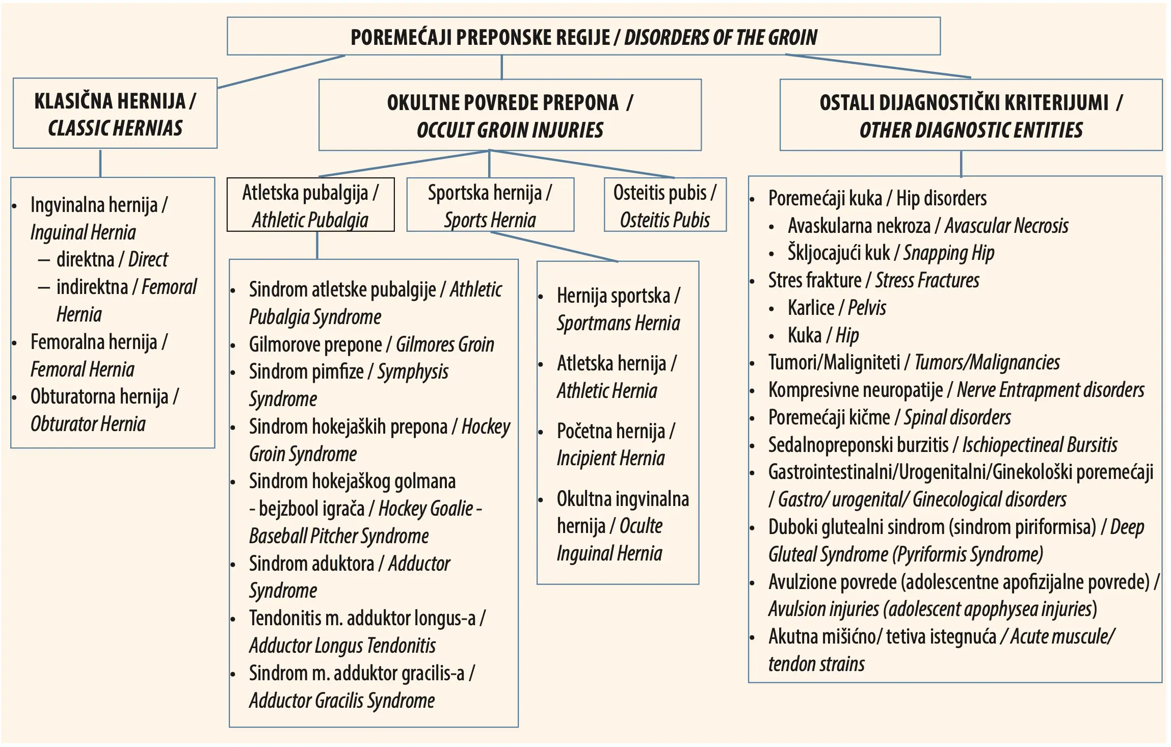
The first step in establishing the diagnosis is taking the appropriate and detailed anamnesis on the reported pain (when it occurs, how long it lasts, the exact localization of the pain, pain propagation, pain intensity, associated symptoms, etc.). The physical examination of athletic pubalgia, as the next important step in the diagnosis of the syndrome, begins with the palpation of the potential injury sites. Palpatory painful sensitivity ca be detected through physical examination at the site of the insertion of the adductors of the thigh, at the insertion of Poupart’s ligament, as well as at the meeting point of the two pubic bones. In the ingenium, lymph nodes can also be palpated and they are extremely painful to the touch. During the contraction of the abdominal wall, when the patient is standing or lying down, it is possible to note the signs of weakness of the oblique and transverse abdominal wall muscles as well as of the abdominal wall aponeurosis – Malgaigne's sign. This sign is represented by the presence of a spindle shaped buldge extending from the pubis to the anterior superior iliac spine (lat. spina iliaca anterior superior). At the same time, this sign indicates the weakness and atonia of the transverse muscle fibers of the anterior abdominal wall [8]. The abduction of the hip joint is limited by the pain arising from the stretching of the adductors. The adduction and abduction of the hip are painful. Active lifting of the leg in external rotation with the knee in full extension is very painful and usually not possible for the patient [10].
Particular muscle provocation tests for adductors of the anterior abdominal wall have been described. The testing of the adductors is of great importance for the physical examination. In this test, the patient is lying supine with the hips in abduction and flexed at 80 degrees. The test is positive if the patient, while attempting to pull the legs against the resistance in the other direction (exerted by the doctor), feels sharp pain in the groin [12]. On the other hand, Kachingwe et al. made a classification comprising the following five clinical signs that may indicate athletic pubalgia: 1) subjective discomfort caused by pain deep in the groin/ pain in the lower abdomen; 2) pain which increases with the increase of exertion (sprinting, crunches/situps) and decreases with rest; 3) palpatory sensitivity of the pubic ramus at the insertion site of the m. rectus abdominis and/or the insertion tendon; 4) pain when resistance is exerted in hip adduction with a flexion of 45 and/or 90 degrees; 5) pain when offering resistance during the rotation of the abdomen [13],[14].
The regional examination is exceptionally important in narrowing down the differential diagnosis. The lumbar segment of the spinal column, the sacroiliac joints, and the hip joints must be functionally tested. At the same time, the symphysis is tested for instability and sensitivity, as pubic bone osteitis is a relatively frequent finding in the population of athletes. Muscle insertions (including adductors, as well as m. rectus femoris, m. sartorius, rectus abdominis) show painful sensitivity to palpation. In order to elicit symptoms, the same muscles are tested for resistance. The patient is examined both in the standing position and when lying down, in order to determine the presence of inguinal or femoral hernia. In female patients a gynecological examination may be necessary as well [15].
Kinesiological analysis has found, in all patients, a weakness of the anterior abdominal wall (especially oblique abdominal muscle weakness), with a manual muscle test score of two out of five (MMT = 2/5). Hip adductors are also weakened in these patients, but primarily due to the lack of activity [8].
Due to all that has been stated so far, but also due to the particular clinical presentation and the overlapping of the symptoms of pubalgia and the symptoms of other conditions involving groin pain, it is useful to carry out one of the diagnostic imaging techniques, in order to exclude other causes of pain [4],[16]. The following techniques may be useful: radiography, ultrasound, magnetic resonance imaging (MR), as well as computed tomography (CT). The following evolution of bone change can be detected on radiographic images: unilateral or bilateral bone proliferation, erosion on the rims of the pubic bones, diastasis symphysis pubis (pubic symphysis separation), symphysis asymmetry, hypertrophy of the pubic crest, the presence of regional loose bodies, demineralization and condensation zones.
With the progression of the disease, the evolution of the signs becomes clearer, they are more easily detected and are in correlation with the symptoms and the findings established during the examination. The radiographic image is similar to the one which can be seen in avascular necrosis of the bone [10]. Ultrasound is 92% reliable in detecting definitive evidence of inguinal hernia and provides good insight into the condition of the soft tissues, as well as into the presence of bursae, calcifications, or tumefactions. Computed tomography helps in identifying the defects in the posterior wall of the inguinal canal and the presence of hernia [7]. Magnetic resonance imaging has excellent diagnostic potential in relation to athletic hernia and may reveal abnormalities in the musculo-fascial layers of the abdominal wall, which closely correlate with the surgeon’s finding on examination. Also, it offers the opportunity of visualizing the edema caused by uneven contractions of the muscles and their insertions on the pubic symphysis [15]. In addition to the described techniques, a reliable but also invasive procedure is laparoscopy, which unites the imaging of the inside of the abdomen with the possibility of surgically treating the detected problem at the same time [13].
DIFFERENTIAL DIAGNOSIS
Although athletes are most commonly young healthy individuals, serious conditions displaying similar symptoms (abdominal aortic aneurism; discus hernia at the level of the L2 vertebra, which produces pain that irradiates into the groin; prostate cancer; metastatic lesions in the structures of the lesser pelvis; ovarian cysts; ectopic pregnancy; compressive fractures of the bodies of the lumbar vertebrae, etc.) mustn’t be overlooked. Establishing the precise diagnosis is important for several reasons. The first and most important one is that, if the diagnosis is serious (such as malignancies or abdominal aortic aneurism) the patient should not waste precious time but be treated immediately. As the symptoms usually seem harmless and usually bearable at the start, athletes do not want to waste time and miss out on important competitions and training sessions. However, due to all the above stated, the diagnosis must not be rushed, nor should certain procedures be omitted, as the consequences of the wrong therapeutic approach may be irreparable. As a part of the differential diagnosis, the following diseases/conditions may be considered: osteitis pubis, osteomyelitis, osteoma, osteochondroma, tuberculosis, sarcoidosis, hemochromatosis, rheumatic disease, traumatic injury to the symphysis, intraarticular/extraarticular pathology of the hip and the intraabdominal space, femoroacetabular impingement (FAI), acetabular labral tear, injury to the adductor muscles, etc. [10],[17].
TREATMENT PRINCIPLES
Only after establishing a definitive diagnosis can treatment be started. There are two treatment approaches – conservative treatment and surgical treatment. Within conservative treatment, resting, i.e., cessation of training is the most important. This measure can last from 3 to 12 months, depending on the severity of the injury and the rate of recovery. Localized and systemic pharmacotherapy combined with physical therapy is applied to treat the inflammation and pain.
PHYSICAL THERAPY AND REHABILITATION PROGRAM
Gradual physical therapy in combination with pharmacotherapy (antiinflammation drugs and myorelaxants) is effective in most cases. Rehabilitation is the treatment of first choice for most of the patients in this group. It most commonly involves physical agents such as magnetotherapy, ultrasound, transcutaneous electrical nerve stimulation, laser therapy, galvanic currents, neuromuscular taping, acupuncture, and kinesitherapy (when pain and the functional status of the patient allow it). However, treatment needs to be individualized and based on the level of fitness of each athlete, the time period necessary for the athlete to return to their sports activities, as well as the duration of the sports season. The treatment primarily comprises of rest, active mobilization of the soft tissue in case of muscle tightening, as well as of articular manipulations of the pelvic, sacroiliac, and hip joints, which can be useful in the alleviation of pain linked to dysfunction [13],[18]. Therapeutic ultrasound treatment, cold tubs, and deep tissue massage of the groin can also be useful.
Next to pain alleviation, one of the first goals is working on the recovery and improvement of the scope of movement. Following this, treatment consists of exercises for strengthening central musculature, which are primarily directed towards the abdomen, lumbar spine and hips. Exercises of stretching and strengthening the adductor muscles, the muscles of the abdominal wall, the m. iliopsoas and m. quadriceps femoris, and of the associated tendons are included. The aim of the treatment is establishing a balance amongst the stabilizer muscles of the hip and pelvis.
The next crucial part of rehabilitation is neuromuscular reeducation, focused on the adductors and abdominal muscles. It starts with controlled contractions of the m. transversus abdominis. Along with these, exercises for pelvic and gluteal muscles are added, as well as exercises for the m. multifidus (postural stabilizer). It is exceptionally important to train the adductors in the open and closed kinetic chain, thus improving proprioception, as well as to train the execution of co-contractions of the postural muscles, in order to establish an equilibrium. Autogenous stretching has a twofold function – relaxation of narrow muscles and support in proprioception [13],[14],[18]. Coordination and stabilization are of vital importance for enabling the patient to resume sports and everyday activities [18]. If these measures do not produce the desired effect, infiltrating local anesthetics and corticosteroids at the site of the pain should be considered. All of the described forms of treatment may be applied in the form of monotherapy, or they may be combined as per the doctor’s instructions. The therapeutic algorithm for pubalgia in athletes, recorded in literature (Figure 2), indicates the different therapeutic approaches that should be considered regarding this syndrome [19].
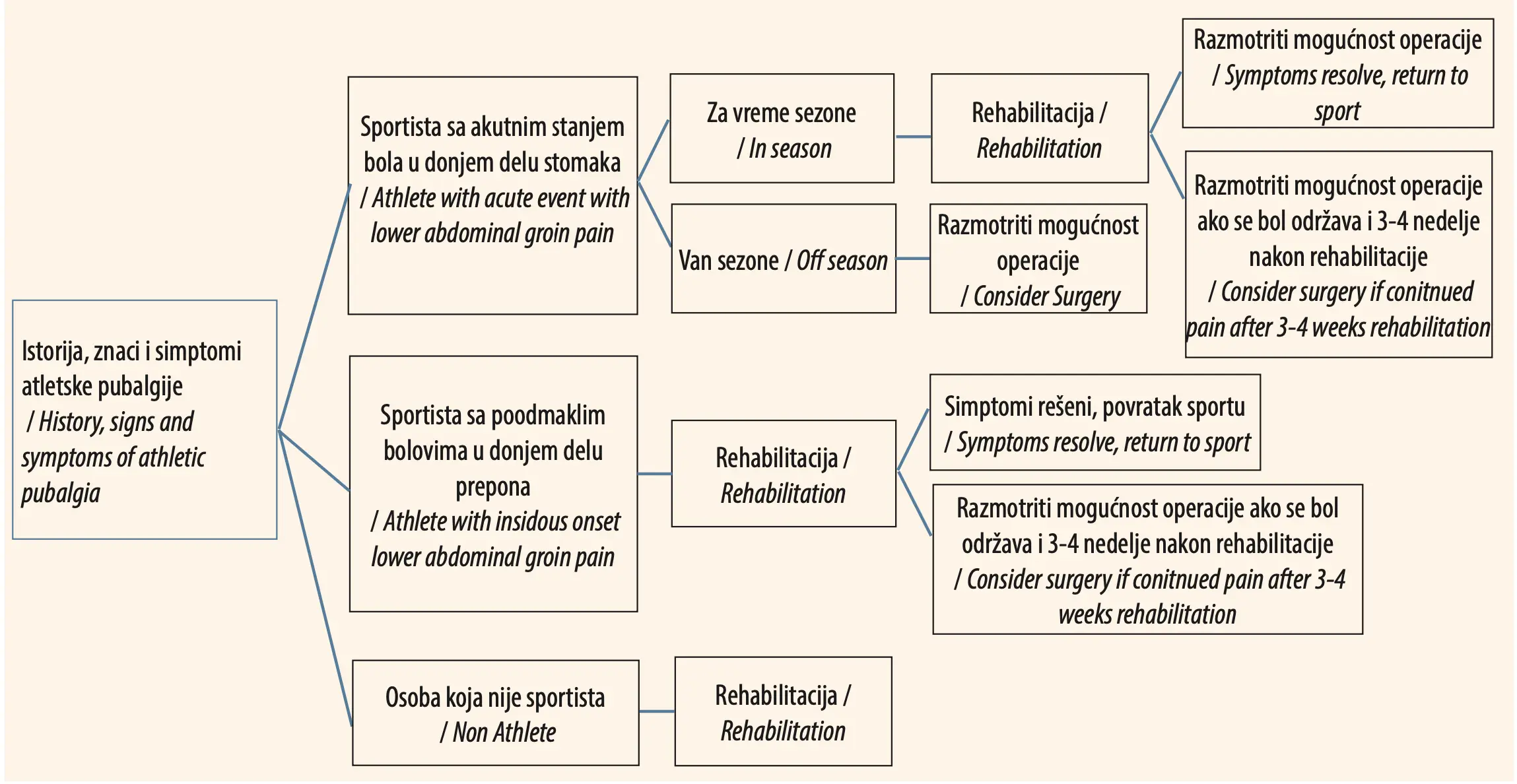
If none of the conservative procedures, applied for at least three months, does not yield the appropriate effect, the most invasive therapeutic approach – surgery, should be considered [20]. There are different surgical techniques for treating pubalgia, and for most of them satisfactory results have been recorded. The principles of surgical treatment include the procedures of strengthening the posterior wall and the fixation of the m. rectus abdominis or its insertion tendon. Another type of surgical treatment is laparoscopic surgery (fixation of a total extraperitoneal mesh behind the pubic bone and/or the posterior wall of the inguinal canal). Paajanen et al. reported that, in their study, laparoscopic surgery of pubalgia in athletes was more efficient than non-operative treatment [21]. After the surgery, the pain decreased after a month, while 90% of all the surgically treated athletes completely returned to their sports activities, after three months.
It is necessary to carry out surgical treatment acting on the principle of the earliest possible intervention, as the results are, in such cases, more favorable. In surgical treatment, it is necessary to balance the forces exerted by both muscular regions, which is known as the method of musculoaponeurotic treatment, used to strengthen the abdominal wall and relieve the insertions on the pubis. One of the best-known techniques of surgical treatment is carried out in keeping with the method – Plastica telmentis abdominalis et canalis inguinalis secundum Bassini cum modificatione Nešović, which enables the balancing of the strength of the pelvifemoral muscles, especially the adductors, and the strength of the anterior abdominal wall muscles. This technique was successfully presented by Dr. Branislav Nešović (Belgrade, Serbia), in 1967, who modified the classical Bassini technique of inguinal hernia surgery [8]. During the surgery it is necessary to close the inguinal canal and create new passageways for neurovascular elements in this region [21].
POSTOPERATIVE REHABILITATION
Rehabilitation after surgery requires gradual patient verticalization and strengthening of the target muscle groups. If recovery goes according to plan, football players return to the football pitch, and the best indicator of recovery is the absence of pain. Recurrence of the syndrome after surgery has practically never been registered, which is why the results achieved can be considered as complete success in the treatment of painful symphysis syndrome [10]. For each patient, especially for an athlete, it is important that cardiovascular endurance remains at the same level or that it improves after repeated testing. To that effect, each session needs to begin with certain cardiovascular exercises. The program of active training is superior in physical therapy treatment [14].
PREVENTION
Due to the complexity of the therapeutic approach to the painful symphysis syndrome, it is necessary to implement preventive measures in athletes, from the earliest age onwards. These preventive measures involve exercises for strengthening anterior abdominal wall muscles on the basis of an everyday training and exercising regimen, with special emphasis on the exercises for oblique and transverse abdominal muscles. Muscle contractions without weights are also very important. If the painful syndrome does develop, it is important to start conservative therapy accompanied by the appropriate physical therapy regimen, as early as possible.
CONCLUSION
Painful symphysis syndrome is a significant clinical state for athletes, with special emphasis on football players. The etiology of the development of pain in the groin stems from the conflict between the uneven muscular strength of neighboring regions – the anterior abdominal wall and the lower extremities, as well as from the disproportion of the strain endured by the different insertions in the symphysis region. As the result of the development of pubalgia, the athlete becomes incapacitated to perform sports activities for a longer period of time. In order to avoid such a state and enable the athlete to actively participate in training sessions and games in an adequate manner, the importance of prevention, as the basis and foundation of protecting the athlete’s health from this painful condition in the long run, must be emphasized. However, if even with all the precautions, pain still develops, the importance of timely and specific diagnostics directing the doctor towards the most effective approach, must not be overlooked.
Informations

-
Keywords:
-
Received:
-
Revised:
-
Accepted:
-
Online first:
-
DOI:
-
Cite this article:

Katarina Vukosavljević
College of Sports and Health
11 Toše Jovanovića Street, 11030 Belgrade, Serbia
E-mail:
1. Elattar O, Choi HR, Dills VD, Busconi B. Groin Injuries (Athletic Pubalgia) and Return to Play. Sports Health. 2016 Jul;8(4):313-23. doi: 10.1177/1941738116653711.[CROSSREF]
2. Meyers WC, Yoo E, Devon ON, Jain N, Horner M, Lauencin C, et al. Understanding “sports hernia” (athletic pubalgia): the anatomic and pathophysiologic basis for abdominal and groin pain in athletes. Operative techniques in sports medicine. 2008. 20(1):33-45.[HTTP]
3. Omar IM, Zoga AC, Kavanagh EC, Koulouris G, Bergin D, Gopez AG, et al. Athletic pubalgia and "sports hernia": optimal MR imaging technique and findings. Radiographics. 2008 Sep-Oct; 28(5):1415-38.[CROSSREF]
4. Dimitrakopoulou A, Schilders E. Sportsman's hernia? An ambiguous term. J Hip Preserv Surg. 2016 Feb 24;3(1):16-22. doi: 10.1093/jhps/hnv083.[CROSSREF]
5. Balconi G. US in pubalgia. J Ultrasound. 2011 Sep;14(3):157-66. doi: 10.1016/j. jus.2011.06.005.[CROSSREF]
6. Cavalli M, Bombini G, Campanelli G. Pubic inguinal pain syndrome: the so-called sports hernia. Surg Technol Int. 2014 Mar;24:189-94.
7. Morales-Conde S, Socas M, Barranco A. Sportsmen hernia: what do we know? Hernia. 2010 Feb;14(1):5-15. doi: 10.1007/s10029-009-0613-z.[CROSSREF]
8. Jevtić M, Vranić K, Zečević M. Rehabilitacija posttraumatskih i ortopedskih bolesnika. Kragujevac-Beograd: Medicinski fakultet univerziteta u Kragujevcu, 1995.
9. Garvey JF, Read JW, Turner A. Sportsman hernia: what can we do? Hernia. 2010 Feb;14(1):17-25. doi: 10.1007/s10029-009-0611-1.[CROSSREF]
10. Nesovic B. The painful symphysis syndrome in athletes and treatment possibilities Reflection on the topic after 20 years of personal experience. Aspetar Sports Med J. 2012 Dec; 3:254–7.
11. Gerhardt MB, Brown JA, Giza E. Occult groin injuries: athletic pubalgia, sports hernia, and osteitis pubis. Practical orthopedics, sports medicine and arthroscopy, ed. DA Johnson and RA Pedowitz 2006. Baltimore, MD: Lippincott Williams & Wilkins, 531-45.
12. Janković S, Delimar D, Hudetz D. Sindrom bolne prepone: The groin pain syndrome. Arh Hig Rada Toksikol. 2001 Dec; 52(4):421-8.
13. Kachingwe AF, Grech S. Proposed algorithm for the management of athletes with athletic pubalgia (sports hernia): a case series. J Orthop Sports Phys Ther. 2008 Dec;38(12):768-81. doi: 10.2519/jospt.2008.2846.[CROSSREF]
14. Ellsworth AA, Zoland MP, Tyler TF. Athletic pubalgia and associated rehabilitation. Int J Sports Phys Ther. 2014 Nov;9(6):774-84.[HTTP]
15. Swan KG Jr, Wolcott M. The athletic hernia: a systematic review. Clin Orthop Relat Res. 2007 Feb;455:78-87. doi: 10.1097/BLO.0b013e31802eb3ea.[CROSSREF]
16. Minnich JM, Hanks JB, Muschaweck U, Brunt LM, Diduch DR. Sports hernia: diagnosis and treatment highlighting a minimal repair surgical technique. Am J Sports Med. 2011 Jun;39(6):1341-9. doi: 10.1177/0363546511402807.[CROSSREF]
17. Koutserimpas C, Ioannidis A, Konstantinidis MK, Makris MC, Antonakopoulos F, Mazarakis A, et al. Insights in clinical examination and diagnosis of Athletic Pubalgia. G Chir. 2020 Jan-Feb;41(1):131-5.
18. Serner A, van Eijck CH, Beumer BR, Hölmich P, Weir A, de Vos RJ. Study quality on groin injury management remains low: a systematic review on treatment of groin pain in athletes. Br J Sports Med. 2015 Jun;49(12):813. doi: 10.1136/ bjsports-2014-094256.[CROSSREF]
19. Cohen B, Kleinhenz D, Schiller J, Tabaddor R. Understanding Athletic Pubalgia: A Review. R I Med J (2013). 2016 Oct 4;99(10):31-5.
20. Kajetanek C, Benoît O, Granger B, Menegaux F, Chereau N, Pascal-Mousselard H, et al. Athletic pubalgia: Return to play after targeted surgery. Orthop Traumatol Surg Res. 2018 Jun;104(4):469-72. doi: 10.1016/j.otsr.2018.01.012.[CROSSREF]
21. Paajanen H, Brinck T, Hermunen H, Airo I. Laparoscopic surgery for chronic groin pain in athletes is more effective than nonoperative treatment: a randomized clinical trial with magnetic resonance imaging of 60 patients with sportsman's hernia (athletic pubalgia). Surgery. 2011 Jul;150(1):99-107. doi: 10.1016/j.surg.2011.02.016.[CROSSREF]