


The importance of multidetector computed tomography in acute stroke detection, monitoring and selection of therapy
- University Clinical Center of Serbia, Center for Radiology and Magnetic Resonance Imaging, Belgrade, Serbia
- University of Belgrade, Faculty of Medicine, Belgrade, Serbia
ABSTRACT
Introduction: A stroke is a sudden, focal nonconvulsive neurological dysfunction which occurs due to vascular damage.
Aim: The aim of the study was monitoring all patients who reported to the Emergency Center due to some neurological deficit, within a particular period of time.
Materials and methods: For a period of one month, we monitored 123 patients who were admitted to the Department of Emergency Diagnostics with some form of neurological deficit and who underwent a native computed tomography (CT) scan and/or angiographic examination of the endocranium. All examinations were performed with the GE BrightSpeed 16 Slice CT scanner (USA). After initial native CT diagnostics, depending on the finding, the examination was either stopped, or the contrast dye was administered and postcontrast examination was performed or CT angiography was carried out.
Results: Most of the patients with vascular neurological deficit were between 50 and 59 years old, while a smaller number of patients was registered in the age group of persons younger than 40 years. Most of the female patients were in the 50 – 59 age group, while most of the male patients were between 40 and 49 years old. Of the 94 patients who developed stroke, 78 (83%) patients had ischemic stroke, while 16 (17%) patients suffered hemorrhagic stroke. There is no statistically significant difference in the occurrence of intracerebral hemorrhage and subarachnoid hemorrhage between the sexes. The localization of ischemic brain infarction was in the vascular territory of the anterior cerebral artery (ACA) – 3.2% of the cases, the middle cerebral artery (MCA) – in 38.9% of the cases, the internal carotid artery (ICA) – in 8.4% of the patients, the basilar artery (BA) – in 13.7% of the cases, the posterior cerebral artery (PCA) – in 7.4% of the patients, the vertebral artery (VA) – 9.5% of the cases, and the supratentorial watershed areas of arterial irrigation – in 11.7% of the patients.
Conclusion: Native CT examination is the golden standard for the triage of patients with acute stroke. The advantages of using a CT scan in the assessment of patients with acute stroke are that it is practical, precise, quick and available. CT imaging has prognostic value as well, as it can predict the response to the administered thrombolytic therapy.
INTRODUCTION
Acute stroke is the second most common cause of death in the world and the first most common cause of disability in developed countries. It is estimated that one person suffers stroke every 45 seconds, while there is one death related to acute stroke every three minutes, in the world. The incidence of stroke in developed countries is 100 – 150 cases per 100,000 population, per year. In Serbia, however, it is significantly higher and is estimated at 300 cases per 100,000 population, per year. Only around a half of the surviving patients remain independent after stroke, while 30% are incapable of living without assistance. A significant number of patients, around 20%, suffer a repeated stroke [1].
A stroke is a sudden, focal nonconvulsive neurological dysfunction which occurs due to vascular damage. The basic characteristic of stroke is sudden onset of neurological symptoms. However, neuro-visualization techniques (CT – computed tomography; MRI – magnetic resonance imaging) can also reveal so called “silent brain infarcts”, which develop without clear clinical manifestation.
Stroke is classified according to the pathology which is at the core of the focal brain damage. Therefore, we recognize two types of stroke – ischemic and hemorrhagic stroke [2]. The occurrence of a headache, vomiting, epileptic seizures, or coma, indicate, with a higher degree of certainty, that the cause of stroke is hemorrhage, however, the only reliable method of definitively determining the type of stroke is scanning the head with a CT scanner.
The contemporary concept of stroke as an emergency, requires a fast, focused, and precise diagnosis, on which not only the type of therapeutic approach is dependent, but also the prognosis of the illness. The recommended method and the method of choice in emergency neurological states is the native CT scan, which enables the exclusion of other, nonvascular causes of neurological deficit and the selection of patients eligible for different emergency therapeutic procedures (e.g., administering thrombolytic and other reperfusion therapy options, performing emergency endovascular procedures) [3].
The aim of the study was monitoring all patients who reported to the Emergency Center due to some neurological deficit, within a particular period of time. Based on the obtained results, we can draw conclusions on the advantages and disadvantages of early multidetector computed tomography (MDCT) examination within the diagnostics of acute stroke.
MATERIALS AND METHODS
During the period between December 1, 2017 and January 1, 2018, we monitored 123 patients who were admitted to the Department of Emergency Diagnostics of the University Clinical Center of Serbia with some form of neurological deficit and who underwent a native computed tomography (CT) scan and/or angiographic examination of the endocranium. All examinations were performed with the GE BrightSpeed 16 Slice CT scanner (USA). After initial native CT diagnostics, depending on the finding, the examination was either stopped, or the contrast dye was administered and postcontrast examination was performed or CT angiography was carried out.
The results were processed with methods of descriptive (mean, median, mode) and analytical (X2 test) statistics.
RESULTS
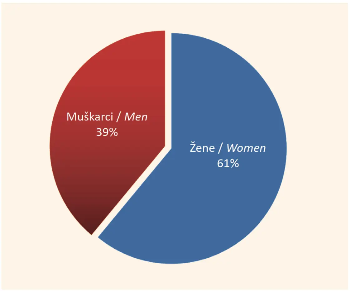
During the period between December 1, 2017 and January 1, 2018, 123 patients were admitted to the Department of Emergency Diagnostics of the University Clinical Center of Serbia with some form of neurological deficit. Of the 123 patients, in 20 patients nonvascular pathology was discovered on the CT scan. As presented in Figure 1, the female sex was dominant, at a ratio of 1.5:1 (Figure 1).
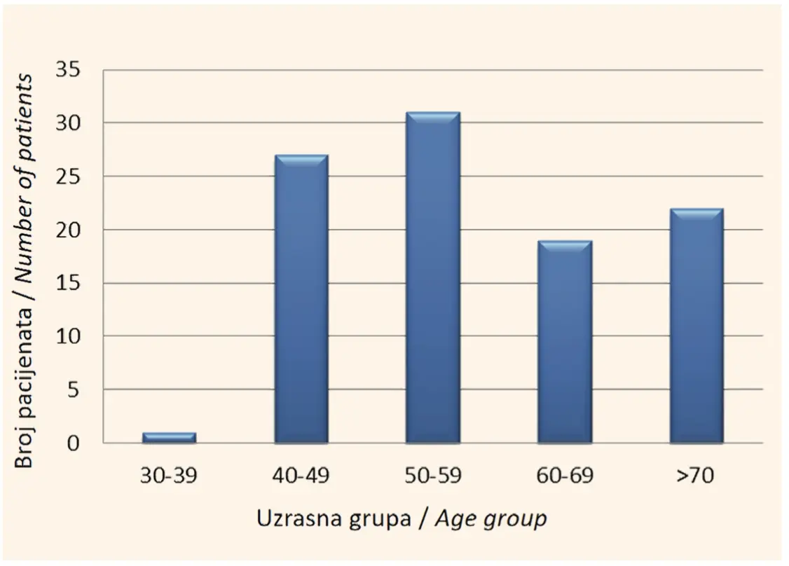
The youngest patient was 35 years old, while the oldest patient was 94 years old. It has been observed that the greatest number of patients were in the 50 – 59 age group, followed by patients in their forties, and then those in their seventies and their sixties, with a similar frequency amongst the said groups, while the smallest number of patients was registered in the group of patients younger than 40 years (Figure 2).
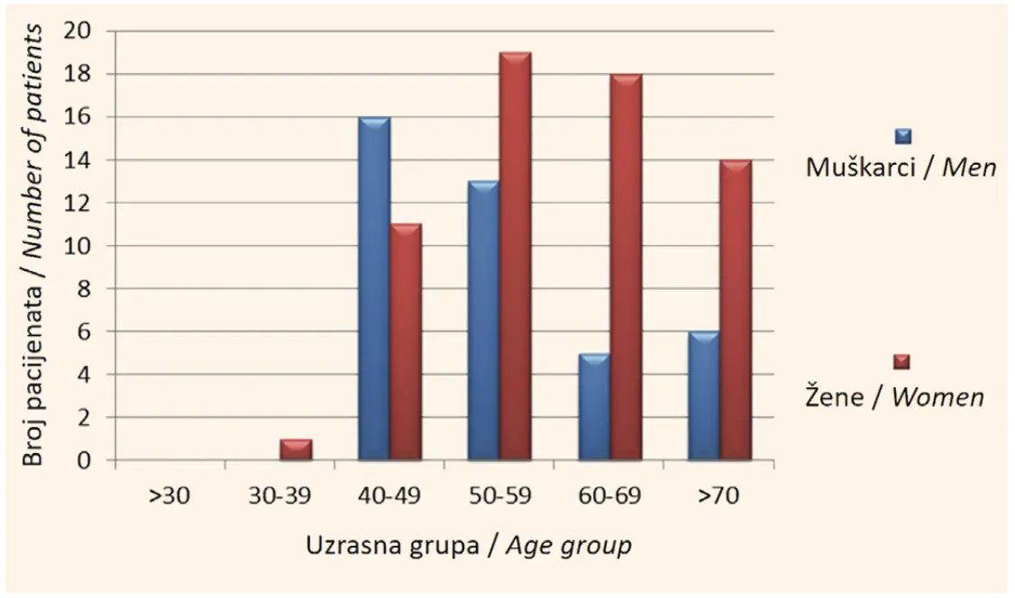
The greatest number of female patients was in the age group 50 – 59 years, while the greatest number of male patients belonged to the age group 40 – 49 years (Figure 3).
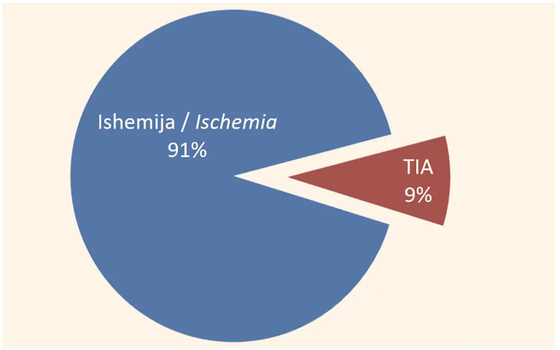
Of the 103 patients with a neurological deficit of vascular etiology, 94 (91%) patients developed stroke. In the remaining 9%, the deficit in question was transient ischemic attack (TIA) (Figure 4).
Of the 94 patients who suffered stroke, 78 (83%) patients sustained ischemic stroke, while 16 (17%) patients had hemorrhagic stroke (Figure 5).

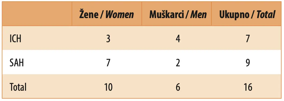
Of the 16 patients with hemorrhagic stroke, there were 10 (62.5%) women, while 6 (37.5%) patients were men. Intracerebral hemorrhage (ICH) was found in 7 (43.75%) patients, of whom 3 were women, while 4 were men. A total of 9 (56.25%) patients suffered subarachnoid hemorrhage (SAH) – 7 women and 2 men (Table 1). There was no statistically significant difference in the occurrence of ICH or SAH between the sexes, p = 0.152.
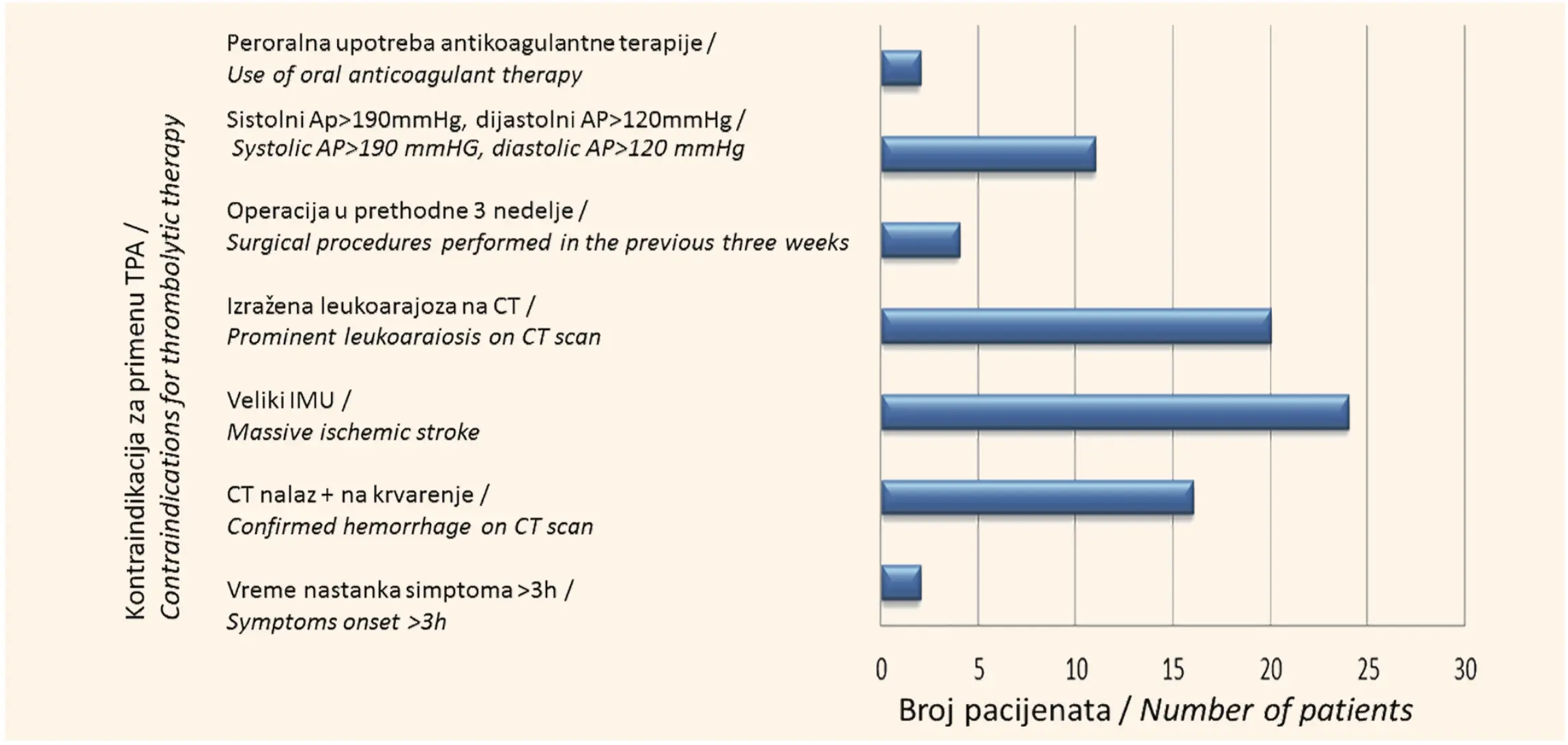
Of all the patients with vascular etiology, thrombolytic therapy was administered in 24 (23.3%) persons, while 79 (76.7%) patients had contraindications for the application of this therapy. The most frequent contraindications were a developed ischemic zone visible on CT (in 25.3% of the cases) and intracerebral hemorrhage visible on CT (in 20.3% of the cases). After intravenous administration of thrombolytic therapy, intracerebral hemorrhage with a consequent lethal outcome developed in 5 (0.2%) patients (Figure 6).
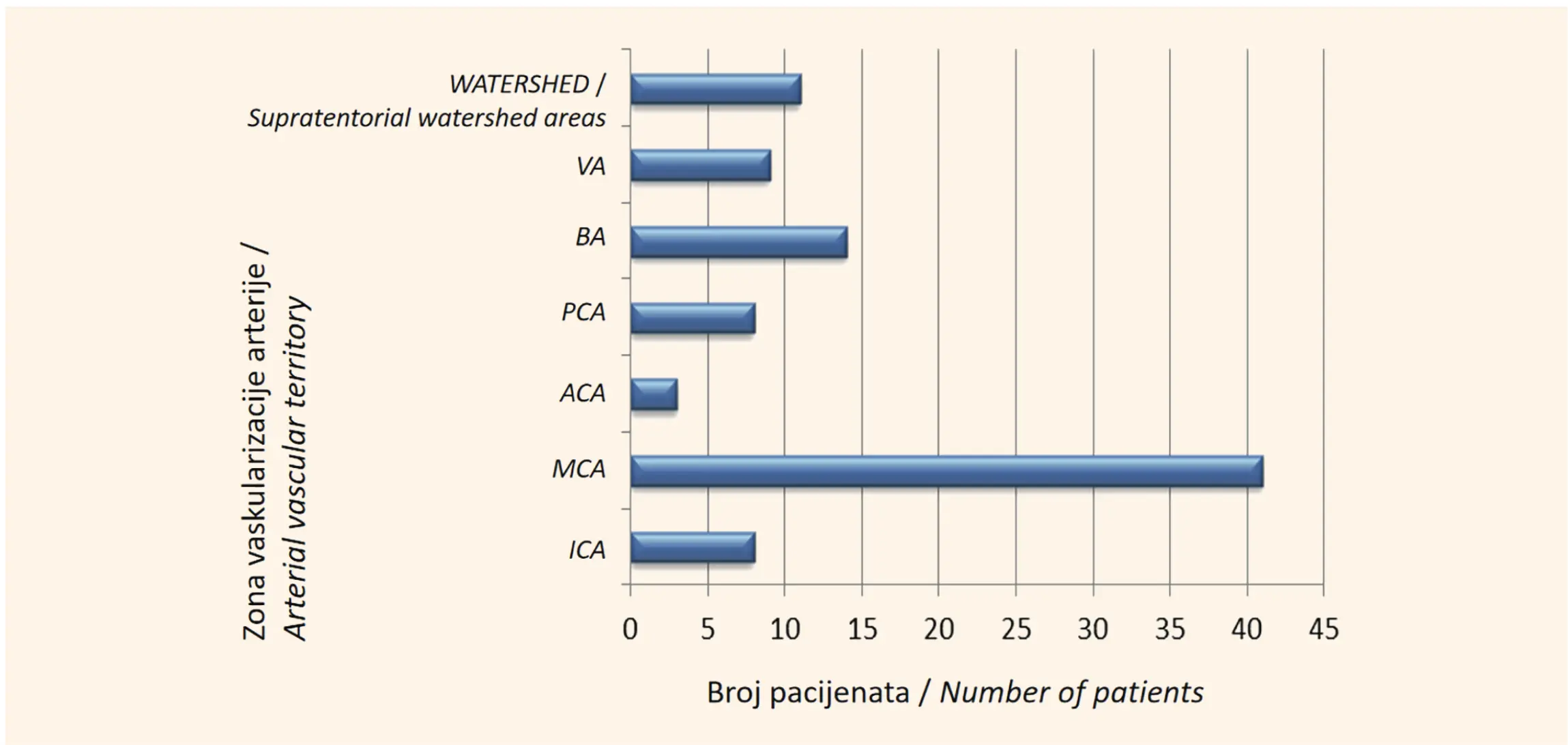
The localization of ischemic brain infarction was in the vascular territory of the anterior cerebral artery (ACA) – 3.2% of the cases, the middle cerebral artery (MCA) – in 38.9% of the cases, the internal carotid artery (ICA) – in 8.4% of the patients, the basilar artery (BA) – in 13.7% of the cases, the posterior cerebral artery (PCA) – in 7.4% of the patients, the vertebral artery (VA) – 9.5% of the cases, and the supratentorial watershed areas of arterial irrigation – in 11.7% of the patients (Figure 7).
DISCUSSION
Of the 123 patients with some form of neurological deficit who were included in the study, 103 patients had a deficit of a vascular origin. Of those 103 patients, 61% were female. Appelros et al. showed the opposite results in their study [1],[2],[3]. The greatest number of female patients was in the age group between 50 and 59 years, followed by patients in their sixties and those in their seventies, while the percentage of female patients who were younger than 50 and suffering from some form of neurological deficit was significantly lower. This can be explained by the fact that women in menopause are at a higher risk of developing cardiovascular disease [4]. Also, in women, 10 years after the onset of menopause, the risk of stroke doubles. The decerase in the level of endogenous estrogen by 60% leads to an excess of androgens, which may lead to increased risk factors for the development of cardiovascular diseases. On the other hand, the greatest number of male patients was in the age group 40 – 49 years, which may indicate risk factors related to lifestyle, which include smoking, excess alcohol intake, drug abuse, physical inactivity and obesity, an unhealthy diet, and stress [5]. Also, hypertension, elevated cholesterol levels, diabetes, and cardiac diseases, lead to stroke.
Acute ischemic stroke (AIS) occurs as the result of blood vessel occlusion, either by a thrombus or embolus. This type of stroke is significantly more frequent and is diagnosed in 83% of patients, while acute hemorrhagic stroke, which can have two types – intracerebral hemorrhage (ICH) and subarachnoid hemorrhage (SAH), is diagnosed in the remaining 17% of patients. According to the latest studies, ischemic stroke includes thrombosis, emboli, venous thrombosis, and systemic hypoperfusion. The most frequent shared risk factor for ICH and SAH are hypertension, smoking and alcohol abuse [6].
The present study included 37.5% of men and 62.5% of women with hemorrhagic stroke, but we did not find a highly statistically significant difference in the occurrence of ICH and SAH between the sexes. Other studies state that the distribution by gender in hemorrhagic stroke is similar, however, probably due to the link between the occurrence of SAH and ICH and pregnancy and puerperium, but also with the use of oral contraceptives, most studies report a higher frequency of these entities in female patients [7]. Some authors, however, report between 1.4 and 2.0 times more frequent cerebral hemorrhage in men [6].
The obtained results indicate a higher frequency of ischemic infarction in the watershed area of the carotid artery vascular territory (54.7%), as compared to the arteries in the watershed area of the vertebrobasilar territory (45,3%). Infarctions in the vascular territory of the middle cerebral artery (MCA) are the most common type of stroke, which has been demonstrated in this study. They may be superficial (involving the cortex and white matter), deep (involving the basal ganglia, internal capsule, and deep white matter), and combined [8]. These infarctions are most commonly caused by cardioembolism, thrombosis of the internal carotid artery (ICA), dissection, or embolism.
All 24 candidates for thrombolytic therapy had thrombotic masses present in the large arteries of the brain, while in 11 candidates for thrombolytic therapy, who had a positive sign of hyperdense artery on the native CT scan, angiographic examination revealed the presence of a thrombus, i.e., a filling defect. The main clinical role of CT angiography in acute stroke is excluding thrombolytic therapy in patients with embolic stroke who do not have large occlusions of the blood vessels, as is the case in lacunar infarct, transient ischemic attack (TIA), migraines, hypoglycemia. Computed tomography angiography (CTA) is very precise in diagnosing intracranial thrombi. In a study including 44 candidates for thrombolysis, who had undergone both CTA and digital subtraction angiography (DSA) within the diagnostic algorithm, CTA showed high sensitivity (98.4%) and specificity (98.4%) in detecting large thrombi in large blood vessels [9]. Additionally, the degree and level of occlusion are important factors in planning the treatment in acute stroke. CTA is also used for serial follow-up of patients with proven occlusions of the internal carotid artery (ICA).
CONCLUSION
Stroke occurs as the result of changes developing in the blood vessels of the brain. Around 80% of strokes are the result of the interruption of blood supply to the brain (ischemic stroke), while around 20% of strokes occur due to hemorrhage in the brain (hemorrhagic stroke).
Despite new technological advances in neuroimaging techniques and protocols for the assessment and treatment of patients with cerebrovascular diseases, native CT remains the golden standard for the tirage of patients with acute stroke. The advantages of using a CT scan in the assessment of patients with acute stroke are that it is practical, precise, quick and available. CT imaging has prognostic value as well, as it can predict the response to the administered thrombolytic therapy. CT angiography is precise in diagnosing intracranial vascular occlusion in patients with symptoms of stroke, within the first 6 hours. Data obtained with CT angiographic examination are valuable, as they enable quick diagnostics and appropriate treatment within a short period of time. CTA is a quick, simple, and precise diagnostic method, which provides enough information to the neurologist with regards to the vascular passability of the affected artery.
Informations

-
Keywords:
-
Received:
-
Revised:
-
Accepted:
-
Online first:
-
DOI:
-
Cite this article:

Kristina Davidović
Center for Radiology and Magnetic Resonance Imaging, University Clinical Center of Serbia
2 Pasterova Street, 11000 Belgrade, Serbia
E-mail:
1. Appelros P, Stegmayr B, Terént A. Sex differences in stroke epidemiology: a systematic review. Stroke. 2009 Apr;40(4):1082-90. doi: 10.1161/STROKEAHA.108.540781.[CROSSREF]
2. Ayala C, Croft JB, Greenlund KJ, Keenan NL, Donehoo RS, Malarcher AM, et al. Sex differences in US mortality rates for stroke and stroke subtypes by race/ethnicity and age, 1995-1998. Stroke. 2002 May;33(5):1197-201. doi: 10.1161/01.str.0000015028.52771.d1.[CROSSREF]
3. Khaw KT. Epidemiology of stroke. J Neurol Neurosurg Psychiatry. 1996 Oct;61(4):333-8. doi: 10.1136/jnnp.61.4.333.[CROSSREF]
4. Lisabeth L, Bushnell C. Stroke risk in women: the role of menopause and hormone therapy. Lancet Neurol. 2012 Jan;11(1):82-91. doi: 10.1016/S1474- 4422(11)70269-1.[CROSSREF]
5. Turanjanin N. Učestalost zloupotrebe alkoholnih pića kod bolesnika sa ishemijskim moždanim udarom. Acta medica Medianae. 2012;51(2):5-10.[HTTP]
6. Simić D, Jovićević M, Slankamenac P, Žarkov M, Božić K. Učestalost i faktori rizika kod hemoragičnog moždanog udara. Aktuelnosti iz neurologije, psihijatrije i graničnih područja. 2008;16(1-2):19-29.
7. Pekmezovic T, Tepavcevic DK, Jarebinski M, Kostic M, Bumbasirevic L. Stroke mortality in Belgrade, Serbia: age, period, and cohort analyses. Cerebrovasc Dis. 2007;24(2-3):191-5. doi: 10.1159/000104476.[CROSSREF]
8. Bogousslavsky J, Caplan LR. Stroke syndromes, 2nd ed. Cambridge University Press, Cambridge, 2001.[HTTP]
9. Lev MH, Farkas J, Rodriguez VR, Schwamm LH, Hunter GJ, Putman CM, et al. CT angiography in the rapid triage of patients with hyperacute stroke to intraarterial thrombolysis: accuracy in the detection of large vessel thrombus. J Comput Assist Tomogr. 2001 Jul-Aug;25(4):520-8. doi: 10.1097/00004728- 200107000-00003.[CROSSREF]
- Figure 1. Number of patients with symptoms of stroke, by sex
- Figure 2. Number of patients, by age groups
- Figure 3. Number of patients, by age groups, in relation to sex
- Figure 4. Number of patients with neurological symptoms with a vascular etiology
- Figure 5. Ratio of patients with ischemic stroke vs. patients with hemorrhagic stroke
- Table 1. Frequency of the occurrence of the types of hemorrhagic stroke, by sex
- Figure 6. Number of patients in relation to the causes of contraindications for the application of thrombolytic therapy
- Figure 7. Number of patients in relation to the localization of ischemic stroke