


Absence of the celiac trunk: a case report
- Institute for Public Health Pančevo, Pančevo, Serbia
- General Hospital Pančevo, Pančevo, Serbia
- University Hospital Center "Dr Dragiša Mišović - Dedinje", Belgrade, Serbia
ABSTRACT
Introduction: The celiac trunk is an arterial trunk that has been the subject of numerous anatomical and radiological studies. From its origin, at the front of the abdominal aorta, the trunk extends for 1.5 – 2 cm and then divides into three branches: the left gastric artery, the common hepatic artery and the splenic artery. This type of trifurcation was first described by Haller, in 1756, and is considered the most common anatomical pattern. Anatomical variations of the celiac trunk are numerous, both in terms of branching and dimensions. The complete absence of the celiac trunk is a rare anatomical variation, wherein all three branches arise directly and independently from the abdominal aorta.
Case report: A 63-year-old oncology patient was referred to the radiology department for an MR examination of the abdomen, in order to differentially diagnose changes in the adrenal glands. During the examination, complete absence of the celiac trunk was observed. The left gastric artery, splenic artery, and common hepatic artery arose directly and independently from the abdominal aorta.
Conclusion: A variation of this type is mostly asymptomatic and has no negative effect on the blood supply of visceral organs. In literature, the prevalence of this vascular pattern ranges from 0.1% to 4%. Knowledge of vascular variations in the abdomen is very important during surgical, diagnostic and endovascular procedures.
INTRODUCTION
The celiac trunk (truncus coeliacus; CT) or Haller’s tripod (tripus Halleri), arises from the abdominal aorta as its first unpaired lateral visceral branch. It vascularizes all abdominal organs situated in the supramesocolic space of the abdominal cavity – the abdominal segment of the esophagus, the gaster, a part of the duodenum and of the pancreas, the liver with bile ducts, the spleen, as well as the peritoneal attachments of these organs.
The celiac trunk is an anterior branch of the abdominal aorta, arising from this artery at the level of the intervertebral disk located between the twelfth thoracic vertebra and the first lumbar vertebra, or less commonly, at the level of the superior margin of the first lumbar vertebra. Very rarely, it is located above or below the said vertebrae. Due to atherosclerotic changes and the elongation of the aorta, the celiac trunk branches off at a lower point more often in the elderly, than in younger persons [1].
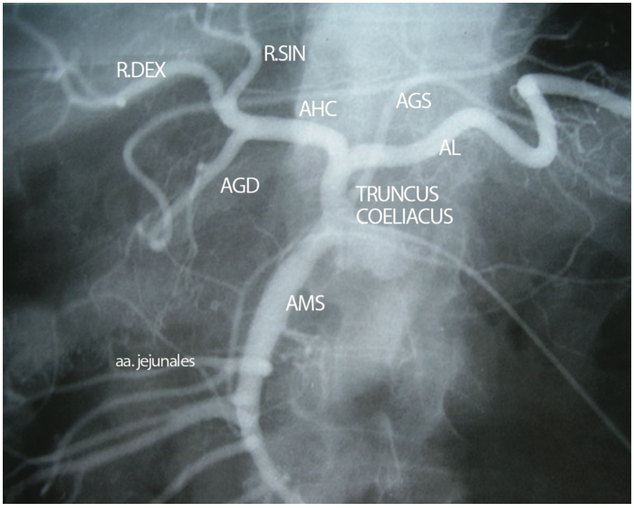
From its proximal end, on the anterior aspect of the abdominal aorta, the celiac trunk extends 1.5 – 2 cm, before dividing into three branches: the left gastric artery (a. gastrica sinistra – AGS), the common hepatic artery (a. hepatica communis – AHC), and the splenic artery (a. lienalis – AL).
This typical trifurcation of the celiac trunk was first described by Haller, in 1756, which is why it is also known as Haller’s tripod (tripus Halleri), and is considered to be the normal, most common form (Figure 1).
In addition to these three typical branches, other, i.e., unusual branches of the celiac trunk can often be seen, such as: the dorsal pancreatic artery (a. pancreatica dorsalis), the left inferior phrenic artery (a. phrenica inferior sinistra) or the right inferior phrenic artery (a. phrenica inferior dextra), the middle colic artery (a. colica media), as well as the a. colica media accesoria [2].
For centuries after Haller’s discovery, the arterial variations of the aorta and of the celiac trunk have attracted the attention of anatomists, clinicians, and radiologists, primarily because of their great significance in many surgical procedures. The celiac trunk is one of the best documented arterial trunks; it has been studied by many researchers and many studies have been performed, involving an impressive number of samples, related to this anatomical structure [3].
The anatomical variations of the celiac trunk are numerous, both as to the way that it branches further and as to its dimensions. The terminal end of the celiac trunk may be in the form of a trifurcation, or one of the three arteries it branches into may arise as a separate lateral branch (this is most commonly the left gastric artery), while the remaining two arteries branch off together, i.e., they form a bifurcation. Each one of the said terminal arteries may arise directly from the abdominal aorta. The celiac trunk and the superior mesenteric artery may branch off via a joint trunk from the aorta, thus creating the celiac-mesenteric trunk. Also, each of the terminal branches of the celiac trunk may form a common trunk with the superior mesenteric artery. Additional, i.e., accessory or replaced hepatic arteries may stem from the celiac trunk, in which cases the celiac trunk has, not just three, but four or even five branches [4].
The first classification of the anatomical variations of the celiac trunk and the superior mesenteric arteries was published by Adachi, in 1928, in the form of a comparative study, wherein he presented the results of his very precise investigation of the anatomical variations of the prehepatic arterial system, performed on 150 cadavers of different age [5]. According to this classification, the celiac trunk has several anatomical variations: hepato-gastro-splenic, hepato-splenic, gastro-splenic, celiac-mesenteric, hepato-spleno-mesenteric, and hepato-mesenteric trunk.
The absence of the celiac trunk was not described in Adachi’s classification. In 1997, Uflacker classified eight celiac trunk types, including all the previously established types, but with two more additional variants – the absence of the celiac trunk (CT agenesis) and the celiac-colic trunk [6].
Subsequent classifications additionally systematized the anatomical variations of the celiac trunk. Thus, Morita’s classification defines five types of variations for the celiac trunk and four types (with ten forms) for the celiac-mesenteric trunk - (I) the celiac trunk, (II) the hepato-splenic trunk, (III) the gastro-splenic trunk, (IV) the hepato-gastric trunk, and (V) the absence of the celiac trunk [7].
The absence of the celiac trunk is defined as an anatomical variant wherein the left gastric artery, the splenic artery, and the common hepatic artery arise directly and independently from the abdominal aorta. This anatomical vascular variant was first described by Saint-Hilaire, in 1832. The anatomical variations of the celiac trunk are the result of developmental changes to the ventral splanchnic arteries. The splanchnic arteries are formed by the fusion of the paired ventral segmental arteries. It is believed that, when the celiac trunk is formed, segmental arteries fuse via longitudinal anastomoses. The regression of the longitudinal anastomoses and the persistence of the root of the segmental arteries later result in the absence of the celiac trunk [8].
CASE PRESENTATION
A 63-year-old patient, with a history of oncological disease (three years prior to the examination described in this paper, the patient had undergone right upper lobectomy and partial resection of the right third and fourth ribs, due to squamous cell lung carcinoma), was referred to the radiology department for abdominal magnetic resonance imaging (MRI), for the purpose of establishing a differential diagnosis of lesions in the suprarenal glands, observed during previous computed tomography (CT) examinations.
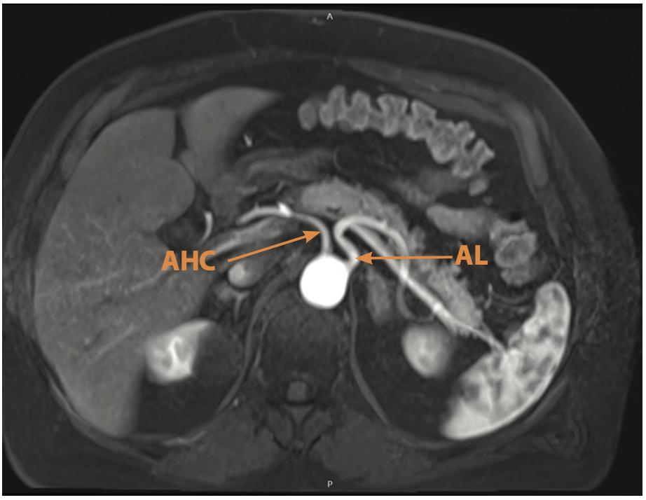
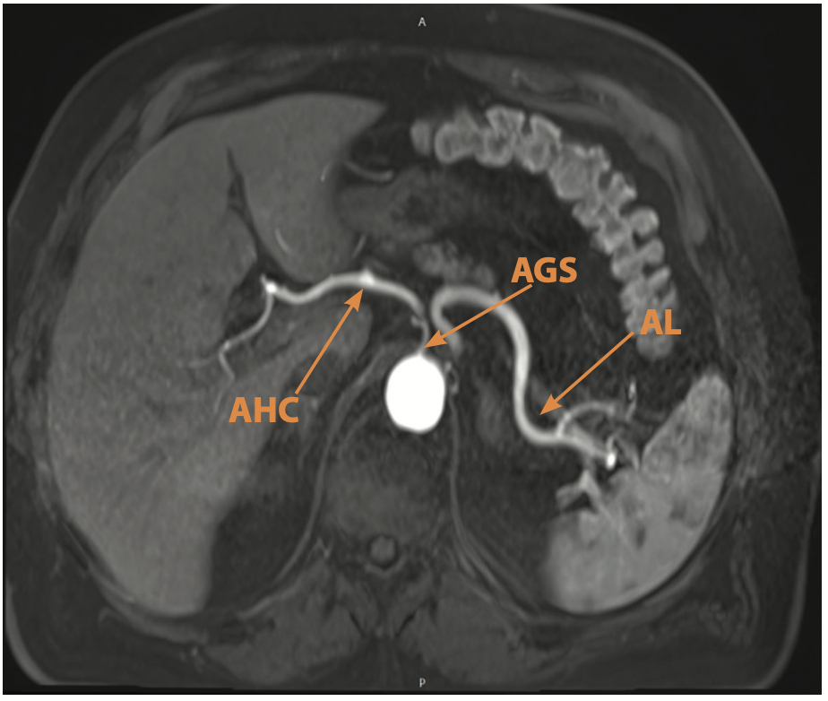
On MRI examination, complete absence of the celiac trunk was observed, while the left gastric artery, the splenic artery, and the common hepatic artery arose, directly and independently, from the abdominal aorta (Figure 2 and Figure 3).
DISCUSSION
The absence of the celiac trunk is a rare anomaly, and, in existing literature, authors have reported a varying prevalence of this aberration.
According to Iacob et al., there have been only 31 cases of reported celiac trunk absence worldwide. Around one third of these cases were detected in studies where modern radiological techniques had been applied, while the remainder of these variations were detected during anatomical dissections [9].
The data from a study by Higashi et al. illustrate to what extent this variation is indeed rare. The authors reported that, in Japan, until the year 1995, only six such cases had been reported [10].
In their study, Vandamme and Bonte reported the presence of such an anatomical form of the celiac trunk in only 0.1% [11] of the cases. Torres et al. obtained the same result (0.1%), based on 1,569 CT scans [12].
According to Matusz et al., agenesis of the celiac trunk was observed in only 0.19% of the cases, which was determined on the basis of a large series of 10,750 cases from nineteen studies, which involved anatomical dissections, surgical procedures, and radiological studies [13]. Araujo et al. concluded that that the absence of the celiac trunk trifurcation was rare, as they observed it in only 0.2% of the population, in their paper [14].
The meta-analysis performed by Whitely et al., in 2020, included 36 studies, which investigated the variations of the celiac trunk with multidetector computed tomography (MDCT). A total of 17,391 subjects, from fourteen countries and four continents, was included in this meta-analysis, and the overall calculated prevalence of anatomical variations of the celiac trunk was 10.85%. The prevalence of the absence of the celiac trunk was 0.28% [15].
In a systematic review of the anatomical variations of the celiac trunk, Panagouli et al. searched through four databases and collected 36 studies that had been performed by that time, with a total of 12,196 cases. They reported finding the typical trifurcation of the celiac trunk in 89.42% of cases, and also concluded that the least frequently found variation was indeed the absence of the celiac trunk, found in only 0.38% of the cases [16].
On the basis of data obtained from 240 CT angiography images, Kurcz et al. concluded that the prevalence of this anatomical variation was slightly higher, amounting to 0.42% [17].
Bergman et al. published a meta-analysis on celiac trunk arterial variations and, based on the data of eight authors who had researched 756 samples, they assessed the prevalence of the absence of the celiac trunk to be 0.4% [18].
In their study, involving a total of one hundred patients who had undergone MDCT angiography of the abdominal aorta for different medical reasons, Ugurel et al. recorded the absence of the celiac trunk in only one case (1%) [19].
The same percentage was recorded by Cankal et al, in their study. Out of a total of 200 patients diagnosed with colorectal carcinoma, who had undergone MSCT imaging for the purpose of surgical procedure and follow-up of their oncological disease, the absence of the celiac trunk was registered in 1% of the cases [20].
In a Japanese study by Shoumura et al., which involved 184 cadavers, the absence of the celiac trunk was found in 2% of the cases [21].
In a cadaveric study, published by Olewnik et al., in 2017, the prevalence of celiac trunk agenesis was 2.5% [22].
In a morphometric study of the celiac trunk, from 2013, based on the data obtained from the dissection of 77 adult cadavers, Venieratos et al. found the said variation in 2.6% of cases [23].
In a study of anatomical variations of the celiac trunk, performed on cadavers, Pushpalatha et al. found that in two (4%), out of a total of 50 cases, the celiac trunk was absent, while all three major branches independently arose from the abdominal aorta [24].
In a study performed in India, on 50 patients who had undergone CT angiography due to medical or surgical indications, Garima et al. also reported a 4% prevalence of this variation [25].
Prakash et al. found, in a sample of 50 cadavers, two (4%) cases with all three celiac trunk arteries arising separately and directly from the abdominal aorta, while in a total of 14% of the cadavers, the presence of some form of anatomical variation of the celiac trunk was discovered [26].
The absence of the celiac trunk is usually asymptomatic. This type of variation most commonly has no negative effect on blood supply to the visceral organs. In fact, the independent origin of the common hepatic, left gastric and splenic artery, directly from the abdominal aorta, may even prevent severe visceral ischemia, which occurs in case of massive thromboembolism of the celiac trunk [27].
However, in a certain number of cases, this variation may lead to the development of symptoms. Mehrnahad et al. described a female patient in whom a CT scan revealed the absence of the celiac trunk, with the common hepatic artery and the splenic artery arising directly from the abdominal aorta. The narrowing and angulation of these blood vessels occurred because they were being compressed by the median arcuate ligament (ligamentum arcuatum mediale), which resulted in the patient having a long history (since early youth) of abdominal pain, which was particularly pronounced postprandially [28].
Understanding the variations regarding the origin, path, and patterns of CT branching is not only of interest anatomically and embryologically, but also of great clinical significance, since these variations may be the cause of pathological conditions. The understanding of these variations is also significant when dealing with patients undergoing diagnostic angiography because of gastrointestinal bleeding, as well as before surgical procedures. Recognizing anatomical variants becomes an obligatory step in planning surgical interventional procedures [29].
Consequently, knowledge of the anatomical variations of the CT is important for surgeons in liver transplants, in laparoscopic surgery, in radiological abdominal procedures, as well as in cases of abdominal injury. Also, knowledge of the variations of the celiac trunk and of its complete absence may be useful in planning and performing radiological procedures, such as celiacography and chemoembolization of liver tumors. In such situations these arterial variations are very important and must not be overlooked. They significantly increase the difficulties and risk in radical gastrectomy [30].
In recent years, MDCT has been used for researching vascular anatomy for scientific and diagnostic purposes. Such studies provide much larger samples than the traditional cadaveric studies [15]. The authors of such a radiological study, which included 126 angiography images, in Turkey, in the period between 2014 and 2016, stated that, in their research of celiac trunk morphometric characteristics and branching patterns, they did not find a single individual with complete absence of the celiac trunk [31]. A similar study, carried out in India, which was based on fifty angiography images of the abdominal aorta, also found no such cases [32]. Koshariya et al. also failed to find this variation amongst fifty cadavers, which speaks to the fact that this is a very rare anatomical variation requiring a larger sample in order to be detected [33].
A certain number of persons with an absent celiac trunk is discovered accidentally, outside targeted studies, during the application of modern radiological diagnostic techniques. Thus, in their case study, Karamanidi et al. [34] described a sixty-nine-year-old female patient, while Van den Broecke et al. [8] described a patient with Ewing sarcoma, in whom this variation was observed upon CT examination of the abdomen. In our study, the absence of the celiac trunk was, in fact, also an incidental finding, observed during MRI examination of the abdomen.
The results of the study carried out in South Africa, in 2020, indicate that the configuration and anatomical variations of the celiac trunk may also be partially genetically predetermined characteristics, which definitely opens new avenues for further research [35].
CONCLUSION
It is important to stress that anatomical diversity and the presence of different anatomical variations are more of a rule rather than an exception in living beings, and it is of vital importance that this concept should be adopted, primarily in different surgical fields, as well as in radiology.
The absence of the celiac trunk is a rare anatomical variation, and in literature its prevalence ranges from 0.1% to 4%. A variation of this type is most commonly asymptomatic and does not have a negative impact on the blood supply of visceral organs.
Understanding this and other anatomical variations of the celiac trunk is of great importance in surgery – during liver transplants, in laparoscopic procedures, in radiological abdominal interventions, in penetrating abdominal injuries, as well as in radiological procedures (celiacography and chemoembolization of liver tumors). In these areas, the understanding of arterial variations is essential and must not be overlooked. Amongst other things, these anatomical variations significantly increase the difficulties in performing radical gastrectomy as well as its risks.
Informations
-
Keywords:
-
Received:
-
Revised:
-
Accepted:
-
Online first:
-
DOI:
-
Cite this article:

Perović Aleksandra
Institute for Public Health Pančevo
2 Pasterova Street, 26000 Pančevo, Serbia
E-mail:
1. Nikolić V, Keros P. Klinička anatomija abdomena. Medicinska biblioteka Zagreb; 2000.
2. Dušanović A. Arterijska vaskularizacija jetre i njen klinički značaj. Magistarski rad. Beograd: Medicinski fakultet Univerziteta u Beogradu, 1996.
3. Paraskevas GK, Raikos A. Multiple aberrant coeliac trunk ramifications. Singapore Med J. 2011 Jul;52(7):e147-9.
4. Stojanović M. Aberantne jetrine arterije i njihov značaj. Magistarski rad. Beograd: Medicinski fakultet Univerziteta u Beogradu, 2008.
5. Adachi B. Das Arteriensystem der Japaner. Kyoto: Verlag der Kaiserlich-Japanichen Universitat zu Kyoto. Tokyo: Kenyusha Press, 1928.
6. Uflacker R (1997). Atlas of Vascular Anatomy: An Angiographic Approach. Baltimore: W i l l i a m s & Wilkins; 2007. [CROSSREF]
7. Morita M. Reports and conception of three anomalous cases on the area of the celiac and the superior mesenteric arteries. Igaku Kenkyu. 1935; (9): 159-72.
8. Van den Broecke M, Leenknegt B, Delrue L. Absence of the Celiac Trunk. J Belg Soc Radiol. 2019 Feb 1;103(1):15. doi: 10.5334/jbsr.1732. [CROSSREF]
9. Iacob N, Sas I, Joseph SC, Pleş H, Miclăuş GD, Matusz P, et al. Anomalous pattern of origin of the left gastric, splenic, and common hepatic arteries arising independently from the abdominal aorta. Rom J Morphol Embryol. 2014;55(4):1449-53.
10. Higashi N, Hirai K. [A case of the three branches of the celiac trunk arising directly from the abdominal aorta]. Kaibogaku Zasshi. 1995 Aug;70(4):349- 52. Japanese.
11. Vandamme JP, Bonte J. The branches of the celiac trunk. Acta Anat (Basel). 1985;122(2):110-4. doi: 10.1159/000145991. [CROSSREF]
12. Torres K, Staśkiewicz G, Denisow M, Pietrzyk Ł, Torres A, Szukała M, et al. Anatomical variations of the coeliac trunk in the homogeneous Polish population. Folia Morphol (Warsz). 2015;74(1):93-9. doi: 10.5603/FM.2014.0059. [CROSSREF]
13. Matusz P, Miclaus GD, Ples H, Tubbs RS, Loukas M. Absence of the celiac trunk: case report using MDCT angiography. Surg Radiol Anat. 2012 Dec;34(10):959-63. doi: 10.1007/s00276-012-0989-9. [CROSSREF]
14. Araujo Neto SA, Franca HA, de Mello Júnior CF, Silva Neto EJ, Negromonte GR, Duarte CM, et al. Anatomical variations of the celiac trunk and hepatic arterial system: an analysis using multidetector computed tomography angiography. Radiol Bras. 2015 Nov-Dec;48(6):358-62. doi: 10.1590/0100- 3984.2014.0100. [CROSSREF]
15. Whitley A, Oliverius M, Kocián P, Havlůj L, Gürlich R, Kachlík D. Variations of the celiac trunk investigated by multidetector computed tomography: Systematic review and meta-analysis with clinical correlations. Clin Anat. 2020 Nov;33(8):1249-62. doi: 10.1002/ca.23576. [CROSSREF]
16. Panagouli E, Venieratos D, Lolis E, Skandalakis P. Variations in the anatomy of the celiac trunk: A systematic review and clinical implications. Ann Anat. 2013 Dec;195(6):501-11. doi: 10.1016/j.aanat.2013.06.003. [CROSSREF]
17. Kurcz J, Nienartowicz E, Słonina J, Arcareki JG, Oron KM. The usefulness of CT-angiography in detecting anatomical variants of arteries arising from the abdominal aorta and aortic arch. Adv Clin Exp Med. 2007; 16: 751–60. Polish.
18. Bergman RA, Afifi AK, Miyauchi R. Illustrated Encyclopedia of Human Anatomic Variation: Opus II: Cardiovascular System: Arteries: Abdomen. Anatomy Atlases. [Internet]. [pristupljeno: 25. 9. 2022.]. Dostupno na: http:// www.anatomyatlases.org/AnatomicVariants /Cardiovascular /Text/series/ coeliacTrunks.html. 25. 09. 2022. prist. [HTTP]
19. Ugurel MS, Battal B, Bozlar U, Nural MS, Tasar M, Ors F, et al. Anatomical variations of hepatic arterial system, coeliac trunk and renal arteries: an analysis with multidetector CT angiography. Br J Radiol. 2010 Aug;83(992):661-7. doi: 10.1259/bjr/21236482. [CROSSREF]
20. Cankal F, Kaya M, Guner MA. Evaluation of Celiac Trunk, Hepatic Artery Variations, and Their Collateral Arteries by Multi-Slice Computed Tomography. Sisli Etfal Hastan Tip Bul. 2021 Jul 2;55(2):217-23. doi: 10.14744/ SEMB.2021.90698. [CROSSREF]
21. Shoumura S, Emura S, Utsumi M, Chen H, Hayakawa D, Yamahira T, et al. [Anatomical study on the branches of the celiac trunk (IV). Comparison of the findings with Adachi's classification]. Kaibogaku Zasshi. 1991 Oct;66(5):452-61. Japanese.
22. Olewnik Ł, Wysiadecki G, Polguj M, Waśniewska A, Jankowski M, Topol M. Types of coeliac trunk branching including accessory hepatic arteries: a new point of view based on cadaveric study. Folia Morphol (Warsz). 2017;76(4):660-7. doi: 10.5603/FM.a2017.0053. [CROSSREF]
23. Venieratos D, Panagouli E, Lolis E, Tsaraklis A, Skandalakis P. A morphometric study of the celiac trunk and review of the literature. Clin Anat. 2013 Sep;26(6):741-50. doi: 10.1002/ca.22136. [CROSSREF]
24. Pushpalatha K, Deepa Bhat, NM Shama Sundar. A study of anatomical variations in the origin, length and branches of celiac trunk and its surgical significance. Int J Anat Res. 2016; 4 (1):1781-88. [CROSSREF]
25. Garima S, Shrivastava AK. Morphometry of celiac trunk: a multidetector computed tomographic angiographic study. JASI. 2013; 62:23-7. [CROSSREF]
26. Prakash, Rajini T, Mokhasi V, Geethanjali BS, Sivacharan PV, Shashirekha M. Coeliac trunk and its branches: anatomical variations and clinical implications. Singapore Med J. 2012 May;53(5):329-31.
27. Tihan D, Babacan S, Ergin T, Teomete U, Dandin Ö, Kafa I. Absence of the celiac trunk in a rectal cancer patient: case report. Anatomy. 2016; 10 (3):235–8. [CROSSREF]
28. Mehrnahad M, Hasanian M, Mosahar Mehrnahad M. Median Arcuate Ligament Syndrome with Common Hepatic and Splenic Arteries Directly Originating from the Abdominal Aorta: A Case Report. Mod Care J. 2022; 19 (4):1-4. [CROSSREF]
29. Dilli Babu E, Poonam K. Coeliac trunk variations: review with proposed new classification. Int J Anat Res. 2013; (3):165-70.
30. Badagabettu SN, Padur AA, Kumar N, Reghunathan D. Absence of the celiac trunk and trifurcation of the common hepatic artery: a case report. J Vasc Bras. 2016 Jul-Sep;15(3):259-62. doi: 10.1590/1677-5449.004016. [CROSSREF]
31. Odabaşıoğlu ME, Cihan ÖF, Yılmaz MT. Investigation of the Coeliac Trunk Morphometry with Multidetector Computed Tomography Angiography. Eur J Ther. 2021; 27 (1): 55-65. [CROSSREF]
32. Hafezji HM, Gupta DS. A study of morphometric variations of celiac trunk using computed tomographic angiography. Journal of The Anatomical Society of India. 2016; 3 (1) :86-90. [CROSSREF]
33. Koshariya M, Khare V, Songra MC, Shukla S, Gupta A. Anomalous Anatomical Variations of Coeliac Trunk: A Cadaveric Study. Cureus. 2021 Oct 28;13(10):e19108. doi: 10.7759/cureus.19108. [CROSSREF]
34. Karamanidi M, Chrysikos D, Samolis A, Protogerou V, Fourla N, Michalis I, et al. Agenesis of the coeliac trunk: a case report and review of the literature. Folia Morphol (Warsz). 2021;80(3):718-21. doi: 10.5603/FM.a2020.0093. [CROSSREF]
35. Pillay D, Nischk S, Ndou R. Variations in the configuration of the celiac trunk among South Africans: Clinical relevance. Int. J. Morphol. 2020; 38 (6):1662-7. [CROSSREF]
- Figure 1. Celiac trunk (CT), typical trifurcation: common hepatic artery (a. hepatica communis – AHC), splenic artery (a. lienalis – AL), and left gastric artery (a. gastrica sinistra – AGS) (Source: Stojanović M. Aberantne jetrene arterije i njihov značaj (Aberrant hepatic arteries and their significance), MA thesis)
- Figure 2. Independent roots of the common hepatic artery (a. hepatica communis – AHC) and the splenic artery (a. lienalis – AL), arising directly from the abdominal aorta Source: private papers
- Figure 3. Independent root of the left gastric artery (a. gastrica sinistra – AGS), arising directly from the abdominal aorta Source: private papers