


Use of negative pressure in wound therapy
- University Hospital Medical Center Bežanijska Kosa, Belgrade, Serbia
- University of Belgrade, Faculty of Medicine, Belgrade, Serbia
ABSTRACT
The use of negative pressure in wound therapy is an innovative therapeutic method that accelerates the wound healing process. This technique is applied to various types of acute and chronic wounds and can shorten their healing time and improve the quality of the scars. Negative pressure therapy acts on the wound by improving blood circulation, reducing swelling, promoting the growth of granulation tissue, reducing the risk of infection, and improving the quality of scar formation. At the molecular level, negative pressure increases the expression of cytokines and growth factors that promote angiogenesis, extracellular matrix remodeling, and tissue regeneration.
Negative pressure therapy, as one of the techniques for temporary abdominal closing, is used in situations where the abdominal cavity must remain open for some time, in complex injuries, infections, and where there is a risk of compartment syndrome. In recent years, endoscopic vacuum therapy, a minimally invasive modality of treatment in colorectal surgery and surgery of the upper gastrointestinal tract, has emerged.
The possibilities of this therapy have not been completely exhausted. In addition to its multiple applications, it has been shown that negative pressure therapy reduces treatment costs, shortens the duration of hospital stay, and enables faster patient recovery.
INTRODUCTION
In surgery, appropriate management of complicated wounds is crucial for patient recovery. Acute and chronic wounds are present in 1% of the population [1]. Factors like infection and the presence of comorbidities, such as diabetes and occlusive peripheral arterial disease, adversely affect wound healing.
Vacuum has been in use in medicine for a number of years, however, in the past twenty years, this method has begun to be routinely applied in the treatment of complicated surgical wounds. In the early nineties, practically at the same time, two groups of researchers in Germany and USA began to apply negative pressure in wound therapy (negative pressure wound therapy – NPWT), i.e., vacuum (vacuum assisted closure – VAC) [2].
VAC is a non-invasive, active wound care therapy that uses controlled, localized negative pressure. Through this mechanism, VAC promotes the healing of acute and chronic wounds, prevents complications, and reduces treatment time.
Initially, vacuum was applied in surgery in the form of various aspiration drains used for evacuating secretions from postoperative wounds [3]. The modern application of negative pressure, i.e., the expanded indications for its use in wound therapy, have led to the fact that it is being increasingly used in everyday practice worldwide.
WOUND THERAPY WITH CONTROLLED NEGATIVE PRESSURE
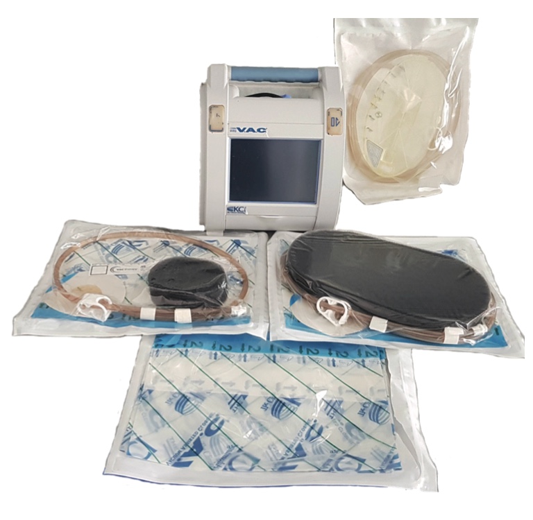
The use of negative pressure in wound healing is an innovative therapeutic method that accelerates the wound healing process. This technique can be applied in different types of surgery [4],[5],[6],[7] to treat different types of acute and chronic wounds. Wound therapy with controlled negative pressure is a technique based on the principle of suction, which evacuates excess exudate from the wound using subatmospheric pressure. This effect is achieved by using a prefabricated set consisting of a polyurethane sponge, a polyurethane film, a plastic tube and an apparatus that creates a vacuum and collects the evacuated liquid in a canister [8]. The use of this technique in wound care has increased dramatically over the past ten years. There are a large number of studies that analyze the influence of controlled negative pressure on the wound healing process [9].
Namely, it has been proven that vacuum therapy is successful in the treatment of diabetic, chronic venous and arterial ulcers, wound dehiscence, pressure ulcers, as well as in abdominal surgery for abdominal wall defects, trauma, abdominal compartment syndrome, acute pancreatitis, necrotic fasciitis, for the treatment of burns, and wounds in open fractures of long bones, while further research is necessary to evaluate its effectiveness in the treatment of other wounds [10],[11],[12]. Furthermore, the application of vacuum has significantly increased the rate of successfully transplanted skin grafts.
Contraindications for the use of this type of therapy are the following: presence of necrotic tissue, untreated osteomyelitis, uncontrolled enterocutaneous fistulas, the presence of malignancy in the wound, direct exposure of intestinal anastomoses and blood vessels, as well as acute bleeding. Caution is required if patients are on anticoagulation therapy or therapy with platelet aggregation inhibitors.
The prefabricated VAC system set consists of polyurethane foam, i.e., a sponge for the wound, a self-adhesive transparent film and a linking drainage tube, which connects the wound to a vacuum device that produces negative pressure (Figure 1). The algorithm for applying the VAC system is presented in Figure 2. Before placing the VAC system, the wound should be thoroughly cleaned of necrotic tissue, pus or other contaminants. Also, the depth and type of wound should be assessed in order to select the appropriate VAC system as well as the appropriate size of the sponge.
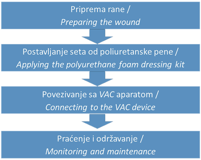
The polyurethane foam as a dressing is applied to the wound in such a way that it completely fills it up and constantly draws fluid from the wound thus promoting healing and recovery. Before placing the polyurethane sponge in the wound cavity, it is necessary to cut it to fit the shape and size of the wound. After placing the sponge over the wound, a polyurethane film is placed on the surrounding healthy skin. In the central part of the polyurethane film, an opening is formed with sterile scissors, through which the cavity of the wound, filled with the polyurethane sponge, is connected to the surgical vacuum apparatus via a polyurethane hose. After bandaging and checking that the wound is hermetically sealed, negative pressure is applied. If there is an air leak, the desired subatmospheric pressure cannot be achieved, nor can adequate drainage of the liquid from it. Any leaking of air is registered by the device and, if there is a problem, the device sounds an alarm.
The negative pressure applied can be continuous or intermittent, depending on the type of wound and the protocol being applied. Polyurethane foam, which completely fills the wound cavity, evenly and continuously drains secretions from all parts of the wound as a result of the action of the vacuum. Also, since the sponge and the skin are covered with foil and are also under the effect of the vacuum, the edges of the wound are drawn inward, which reduces the surface area of the wound. The pressure level depends on the device used – it can vary between -200 mmHg and -75 mmHg, depending on the material, the patient’s tolerance level and the type of wound being treated [13],[14].
For uninfected wounds, it is recommended to replace the sponge with a new one every 48 hours to 72 hours, i.e., more than three times a week. In infected wounds, greater caution and more frequent monitoring are necessary. In such cases, it is recommended that the intervals for the replacement of the sponge in the wound should be significantly shorter, and that the wound should be examined by a medical professional each time the dressing is being changed. Treatment should be discontinued when complete wound closure occurs. If there is no progress in wound healing within two weeks, the therapy should be discontinued. Petite and older patients should be constantly monitored for potential signs of excessive fluid loss and dehydration. It is also necessary to constantly monitor patients who have wounds with abundant exudation or wounds that are large, as compared to the body type and weight of the patient.
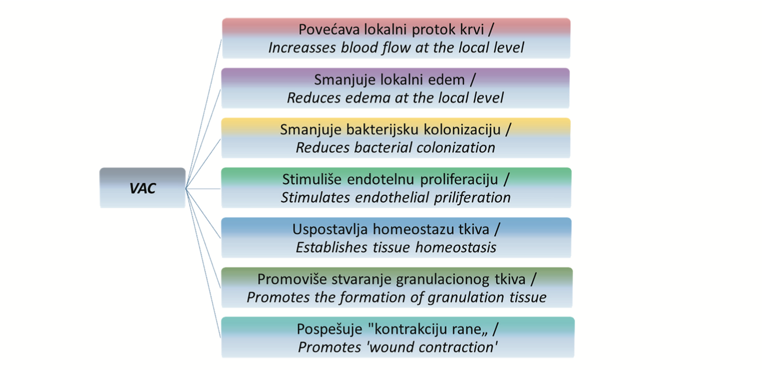
VAC achieves its effect through the following mechanisms: it promotes the formation of granulation tissue, increases local blood flow, reduces bacterial colonization, establishes tissue homeostasis, reduces local edema, stimulates endothelial proliferation, and reduces the wound surface through ‘wound contraction’ (Figure 3).
In most studies conducted so far, the effectiveness of vacuum in wound healing has been measured by the rate of granulation tissue formation. It has been proven that granulation tissue formation increased by an average of two to five times with the use of this method [15]. At the microvascular level, the vacuum exerts its positive effects through the process of angiogenesis, as well as by increasing the diameter of already existing blood vessels [16]. Namely, mechanically, the vacuum causes an increase in volume in the capillary network, stimulating endothelial proliferation and angiogenesis. Morykwas et al. measured the blood flow in tissue and showed that the blood flow increases by four times at 125 mmHg, and that at a pressure of 400 mmHg and more, it actually stops [17]. Also, tissue analyses under the electron microscope have shown restoration of the basal membrane in the capillaries and a reduction of intercellular space, after the application of vacuum. Overall, it has been proven that this technique optimizes blood flow, reduces tissue edema at the local level, and removes excess fluid from the wound cavity, thus leading to faster healing [18]. All these physiological changes facilitate the removal of bacteria from the wound. In addition, the application of subatmospheric pressure changes the cytoskeleton of cells in the wound cavity, leading to the triggering of intracellular signaling cascades that increase the rate of cell division and consequently the formation of granulation tissue. At the molecular level, under the influence of negative pressure, there is an increased expression of cytokines and growth factors that promote angiogenesis, remodeling of the extracellular matrix, and the formation of granulation tissue [19]. The combination of all these mechanisms makes the VAC device an extremely potent and versatile tool in wound healing.
NEGATIVE PRESSURE WOUND THERAPY IN AN OPEN ABDOMEN
Temporary abdominal closure (TAC) is an important surgical technique in the treatment of a wide range of complex injuries, in the treatment of sepsis, as well as in the management of abdominal conditions where there is a need for repeated laparotomy [20].
After a surgical procedure due to trauma, in complications after surgery, in case of infection, increased intra-abdominal pressure, or compartment syndrome, final closure of the abdominal fascia and abdominal wall immediately after laparotomy may be technically impossible or undesirable, and the abdominal cavity must remain open to allow access for additional procedures [21].
Keeping the abdomen open has a number of advantages, such as the following: easier re-access, an open ‘window’ to the abdominal cavity (in case of trauma, damage control), control of the contents of the abdominal cavity, reduction of the risk of intra-abdominal hypertension and compartment syndrome, and preservation of the fascia necessary for the closure of the abdominal cavity. On the other hand, prolonged open abdomen (OA) can contribute to the rapid development of complications, including infections, sepsis, and fistula formation.
Several techniques for temporary closure of the abdominal cavity are described in literature, including the following: the ‘Bogota bag’, the ‘Wittmann patch’, ‘Barker’s vacuum pack’, as well as negative pressure wound therapy (NPWT) [22]. Over the past decade, negative pressure wound therapy (NPWT) has been recognized as a useful technique and is now widely used in the clinical setting [23],[24]. However, although it is a widely adopted method in the management of the open abdomen, the heterogeneity of clinical cases and the quality of available studies made it difficult to reach a definitive conclusion about the superiority of NPWT, as compared to other methods. The heterogeneity related to different ways of treating the open abdomen, the diversity of patient groups, and the lack of wound classification. In order to systematize the data and introduce a standardized nomenclature, it was necessary to introduce a classification system. It was introduced by Björck, in 2009 (it was then revised by the same author in 2016) [25],[26]. The goals of the classification were to establish standardized treatment protocols and facilitate the comparison of results in scientific research.
In a systematic literature review from 2014, which included relevant studies related to each of the aforementioned TAC methods, recommendations for the treatment of the open abdomen were published and issues related to the comparison of NPWT treatment with other TAC methods were reevaluated (the success of fascia closure, mortality, and fistula formation). The majority were primary fascial closures that were performed using commercial NPWT sets in combination with the ‘dynamic closure’ technique (using mesh or placing retention sutures). In non-septic patients, the percentage of successful closure was 72%, and with ‘dynamic closure’ it was 81%. For other TAC techniques, the percentage was lower – 68% for the Whitman patch, and 58% for self-made NPWT. In septic patients, the success rate of closure when using the NPWT set was 48%, with ‘dynamic closure’ the success rate was 74.6%, with the Bogota bag it was 27%, and with self-made NPWT it was 35%. Mortality ranged from 12% – 25%, in non-septic patients, while in septic patients it was between 22% and 40%. The study showed that mortality depended more on the current status of the patient than on the effect of the use of different TAC methods. The previously existing controversy regarding the association of NPWT with fistula formation was refuted [27].
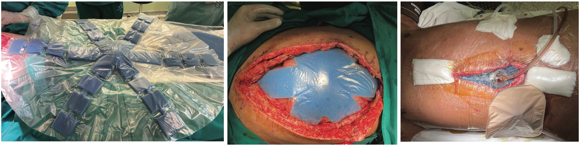
The most commonly used commercial negative pressure sets are Barker’s vacuum pack (i.e., Barker’s vacuum packing technique – BVPT) and VAC, KCI USA (ABTheraTM OA NPT). The ABTheraTM OA NPT set, which is most often used in Serbia, consists of a visceral protective film that is placed around and over the intestine, and under the peritoneum (Figure 4). After that, a perforated sponge of the appropriate size is placed and covered with a semi-occlusive adhesive foil. A small part of the adhesive film and the sponge underneath it is excised. In this spot, a connector with a drainage pipe is placed, which is hooked to the canister, i.e., the device for negative pressure.
As opposed to superficial wounds, where a better effect has been observed with the use of intermittent negative pressure, in the case of an open abdomen, continuous pressure with the initial value of -125 mmHg is recommended. In case of acute bleeding due to coagulopathies or the application of anticoagulation therapy, it is recommended to start with negative pressure values ranging from -25 mmHg to -55 mmHg, with close monitoring of the canister content.
This system achieves positive effects by reducing edema and removing exudate from the space between the abdominal organs, intestinal loops, and the anterior abdominal wall, as well as by removing infectious agents and inflammatory mediators. All of this, as previously stated, contributes to the reduction of intra-abdominal pressure, it reduces the possibility of compartment syndrome and respiratory distress syndrome. Also, it contributes to the reduction of adhesion formation between the intestines and the anterior abdominal wall, as well as the approximation of the fascial edges towards the midline, and thus enables delayed tension free closure of the abdomen [24],[26],[27].
Negative pressure therapy has proven to be an effective and reliable method of temporary abdominal closure, both because of its previously mentioned positive effects, as well as because of the shortening of the length of hospital stay and reducing the total treatment costs.
ENDOSCOPIC VACUUM THERAPY
Endoscopic vacuum therapy (EVT) is a relatively new, minimally invasive treatment modality in colorectal and upper gastrointestinal surgery.
Endoscopic vacuum therapy in colorectal surgery
Although significant progress has been made in the field of colorectal surgery in the past few decades, anastomotic failure remains one of the most difficult complications, due to its association with a significant morbidity and mortality rate, affecting 5% – 15% of patients with colorectal anastomosis [28].
Anastomotic failure can be caused by various factors, which can be divided into those related to the operating technique itself and those related to the characteristics of the patient. If we assume that the operation itself was performed flawlessly, the patient-related factors can be divided into modifiable risk factors and non-modifiable risk factors [29]. Modifiable risk factors include alcohol, smoking, obesity, and some medications. Non-modifiable risk factors include sex, age, diabetes, and factors related to the tumor itself [29],[30].
One of the newly introduced methods for treating complications of anastomotic failure is endoscopic vacuum therapy. Endoscopic vacuum therapy in colorectal surgery was first described in 2008, in a study by Weidenhagen et al., in the treatment of perianastomotic abscess caused by anastomotic dehiscence after low anterior rectal resection due to cancer [31]. The basis of this method is the placement of a polyurethane sponge inside the abscess cavity caused by the dehiscence of the anastomosis, with the help of a flexible endoscope.
A drainage tube attached to the sponge exits transanally and is connected to a vacuum system. The intraabscess position and sponge replacement every two to three days is controlled with an endoscope. Compared to open wounds, in this case, hermetic closure is not required to achieve the desired subatmospheric pressure. Neither sedation nor anesthesia is required to perform endoscopic vacuum therapy.
In the past several years, indications for the use of endoscopic vacuum therapy after rectal or rectosigmoid resection have been extended to its use after resection, due to diverticulitis, ulcerative colitis, iatrogenic or traumatic perforations of the colon. Generalized peritonitis is a contraindication for EVT.
Transanal drainage achieves efficient and continuous emptying of the abscess cavity while simultaneously bypassing the physiological obstruction of the anus without additional trauma to the patient. The return of fluid from the lumen of the colon through wound dehiscence to the lumen of the abscess cavity is blocked. Negative pressure leads to uniform mechanical reduction of the volume of the abscess cavity as the result of uniform and controlled topical application to each point inside the cavity. Not only is the enlargement of the abscess cavity and the occurrence of other complications avoided, but practically complete resolution of the abscess is also achieved. Positive effects such as the formation of granulation tissue and reduction of wound size have already been described earlier regarding the application of vacuum in the treatment of open wounds. According to some authors, the use of endoscopic vacuum therapy in the treatment of anastomotic dehiscence was successful in 90% of cases, the therapy, on average, lasted 23 days, while the average number of sponges used per patient was seven [32]. Among the side effects of therapy, occasionally, minor bleeding may appear after the removal of the polyurethane sponge, which usually stops spontaneously. Stenosis has also been reported in a small number of patients.
In some centers for colorectal surgery where this method is applied, EVT has become the therapy of choice for the treatment of anastomotic dehiscence after rectal resection. Considering that it is a relatively new procedure, new studies are needed that will further valorize the advantages of this method.
Endoscopic vacuum therapy in upper gastrointestinal surgery
Endoscopic vacuum therapy is mainly used in upper gastrointestinal tract surgery to treat dehiscences of esophago-gastric anastomoses and esophageal perforation, which was presented for the first time by Loske and Müller at a surgical meeting in Germany, in 2007 [33], while the first publication regarding this topic was published in 2008. It presents cases of successful treatment of anastomotic failure after esophagectomy [34]. The principles of vacuum application are the same as in the previously described areas. Depending on the patient’s condition, EVT is performed in the operating theatre under general anesthesia or under analgosedation.
The effectiveness of the therapy is measured by the reduction of the extramural cavity, the amount of secretion, and the formation of granulation tissue surrounding the polyurethane sponge. According to some studies, the success rate of endoscopic vacuum therapy in the repair of anastomotic dehiscence after esophagectomy, gastrectomy or esophageal perforation was 90% in more than 200 subjects. Although there are no randomized controlled studies in the currently available literature, based on retrospective reviews published so far, EVT has been shown to be superior to stent therapy [35],[36].
CONCLUSION
Negative pressure therapy is an advanced medical procedure that can be used to treat various types of acute and chronic wounds. It has proven to be particularly valuable as a technique for temporary abdominal closure.
Despite numerous contradictiory studies and findings, a large number of studies have shown numerous advantages of this therapy, its effectiveness and reliability. In recent years, the indications for the application of negative pressure have expanded, and endoscopic vacuum therapy has also emerged.
It has also been shown that negative pressure therapy reduces treatment costs, shortens the duration of hospital stay, and facilitates faster patient recovery.
The possibilities of applying negative pressure have not been completely exhausted. It is believed that future new ideas and technological progress will lead to an even wider application of this method in medicine.
Informations

-
Keywords:
-
Received:
-
Revised:
-
Accepted:
-
Online first:
-
DOI:
-
Cite this article:

Slobodan Todorović
University Hospital Medical Center Bežanijska Kosa
Žorža Matea Street, 11000 Belgrade, Serbia
E-mail:
1. Graham ID, Harrison MB, Nelson EA, Lorimer K, Fisher A. Prevalence of lowerlimb ulceration: a systematic review of prevalence studies. Adv Skin Wound Care. 2003 Nov;16(6):305-16. doi: 10.1097/00129334-200311000-00013. [CROSSREF]
2. Agarwal P, Kukrele R, Sharma D. Vacuum assisted closure (VAC)/negative pressure wound therapy (NPWT) for difficult wounds: A review. Journal of clinical orthopaedics and trauma. 2019 Sep 1;10(5):845-8. doi: 10.1016/j. jcot.2019.06.015. [CROSSREF]
3. Drašković M, Leković I, Bjelanović Z, Mišović S, Kozarski J. Primena vakuuma u lečenju komplikacija hirurških rana. Vojnosanitetski pregled. 2011 Okt;68(10):846. doi: 10.2298/VSP1110846D. [CROSSREF]
4. Ivanovic N, Granic M, Randjelovic T, Todorovic S. Fragmentation of axillary fibrofatty tissue during dissection facilitates preservation of the intercostobrachial nerve and the lateral thoracic vein. Breast. 2008 Jun;17(3):293-5. doi: 10.1016/j.breast.2007.11.006. [CROSSREF]
5. Tošković B, Bilanović D, Resanović A, Todorović S, Mrda D, Crnokrak B, et al. Management of major bile duct injuries following laparoscopic and open cholecystectomy-single center experience. Srpski arhiv za celokupno lekarstvo. 2019;147(7-8):422-6. doi:10.2298/SARH190206030T. [CROSSREF]
6. Todorovic S, Toskovic B, Gačić J, Karaca H, Colakovic N. An Unusually Long Appendix: A Case Report and Literature Review. Open Access Macedonian Journal of Medical Sciences (OAMJMS). 2023 Jan 26;11(C):53-6. doi:10.3889/ oamjms.2023.11177. [CROSSREF]
7. Ivanovic N, Granic M, Randjelovic T, Todorovic S. Identification and releasing of the intercostobrachial nerve and the lateral thoracic vein in the beginning of axillary dissection facilitates their preservation. EJC Supplements. 2008;7(6):208. doi:10.1016/S1359-6349(08)70848-6. [CROSSREF]
8. Fogg E. Best treatment of nonhealing and problematic wounds. Journal of the American Academy of PAs. 2009 Aug 1;22(8):46-8. doi:10.1097/01720610- 200908000-00013. [CROSSREF]
9. Fraccalvieri M, Zingarelli E, Ruka E, Antoniotti U, Coda R, et al. Negative pressure wound therapy using gauze and foam: histological, immunohistochemical and ultrasonography morphological analysis of the granulation tissue and scar tissue. Preliminary report of a clinical study. International wound journal. 2011 Aug;8(4):355-64. doi:10.1111/j.1742-481X.2011.00798.x. [CROSSREF]
10. Xie X, McGregor M, Dendukuri N. The clinical effectiveness of negative pressure wound therapy: a systematic review. Journal of wound care. 2010 Nov;19(11):490-5. doi: 10.12968/jowc.2010.19.11.79697. [CROSSREF]
11. Fitzgerald JE, Gupta S, Masterson S, Sigurdsson HH. Laparostomy management using the ABThera™ open abdomen negative pressure therapy system in a grade IV open abdomen secondary to acute pancreatitis. International wound journal. 2013 Apr;10(2):138-44. doi: 10.1111/j.1742- 481X.2012.00953.x. [CROSSREF]
12. Ubbink DT, Westerbos SJ, Nelson EA, Vermeulen H. A systematic review of topical negative pressure therapy for acute and chronic wounds. Journal of British Surgery. 2008 Jun;95(6):685-92. doi: 10.1002/bjs.6238. [CROSSREF]
13. Webb LX. New techniques in wound management: vacuum-assisted wound closure. JAAOS-Journal of the American Academy of Orthopaedic Surgeons. 2002 Sep 1;10(5):303-11. doi: 10.5435/00124635-200209000-00002. [CROSSREF]
14. Orgill DP, Bayer LR. Update on negative-pressure wound therapy. Plastic and reconstructive surgery. 2011 Jan 1;127:105S-15S. doi: 10.1097/ PRS.0b013e318200a427. [CROSSREF]
15. Clare MP, Fitzgibbons TC, McMullen ST, Stice RC, Hayes DF, Henkel L. Experience with the vacuum assisted closure negative pressure technique in the treatment of non-healing diabetic and dysvascular wounds. Foot & ankle international. 2002 Oct;23(10):896-901. doi: 10.1177/107110070202301002. [CROSSREF]
16. Glass GE, Murphy GF, Esmaeili A, Lai LM, Nanchahal J. Systematic review of molecular mechanism of action of negative‐pressure wound therapy. British Journal of Surgery. 2014 Dec;101(13):1627-36. doi: 10.1002/bjs.9636. [CROSSREF]
17. Morykwas MJ, David LR, Schneider AM, Whang C, Jennings DA, Canty C, et al. Use of subatmospheric pressure to prevent progression of partial-thickness burns in a swine model. The Journal of burn care & rehabilitation. 1999 Jan 1;20(1):15-21. doi: 10.1097/00004630-199901001-00003. [CROSSREF]
18. Venturi ML, Attinger CE, Mesbahi AN, Hess CL, Graw KS. Mechanisms and clinical applications of the vacuum-assisted closure (VAC) device. American journal of clinical dermatology. 2005 Jun 1;6(3):185-94. doi: 10.2165/00128071- 200506030-00005. [CROSSREF]
19. Borys S, Ludwig-Slomczynska AH, Seweryn M, Hohendorff J, Koblik T, Machlowska J, et al. Negative pressure wound therapy in the treatment of diabetic foot ulcers may be mediated through differential gene expression. Acta diabetologica. 2019 Jan 28;56(1):115-20. doi: 10.1007/s00592-018-1223-y. [CROSSREF]
20. Worhunsky DJ, Magee G, Spain DA. Challenges in the management of the open abdomen. ICU Director. 2013 Jan;4(1):33-9. doi: 10.1177/1944451612469628. [CROSSREF]
21. Roberts DJ, Jenne CN, Ball CG, Tiruta C, Léger C, Xiao Z, et al. Efficacy and safety of active negative pressure peritoneal therapy for reducing the systemic inflammatory response after damage control laparotomy (the Intra-peritoneal Vacuum Trial): study protocol for a randomized controlled trial. Trials. 2013 Dec;14(1):141. doi: 10.1186/1745-6215-14-141. [CROSSREF]
22. Brock WB, Barker DE, Burns RP. Temporary closure of open abdominal wounds: the vacuum pack. The American Surgeon. 1995 Jan;61(1):30-5.
23. Quyn AJ, Johnston C, Hall D, Chambers A, Arapova N, Ogston S, et al. The open abdomen and temporary abdominal closure systems–historical evolution and systematic review. Colorectal disease. 2012 Aug;14(8):e429-38. doi: 10.1111/j.1463-1318.2012.03045.x. [CROSSREF]
24. Roberts DJ, Zygun DA, Grendar J, Ball CG, Robertson HL, Ouellet JF, et al. Negative-pressure wound therapy for critically ill adults with open abdominal wounds: a systematic review. Journal of Trauma and Acute Care Surgery. 2012 Sep 1;73(3):629-39. doi: 10.1097/TA.0b013e31825c130e. [CROSSREF]
25. Björck M, Bruhin A, Cheatham M, Hinck D, Kaplan M, Manca G, et al. Classification—important step to improve management of patients with an open abdomen. World journal of surgery. 2009 Jun 1;33(6):1154-7. doi: 10.1007/ s00268-009-9996-3. [CROSSREF]
26. Björck M, Kirkpatrick AW, Cheatham M, Kaplan M, Leppäniemi A, De Waele JJ. Amended classification of the open abdomen. Scandinavian Journal of Surgery. 2016 Mar;105(1):5-10. doi: 10.1177/1457496916631853. [CROSSREF]
27. Bruhin A, Ferreira F, Chariker M, Smith J, Runkel N. Systematic review and evidence based recommendations for the use of negative pressure wound therapy in the open abdomen. International Journal of Surgery. 2014 Oct 1;12(10):1105-14. doi: 10.1016/j.ijsu.2014.08.396. [CROSSREF]
28. Bobkiewicz A, Krokowicz L, Banasiewicz T, Borejsza-Wysocki M. Endoscopic vacuum therapy with instillation (iEVT)–a novel endoscopic concept for colorectal anastomotic leak and perianal complications. Videosurgery and Other Miniinvasive Techniques. 2020 Dec 4;15(4):560-6. doi: 10.5114/wiitm.2020.93204. [CROSSREF]
29. Fang AH, Chao W, Ecker M. Review of colonic anastomotic leakage and prevention methods. Journal of Clinical Medicine. 2020 Dec 16;9(12):4061. doi: 10.3390/jcm9124061. [CROSSREF]
30. Ajtic OM, Todorovic S, Diklic M, Suboticki T, Beleslin-Cokic B, Jovcic G, et al. Proliferation and differentiation markers of colorectal adenocarcinoma and their correlation with clinicopathological factors. Turkish Journal of Medical Sciences. 2016;46(4):1168-76. doi: 10.3906/sag-1412-85. [CROSSREF]
31. Weidenhagen R, Gruetzner KU, Wiecken T, Spelsberg F, Jauch KW. Endoscopic vacuum-assisted closure of anastomotic leakage following anterior resection of the rectum: a new method. Surgical endoscopy. 2008 Aug 1;22(8):1818- 25. doi: 10.1007/s00464-007-9706-x. [CROSSREF]
32. Kuehn F, Janisch F, Schwandner F, Alsfasser G, Schiffmann L, Gock M, et al. Endoscopic vacuum therapy in colorectal surgery. Journal of Gastrointestinal Surgery. 2016 Feb 1;20(2):328-34. doi: 10.1007/s11605-015-3017-7. [CROSSREF]
33. Loske G, Müller C. Endoscopic vacuum-assisted closure of upper intestinal anastomotic leaks. Gastrointestinal endoscopy. 2009 Mar 1;69(3):601-2. doi: 10.1016/j.gie.2008.06.058. [CROSSREF]
34. Wedemeyer J, Schneider A, Manns MP, Jackobs S. Endoscopic vacuum-assisted closure of upper intestinal anastomotic leaks. Gastrointestinal endoscopy. 2008 Apr 1;67(4):708-11. doi: 10.1016/j.gie.2007.10.064. [CROSSREF]
35. Hwang JJ, Jeong YS, Park YS, Yoon H, Shin CM, Kim N, et al. Comparison of endoscopic vacuum therapy and endoscopic stent implantation with self-expandable metal stent in treating postsurgical gastroesophageal leakage. Medicine. 2016 Apr;95(16). doi: 10.1097/MD.0000000000003416. [CROSSREF]
36. Schniewind B, Schafmayer C, Voehrs G, Egberts J, von Schoenfels W, Rose T, et al. Endoscopic endoluminal vacuum therapy is superior to other regimens in managing anastomotic leakage after esophagectomy: a comparative retrospective study. Surgical endoscopy. 2013 Oct 1;27(10):3883-90. doi: 10.1007/s00464-013-2998-0. [CROSSREF]