


A giant phyllodes tumor: а case report and literature overview
- The Institute of Oncology and Radiology of Serbia, Belgrade, Serbia
- Breast Surgery Service, Khoo Teck Puat Hospital, Yishun, Singapore
- University of Belgrade, Faculty of Medicine, Belgrade, Serbia
ABSTRACT
Introduction: Phyllodes tumors of the breast are extremely uncommon, both in Serbia and worldwide. Identifying and treating these tumors can be challenging and they often represent a dilemma in diagnosis and treatment.
Case report: We are presenting the case of a 60-year-old female with a large breast tumor that she had had for 15 years. According to the histopathologic diagnosis, it is a borderline phyllodes tumor measuring 18x17x7 cm and weighing 4.6 kg.
Conclusion: An accurate preoperative diagnosis and proper management of borderline phyllodes tumors are crucial for successful surgery and for avoiding any further surgeries. This secondary prevention is critical because phyllodes tumors often recur and have malignant potential.
INTRODUCTION
Breast tumors that belong to the category of phyllodes tumors (PT), and are also referred to as cystosarcoma phyllodes, account for less than 1% of all breast tumors. Their incidence is about 2.1 per one million [1],[2],[3].
Traditionally, phyllodes tumors containing both stromal and epithelial elements are graded into benign, borderline, and malignant categories based on histological parameters. A set of histologic features determines the nature of tumor borders, including the degree of stromal cellularity and atypia, mitotic count, and stromal overgrowth [4],[5]. All forms of phyllodes tumors are regarded as having malignant potential. Phyllodes tumors are usually well-demarcated and may compress the surrounding tissue.
Most malignant phyllodes tumors contain liposarcomatous or rhabdomyosarcomatous elements rather than a fibrosarcomatous component. Counting the number of mitoses can aid in identifying a malignant tumor [6]. Surgery is the only treatment for these patients, so it is crucial to select the appropriate surgical option.
CASE REPORT
A rare case is presented by this case report. The patient was a 60-year-old female who presented with a massive tumor in her left breast, which she had suffered from for 15 years prior to surgery. She had pain in her left breast. Over the final three years, the size of the tumor increased drastically (Figure 1).
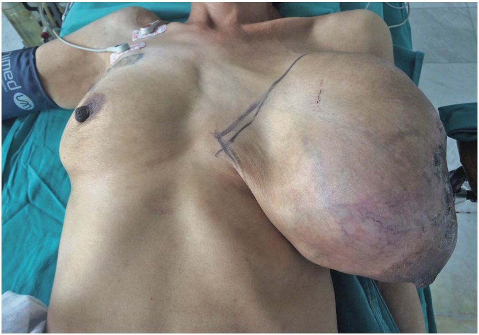
She reported to the Institute of Oncology and Radiology of Serbia, where the diagnostic process was performed, including mammography and ultrasonography. The mammography showed an extremely voluminous left breast. The tumor’s intense shadow occupied the whole breast. Despite the obvious change, the parenchyma of the left breast could not be seen.
A simple mastectomy was performed with no axillary lymph node dissection (Figures 2,3,4). The histopathological examination upon surgery showed a borderline phyllodes tumor of the breast. In the cross-section, almost all the breast tissue was infiltrated by the tumor which was 18x17x7 cm in size. The tumor’s intersection was found to contain numerous central cavities which were necrotic.
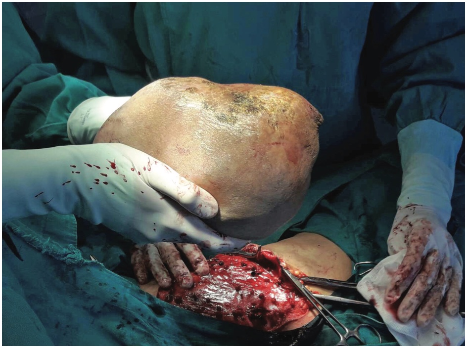
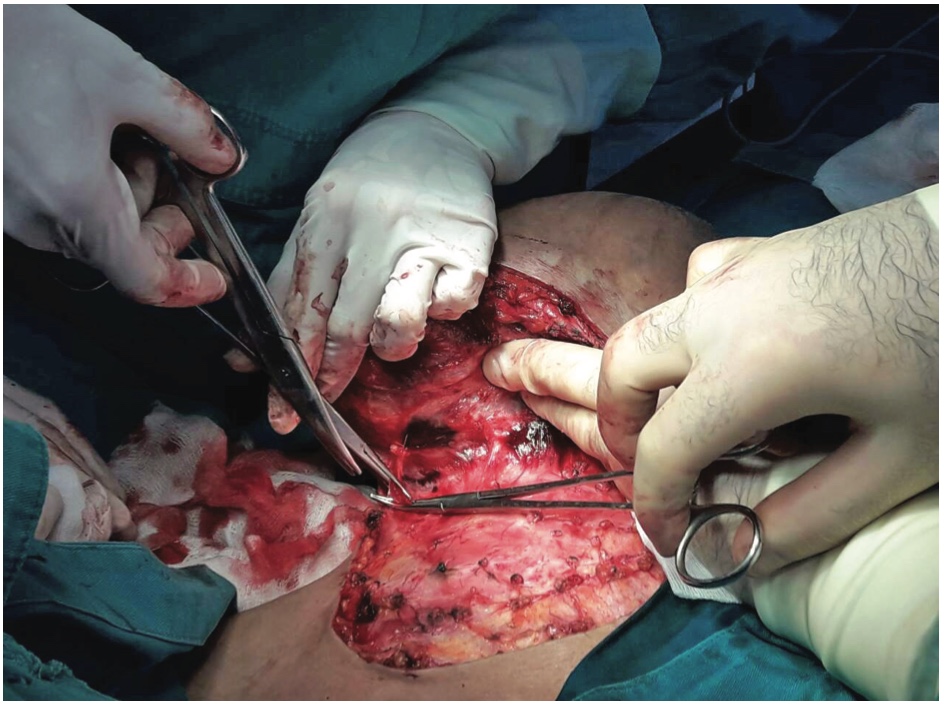
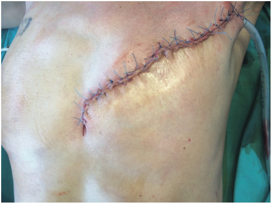
The stroma of the tumor was moderately cellular. It was found to possess a mild pleomorphism and low mitotic index, which was partially hyaline modified. The epithelial component was found to be partly atrophic, and partly of a squamous metaplasia, and focal atypical hyperplasia (Figures 5, 6).


DISCUSSION
Phyllodes tumors (PTs) most commonly occur in females in the 4th or 5th decade of life. This disease is primarily found in women, but there have been a few reports of these tumors in male breasts as well [7],[8]. The cause of these tumors has not been understood yet. Benign phyllodes are more commonly presented as painless breast lumps. The most frequently performed surgery in case of such tumors is a wide local excision [9]. Upon clinical examination, mammography, ultrasonography and FNAC, a preliminary diagnosis is made; however, the final diagnosis is made based on histological findings. Benign tumors can be treated with lumpectomy, but recurrent, malignant, and massive tumors require mastectomy without axillary dissection. Attempts have been made to use adjuvant therapy along with radiotherapy and/or chemotherapy, despite its limited effectiveness [10],[11].
As per the guidelines given by the World Health Organization (WHO), phyllodes tumors are to be divided into benign, borderline, and malignant based on the three main characteristics: 1) the degree of cellular atypia of the stromal tumor; 2) the mitotic activity measured as the number of cells undergoing mitosis per area of tissue, typically counted in 10 high-power fields; and 3) the degree of stromal overgrowth. Tumor necrosis and margin appearance are also considered when categorizing a PT [12],[13].
Local recurrence
Local recurrence rates have been reported to range from 10-40%, with most cases averaging approximately 15%. When local recurrence occurs, it is often due to an incomplete removal of the original PT during the primary surgical treatment. The only independent predictive factor for local recurrence is the surgical margin, according to multivariate analyses. In the majority of patients, such recurrence is found to be isolated and therefore disassociated with the occurrence of distant metastases. Nevertheless, repeated local recurrence does occur in a minority of patients, which is often unrelated to either the histological type of tumor or the extent of the specimen margins. Further wide excision (with a margin of 1 cm) can usually prevent local recurrence. If localized surgery for borderline or malignant tumors is followed by a recurrence at the same site, it may be worth considering mastectomy as a possible option, even though it is not typically necessary. This could help to increase the chances of preventing future recurrences or complications. There is a risk of aggressive local recurrence resulting in widespread chest-wall disease where direct invasion of the underlying lung parenchyma occurs. For PT cases, isolated reports of proper palliation using radiotherapy have also been published [14]. Radiotherapy may have a role in the treatment of PT depending on the number of recurrences, the mitotic index, the size of the tumor, the status of the resection margins, as well as the tumor’s p53 and Ki67 expression [15].
Metastases
About 10% of all PT patients develop distant metastases, wherein approximately 25% of all PT patients eventually experience histologically malignant tumors. Metastases that are far away from the cancer’s site of origin can develop without any signs of the cancer returning locally. The most common sites for metastases are the lungs (66%), bones (28%), and the brain (9%). In rare instances, metastases may also be found in the liver or the heart. The extent of the initial surgery does not affect the risk of metastatic disease. It appears that tumor biology is a better predictor of risk than other factors. Phyllodes tumors that metastasize have a poor prognosis and no long-term survival [10].
Follow-up
It is crucial for patients with phyllodes tumors to have regular follow-ups at 6-month intervals for the first two years upon the initial surgery. This is because these tumors have a high chance of recurrence, particularly if they were not excised using clear margins. Moreover, phyllodes tumors are unpredictable in their growth and metastatic activity. The chances of recurrence are at their highest in the first two years after the surgery. Thereafter follow-ups are to be done on an annual basis. It is crucial to provide thorough instructions to patients on the proper technique of regular breast self-examination. If any abnormality is detected, patients should immediately consult their doctor. It is important to physically examine patients during their follow-up appointments. This allows for thorough monitoring of their condition and progress. If any abnormalities are found, it is recommended to conduct further investigation using ultrasonography, mammography, MRI, and/or tissue biopsy to ensure an accurate diagnosis and prompt treatment [10].
Although literature has thoroughly addressed surgical management of PTs, it can still be made clearer. Some reports or sources have specifically mentioned giant phyllodes tumors. The arbitrary cut off point for designating a PT as a giant phyllodes tumor is 10 cm [16]. Such entities present the surgeon with several unique management problems.
Based on the initial clinical examination, ultrasonography, and mammography, the phyllodes tumor presented here had been conclusively diagnosed prior to surgery. It was decided against performing any type of biopsy of the tumor due to the high risk of bleeding.
Since a malignant transformation of a primarily benign phyllodes tumor is an eventual outcome for any patient, it is vital that surgery is conducted as early as possible before the tumor transforms into a malignant one [17].
For malignant phyllodes tumors (PTs), it may be best to begin with wide excision that leaves a clear margin. However, routine axillary lymph node dissection is not recommended. Patients with tumors that have infiltrating tumor margins, severe stromal overgrowth, atypia, and cellularity have a high risk of metastases, and should be closely monitored [18],[19],[20],[21],[22],[23],[24],[25],[26].
Although the patient in this case report had had a phyllode tumor for 15 years, it did not transform into a malignant tumor and that is the most important thing of all.
During the first year of follow-up, no local recurrence was found.
CONCLUSION
Phyllodes tumor of the breast is a rare but intriguing type of fibroepithelial neoplasm. An accurate preoperative diagnosis and proper surgery are crucial for preventing positive margins, which can lead to local recurrence, metastases, and decreased survival rates. Avoiding new surgeries is also important.
Informations

-
Keywords:
-
Received:
-
Revised:
-
Accepted:
-
Online first:
-
DOI:
-
Cite this article:

Zorka Inić
Institute of Oncology and Radiology of Serbia,
14, Pasterova Street, 11000 Belgrade, Serbia
E-mail:
1. Esposito NN, Mohan D, Brufsky A, Lin Y, Kapali M, Dabbs DJ. Phyllodes tumor: a clinicopathologic and immunohistochemical study of 30 cases. Arch Pathol Lab Med. 2006 Oct;130(10):1516-21. doi: 10.5858/2006-130-1516-PTACAI. [CROSSREF]
2. Yohe S, Yeh IT. “Missed” diagnosis of Phyllodes tumor on breast biopsy: pathological clues to its recognition. Int J Surg Pathol. 2008;16(2):137-42. doi: 10.1177/1066896907311378.
3. Farias-Eisner GT, Small K, Swistel A, Ozerdem U, Talmor M. Immediate implant breast reconstruction with acellular dermal matrix for treatment of a large recurrent malignant phyllodes tumor. Aesthetic Plast Surg. 2014 Apr;38(2):373-8. doi: 10.1007/s00266-014-0283-9. [CROSSREF]
4. Khosravi-Shahi P. Management of non metastatic phyllodes tumors of the breast: review of the literature. Surg Oncol. 2011 Dec;20(4):e143-8. doi: 10.1016/j.suronc.2011.04.007. [CROSSREF]
5. Lakhani SR, Ellis IO, Schnitt S, Tan PH, van de Vijver M. WHO classification of tumours of the breast. Lyon: IARC Press; 2012.
6. Khan SA, Badve S. Phyllodes tumors of the breast. Curr Treat Options Oncol. 2001 Apr;2(2):139-47. doi: 10.1007/s11864-001-0056-y. [CROSSREF]
7. Bapat K, Oropeza R, Sahoo S. Benign phyllodes tumor of the male breast. Breast J. 2002 Mar-Apr;8(2):115-6. doi: 10.1046/j.1524-4741.2002.08209.x. [CROSSREF]
8. Nielsen VT, Andreasen C. Phyllodes tumour of the male breast. Histopathology. 1987 Jul;11(7):761-2. doi: 10.1111/j.1365-2559.1987.tb02690.x. [CROSSREF]
9. Rajendra B, Tubachi P, Prabhu R, Shenoy R. Phyllodes tumor – a clinicopathological study. IOSR J. Dent. Med. Sci. 2016;15(3):33-7. doi: 10.9790/0853-15313337.
10. Pietruszka M, Barnes L. Cystosarcoma phyllodes: a clinicopathologic analysis of 42 cases. Cancer. 1978 May;41(5):1974-83. doi: 10.1002/1097-0142(197805)41:53.0.co;2-c.
11. Moffat CJ, Pinder SE, Dixon AR, Elston CW, Blamey RW, Ellis IO. Phyllodes tumours of the breast: a clinicopathological review of thirty-two cases. Histopathology. 1995 Sep;27(3):205-18. doi: 10.1111/j.1365-2559.1995.tb00212.x. [CROSSREF]
12. Azzopardi JG, Ahmed A, Millis RR. Problems in breast pathology. London: Saunders; 1979. p. 346.
13. Salvadori B, Cusumano F, Del Bo R, Delledonne V, Grassi M, Rovini D, et al. Surgical treatment of phyllodes tumors of the breast. Cancer. 1989 Jun 15;63(12):2532-6. doi: 10.1002/1097-0142(19890615)63:123.0.co;2-q.
14. Mishra SP, Tiwary SK, Mishra M, Khanna AK. Phyllodes tumor of breast: a review article. ISRN Surg. 2013;2013:361469. doi: 10.1155/2013/361469. [CROSSREF]
15. Erhan Y, Zekioglu O, Ersoy O, Tugan D, Aydede H, Sakarya A, et al. p53 and Ki67 expression as prognostic factors in cystosarcoma phyllodes. Breast J. 2002 Jan-Feb;8(1):38-44. doi: 10.1046/j.1524-4741.2002.08008.x. [CROSSREF]
16. Tan PH, Jayabaskar T, Chuah KL, Lee HY, Tan Y, Hilmy M, et al. Phyllodes tumors of the breast: the role of pathologic parameters. Am J Clin Pathol. 2005 Apr;123(4):529-40. doi: 10.1309/U6DV-BFM8-1MLJ-C1FN. [CROSSREF]
17. Kuipers T, Stark GB, Spilker G. Explosive malignant transformation of a primarily benign phyllodes tumor. Eur J Plast Surg, 1992 Sep;15(5):233-6. doi: 10.1007/BF00193661.
18. Chen WH, Cheng SP, Tzen CY, Yang TL, Jeng KS, Liu CL, et al. Surgical treatment of phyllodes tumors of the breast: retrospective review of 172 cases. J Surg Oncol. 2005 Sep 1;91(3):185-94. doi: 10.1002/jso.20334. [CROSSREF]
19. Ofri A, Stuart KE, Chan B, Mak C, Warrier S, Bhadri V, et al. Diagnosis and management of phyllodes tumours for the surgeon: An algorithm. Surgeon. 2022 Dec;20(6):e355-e365. doi: 10.1016/j.surge.2022.01.004. [CROSSREF]
20. Limaiem F, Kashyap S. Phyllodes Tumor of the Breast. [Updated 2023 Jan 1]. In: StatPearls [Internet]. Treasure Island (FL): StatPearls Publishing; 2023 Jan- . Available from: https://www.ncbi.nlm.nih.gov/books/NBK541138/ [HTTP]
21. Ogunbiyi S, Perry A, Jakate K, Simpson J, George R. Phyllodes tumour of the breast and margins: How much is enough. Can J Surg. 2019 Feb 1;62(1):E19-E21. doi: 10.1503/cjs.005718. [CROSSREF]
22. Mustață L, Gică N, Botezatu R, Chirculescu R, Gică C, Peltecu G, et al. Malignant Phyllodes Tumor of the Breast and Pregnancy: A Rare Case Report and Literature Review. Medicina (Kaunas). 2021 Dec 26;58(1):36. doi: 10.3390/ medicina58010036. [CROSSREF]
23. Lissidini G, Mulè A, Santoro A, Papa G, Nicosia L, Cassano E, et al. Malignant phyllodes tumor of the breast: a systematic review. Pathologica. 2022 Apr;114(2):111-20. doi: 10.32074/1591-951X-754. [CROSSREF]
24. Wei Y, Dai Y, Guan Q, Min N, Geng R, Hu H, et al. Predicting the recurrence-free survival of phyllodes tumor of the breast: a nomogram based on clinicopathology features, treatment, and surgical margin. Gland Surg. 2023 Feb 28;12(2):152-64. doi: 10.21037/gs-22-542. [CROSSREF]
25. Li Y, Song Y, Lang R, Shi L, Gao S, Liu H, Wang P. Retrospective study of malignant phyllodes tumors of the breast: Younger age, prior fibroadenoma surgery, malignant heterologous elements and surgical margins may predict recurrence. Breast. 2021 Jun;57:62-70. doi: 10.1016/j.breast.2021.03.001. [CROSSREF]
26. Shin E, Koo JS. Prognostic factors of breast phyllodes tumors. Histol Histopathol. 2023 Aug;38(8):865-78. doi: 10.14670/HH-18-600 [CROSSREF]