


The final outcome in the treatment of Legg-Calve-Perthes disease using the combination of ilizarov method of treatment and tunelisation (Case report)
- Institute for Orthopedic Surgery “Banjica”, Belgrade, Serbia
- Medical Faculty, University of Belgrade, Belgrade, Serbia
- University Children`s Hospital, Belgrade, Serbia
ABSTRACT
Introduction: Legg-Calve-Perthes disease (LCPD) is a common childhood hip disease, affecting approximately 0.8 in 100,000 children. It usually affects children between 4 and 8 years old, and boys are more affected than girls. One hip is initially affected, but bilateral localization is present in 9-15 of the cases. The primary treatment in LCPD focuses on containing the femoral head in the acetabulum, thus preventing mollification and fragmentation. The most common surgical treatment method consists of either femoral or pelvic osteotomy or a combination of both. This paper aims to present a minimally invasive alternative method for treating LCPD.
Case report: We are presenting a case report of a 19-year-old with a lengthy history of illness. His treatment started at the age of 5, and this is an update on his condition 14 years after the surgery.
Conclusion: Hip arthrodiastasis using the Ilizarov circular frame combined with femoral head and neck tunneling is a minimally invasive surgical procedure. From a technical standpoint, this is a relatively demanding procedure that results in shorter in-patient treatment, reduced risk of complications, and cost compared to pelvic and femoral osteotomy.
INTRODUCTION
Legg-Calve-Perthes disease (LCPD) is a common childhood hip disease, affecting approximately 0.8 in 100,000 children [1]. The disease was first described in 1910 by three scientists from three different countries, but its etiology and treatment modalities are still a matter of debate [2]. It usually occurs in children aged 4 to 8 years, and it is more common in boys than girls [1],[2]. One hip is initially affected, but bilateral localization is present in 9-15 cases [1],[2].
Legg-Calve-Perthes disease is an avascular condition that affects the capital femoral epiphysis (head) [1],[3]. The avascular condition is followed by growth retardation of the ossific nucleus. The ossific nucleus is subsequently resorbed, replaced by a new bone, and remodeled according to mechanical properties. After remodeling the femoral head becomes flattened and enlarged. The proximal femoral physis and metaphysis could also be affected, leading to secondary acetabulum changes. The treatment of LSPD remains controversial despite the extensive literature on this topic [3].
The principle of LCPD treatment is the concept of containment of the femoral head in acetabulum, as a protection against a mollified and fragmented femoral head [4],[5]. The containment concept could be attained using conservative methods (like rest or orthosis) or surgical treatment. This principle of treatment in the early stages of LCPD at the age of under 8 is widely accepted.
The treatment of late LCPD (over the age of 8) is still challenging. Late LCPD treated by femoral or pelvic osteotomy, or a combination of both has had poor outcomes [6],[7],[8],[9].
Many authors recommend the concept of hip distraction (arthrodiastasis) as the initial treatment method [10],[11],[12],[13],[14],[15],[16].
Arthrodiastasis does not have an impact on anatomic changes in the hip joint. It decreases the stress on the hip articular surface and enhances the healing of the femoral head [17]. It could also be combined with the femoral neck and head tunneling to improve healing [13].
This study aims to present a case of a minimally invasive alternative method in treating LCPD in a 5-yearold boy and a follow-up on the initial procedure 14 years later.
CASE REPORT
We will present the case report on a 5-year-old boy with long LCPD history. He complained of pain in the left hip and left knee. He had also been limping on the left leg. He had no general signs of infection, night pain, or concomitant diseases. In the clinical findings, he had the limitation of abduction (30 degrees) and internal rotation (5 degrees) in the left hip. He also had a left-side positive Trendelenburg sign.
Legg-Calve-Perthes disease was confirmed after blood analysis and X-ray findings (in AP and ‘frog-like’ position). According to Catarell and Herring classification he was classified as Catarell 3 and Herring B group.
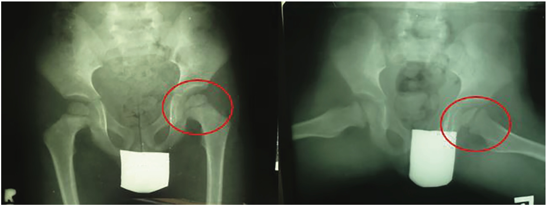
After admission, we started with conservative treatment using percutaneous traction in slight abduction for one week (approximately 10% of the patient’s total weight). The traction was followed by nonweight-bearing rest for one week more. After that, we started the bracing protocol using Atlanta orthosis, in the position of a slight abduction and internal rotation with full-weight bearing on the affected leg. The position of the femoral head (containment) was confirmed by the X-ray (in AP view). After 4 months of conservative treatment, the outcome was poor and the surgical consulting body (3 specialists in orthopedic surgery) decided to perform a surgical treatment.
All modalities of the surgical treatment were presented to the parents, and they approved of this rarely used surgical treatment. This method was performed as a combination of left hip arthrodiastasis using the Ilizarov frame (circular external fixator) and the femoral head and neck tunneling using 2.0 mm Kirschner wires (K wires).
This method of treatment is a minimally invasive surgical method. The main goal of arthrodiastasis using the Ilizarov circular fixator is to distract the hip joint and to reduce the pressure on the fragmented femoral head. On the other hand, the main goal of tunneling using K wires is to increase the blood supply to the femoral neck and head (Figure 2). The healing process of the femoral head will be improved in the outcome.
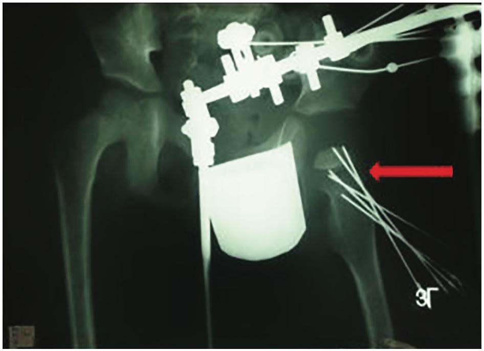
No complications occurred during the surgical procedure and postoperatively. Two days after the surgery, the patient started with physical therapy, passive kinesiotherapy with partial weight bearing on the left leg using crutches. All physical therapy treatments were provided in the hospital during the two months. Six weeks after the surgery K wires were removed from the femoral neck and head. Three months after the surgery, the patient was admitted for a redo-surgery procedure when the Ilizarov circular fixator was extracted. The surgical outcome is presented in Figure 4.
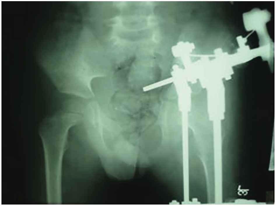
After the second surgical procedure (the Ilizarov frame removal), a short physical therapy treatment with full-weight bearing was provided during the stay; at first, it was supported with crutches during the first two weeks and afterward with full-weight bearing.
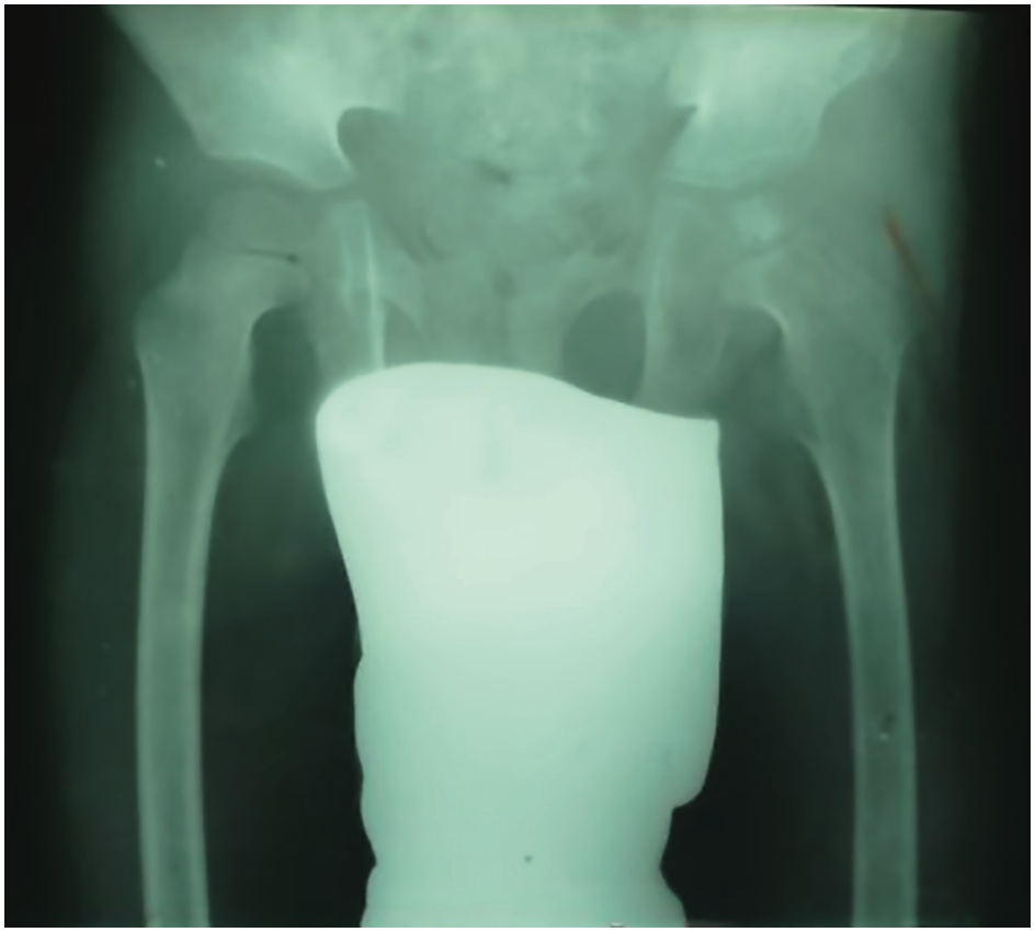
The patient was controlled at 6 weeks, threemonth, and six-month intervals after the second surgery. Figure 5 presents 6-month and 18-month X-ray findings after the second surgery (hardware removal).
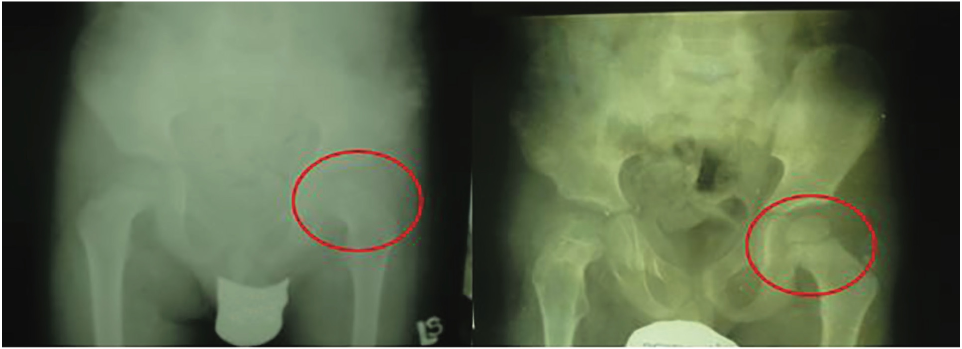
In the clinical outcome, 18 months after the hardware removal the patient had the full range of motion in the left hip and left knee, painlessly walked without any support, and carried out normal physical activities besides jumping.
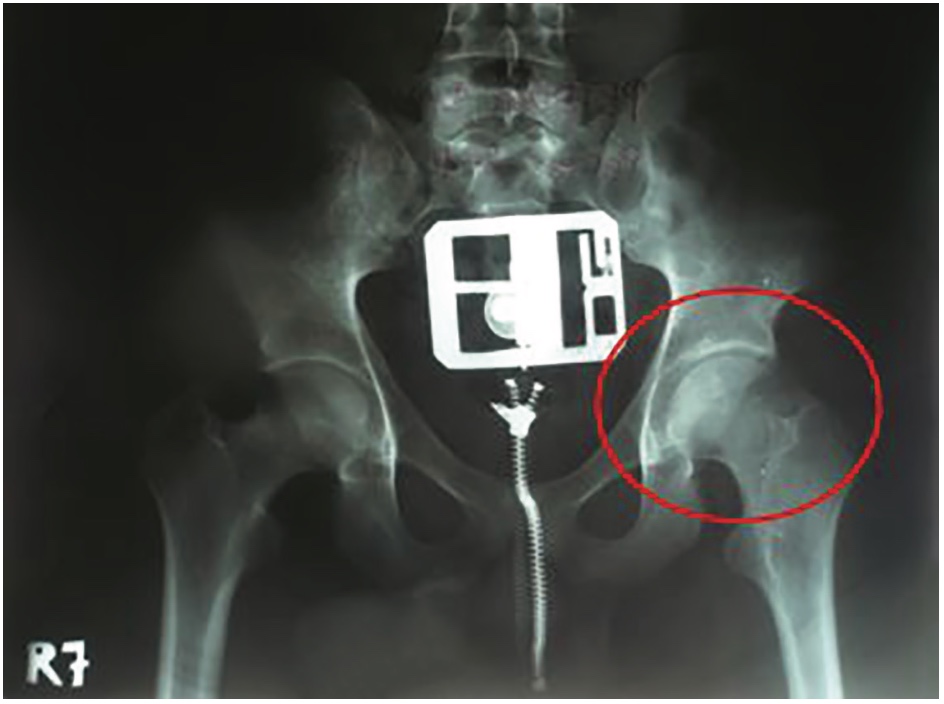
The last follow-up was done when the patient turned 19 (14 years after the first surgery). The X-ray in the AP position presented the congruency of the femoral head without any signs of avascular necrosis. Aesthetic reasons were also essential; the femoral and iliac scars were almost invisible.
DISCUSSION
The treatment modalities of LCPD are various, whether conservative or surgical. Patients under the age of 6 have better outcomes and prognoses despite the method of treatment than patients over the age of 6 [15],[18]. Conservative methods of treatment (physical therapy and orthotics usage) have many advantages as non-invasive methods: no scar formation, no infection risk, and no psychological effects on a child and parents. It takes, on the other hand, longer treatment and good compliance between a doctor, a child, and parents. To conclude, all these factors have to be considered in making a final decision [19].
The most important factors in LCPD treatment are the patient’s age X-ray findings in AP and the “frog-like” position (according to Catarell and Herring classification).
Patients over the age of 6 have better outcomes using the surgical approach in LCPD treatment [16]. According to their studies, some authors concluded that the major factor in LCPD prognosis of the outcome is an X-ray finding, precisely the level of aseptic narcosis of the femoral head [20],[21].
It has been shown that drilling a tunnel affects less than 10% of the total area of the growth cartilage, it does not impair the growth plate and it does not reduce growth in the hip region [22]. Several authors treated patients with arthrodiastasis using monolateral fixator; however, they used a biomechanically weaker fixator method, and large diameter pins for tunneling and their patients did not weight bear during the whole length of treatment [23],[24]. Kocaoglu presented and published results of the Ilizarov apparatus usage in Perthes disease. He used a robust and complex Ilizarov frame at the early stage of the femoral head fragmentation, and his success rate was not very impressive. To maintain the containment of the femoral head it is mandatory to initiate the treatment during the stage of necrosis or early fragmentation stage, thus before the collapse of the femoral epiphysis [25]. Despite the literature review on the gold standard in surgical approach (pelvic osteotomy like Salter, Chiari, or triple pelvic osteotomy), hip arthrodiastasis with tunneling should be considered as an alternative surgical technique. Hip arthrodiastasis and femoral head and neck tunneling should be considered at an early stage of LCPD and the late onset of disease when standard surgical procedures do not give great outcomes [26].
The usage of this technique has gained a full range of painless motion, without secondary disorders or gait disturbances. The risk of lateral subluxation of the femoral head was decreased in X-ray findings, the femoral head congruency was reached without anatomic violation in the hip joint. This technique might also be used in older children and adolescents with poor prognosis (according to X-ray findings) and in contraindications to do pelvic osteotomy.
This LCPD case report presents the most important values in the outcome: age, rating according to X-ray findings (according to Herring and Catarell), and adequate strategy in the treatment approach.
We chose the less invasive and rarely used technique: hip arthrodiastasis using the Ilizarov circular frame in combination with femoral neck and head tunneling. It is a minimally invasive surgery procedure and technically less demanding: shorter patient–in treatment, minor risk of complications, and lower costs in comparison to pelvic and femoral osteotomy. This technique might help treat a minor leg discrepancy, as one of the clinical and radiological signs in the evolution of Perthes disease.
We want to emphasize that arthrodiastasis with tunneling, as a minimally invasive surgery should be considered as one of the first treatment options at an early stage of LCPD when the femoral head still does not have any structural changes and conservative treatment failed.
Informations

-
Keywords:
-
Received:
-
Revised:
-
Accepted:
-
Online first:
-
DOI:
-
Cite this article:

Branislav Krivokapić
Institute for Orthopedic Surgery “Banjica”
28 Mihajla Avramovića Street, 11000 Belgrade, Serbia
E-mail:
1. Hefti F, Clarke NM. The management of Legg-Calvé-Perthes’ disease: is there a consensus? A study of clinical practice preferred by the members of the European Paediatric Orthopaedic Society. J Child Orthop. 2007 Mar;1(1):19- 25. doi: 10.1007/s11832-007-0010-z. [CROSSREF]
2. Nelitz M, Lippacher S, Krauspe R, Reichel H. Perthes disease: current principles of diagnosis and treatment. Dtsch Arztebl Int. 2009 Jul;106(31-32):517- 23. doi: 10.3238/arztebl.2009.0517. [CROSSREF]
3. Heimkes B. Alternative to crutches? Dtsch Arztebl Int. 2010 Jan;107(4):62; author reply 62. doi: 10.3238/arztebl.2010.0062a. [CROSSREF]
4. Herring JA, Kim HT, Browne R. Legg-Calve-Perthes disease. Part II: Prospective multicenter study of the effect of treatment on outcome. J Bone Joint Surg Am. 2004 Oct;86(10):2121-34.
5. Catterall A. Legg-Calvé-Perthes syndrome. Clin Orthop Relat Res. 1981 JulAug;(158):41-52.
6. Joseph B, Nair NS, Narasimha Rao K, Mulpuri K, Varghese G. Optimal timing for containment surgery for Perthes disease. J Pediatr Orthop. 2003 SepOct;23(5):601-6. doi: 10.1097/00004694-200309000-00006. [CROSSREF]
7. Joseph B, Rao N, Mulpuri K, Varghese G, Nair S. How does a femoral varus osteotomy alter the natural evolution of Perthes’ disease? J Pediatr Orthop B. 2005 Jan;14(1):10-5. doi: 10.1097/01202412-200501000-00002. [CROSSREF]
8. Salter RB. Legg-Perthes disease: the scientific basis for the methods of treatment and their indications. Clin Orthop Relat Res. 1980 Jul-Aug;(150):8-11.
9. Sarassa CA, Herrera AM, Carvajal J, Gomez LF, Lopez CA, Rojas AF. Early clinical and radiological outcomes after double osteotomy in patients with late presentation Legg-Calvé-Perthes disease. J Child Orthop. 2008 Dec;2(6):425- 9. doi: 10.1007/s11832-008-0132-y. [CROSSREF]
10. Yoon TR, Rowe SM, Chung JY, Song EK, Mulyadi D, Anwar IB. A new innominate osteotomy in Perthes’ disease. J Pediatr Orthop. 2003 May-Jun;23(3):363-7.
11. Aldegheri, R., Trivella, G., Saleh, M. Articulated Distraction of the Hip. In: De Bastiani, G., Apley, A.G., Goldberg, A. (eds) Orthofix External Fixation in Trauma and Orthopaedics. Springer, London. 2000. https://doi.org/10.1007/978-1-4471-0691-3_57 [HTTP]
12. Aly TA, Amin OA. Arthrodiatasis for the treatment of Perthes’ disease. Orthopedics. 2009 Nov;32(11):817. doi: 10.3928/01477447-20090922-15. [CROSSREF]
13. Cañadell J, Gonzales F, Barrios RH, Amillo S. Arthrodiastasis for stiff hips in young patients. Int Orthop. 1993;17(4):254-8. doi: 10.1007/BF00194191. [CROSSREF]
14. Segev E, Ezra E, Wientroub S, Yaniv M, Hayek S, Hemo Y. Treatment of severe late-onset Perthes’ disease with soft tissue release and articulated hip distraction: revisited at skeletal maturity. Journal of children’s orthopaedics. 2007;1(4):229-35. doi: 10.1007/s11832-007-0046-0. [CROSSREF]
15. Sudesh P, Bali K, Mootha AK, Dhillon MS, Saini R. Arthrodiastasis and surgical containment in severe late-onset Perthes disease: an analysis of 14 patients. Acta Orthop Belg. 2010 Jun;76(3):329-34.
16. Thacker MM, Feldman DS, Madan SS, Straight JJ, Scher DM. Hinged distraction of the adolescent arthritic hip. J Pediatr Orthop. 2005 Mar-Apr;25(2):178-82. doi: 10.1097/01.bpo.0000150811.33920.27. [CROSSREF]
17. Nguyen NA, Klein G, Dogbey G, McCourt JB, Mehlman CT. Operative versus nonoperative treatments for Legg-Calvé-Perthes disease: a meta-analysis. J Pediatr Orthop. 2012 Oct-Nov;32(7):697-705. doi: 10.1097/BPO.0b013e318269c55d. [CROSSREF]
18. Herring JA. The treatment of Legg-Calvé-Perthes disease. A critical review of the literature. J Bone Joint Surg Am. 1994 Mar;76(3):448-58. doi: 10.2106/00004623-199403000-00017. [CROSSREF]
19. Herring JA. Legg-Calvé-Perthes disease at 100: a review of evidence-based treatment. J Pediatr Orthop. 2011 Sep;31(2 Suppl): S137-40. doi: 10.1097/ BPO.0b013e318223b52d. [CROSSREF]
20. Hardesty CK, Liu RW, Thompson GH. The role of bracing in Legg-CalvePerthes disease. J Pediatr Orthop. 2011 Sep;31(2 Suppl): S178-81. doi: 10.1097/BPO.0b013e318223b5b1. [CROSSREF]
21. Fulford GE, Lunn PG, Macnicol MF. A prospective study of nonoperative and operative management for Perthes’ disease. J Pediatr Orthop. 1993 MayJun;13(3):281-5. doi: 10.1097/01241398-199305000-00001. [CROSSREF]
22. Klisić P, Blazević U, Seferović O. Approach to treatment of Legg-Calvé-Perthes disease. Clin Orthop Relat Res. 1980 Jul-Aug;(150):54-9.
23. Boyden EM, Peterson HA. Partial premature closure of the distal radial physis associated with Kirschner wire fixation. Orthopedics. 1991 May;14(5):585-8. doi: 10.3928/0147-7447-19910501-13. [CROSSREF]
24. Aldegheri R, Trivella G, Saleh M. Articulated distraction of the hip. Conservative surgery for arthritis in young patients. Clin Orthop Relat Res. 1994 Apr;(301):94-101.
25. Kocaoglu M, Kilicoglu OI, Goksan SB, Cakmak M. Ilizarov fixator for treatment of Legg-Calvé-Perthes disease. J Pediatr Orthop B. 1999 Oct;8(4):276-81.
26. Kucukkaya M, Kabukcuoglu Y, Ozturk I, Kuzgun U. Avascular necrosis of the femoral head in childhood: the results of treatment with articulated distraction method. J Pediatr Orthop. 2000 Nov-Dec;20(6):722-8. doi: 10.1097/00004694-200011000-00005. [CROSSREF]