


The interaction between mesenchymal stem cells and macrophages in the inflammatory microenvironment
- University of Belgrade, Institute for Medical Research, Group for Neuroendocrinology, Belgrade, Serbia
- University of Belgrade, Institute for Medical Research, Group for Nutritional Biochemistry and Dietology, Belgrade, Serbia
ABSTRACT
Mesenchymal stem cells (MSCs) are cells with significant therapeutic potential in regenerative medicine, primarily due to their capacity for self-renewal and differentiation into various cell types. The immunomodulatory properties of these cells enable them to regulate the inflammatory process, depending on their interaction with immune system cells. Macrophages, which are part of the innate immune system, can exhibit a pro-inflammatory M1 or an anti-inflammatory M2 phenotype, thereby influencing the immune response and tissue homeostasis. Mesenchymal stem cells affect macrophage polarization, thus modulating their effector functions. Conversely, macrophages can influence the differentiation potential of mesenchymal stem cells and their immunoregulatory function. The interaction between mesenchymal stem cells and macrophages, as well as their interaction with various immune and non-immune cells, highlights the complexity of processes within the inflammatory microenvironment. The interaction between these cells is essential for the tissue repair process. This brief review aims to analyze the importance of the interaction between mesenchymal stem cells and macrophages in certain inflammation-associated diseases, with special emphasis on the key role of the inflammatory microenvironment and the therapeutic potential of these cells.
INTRODUCTION
Mesenchymal stem cells (MSCs) are multipotent stem cells that possess self-renewal and differentiation capacities. Although MSCs can be obtained from almost all tissues within the human body, the main sources are the bone marrow and adipose tissue. MSCs can differentiate into multiple cell lineages, including osteoblasts, chondrocytes, myocytes, and adipocytes, which enables their applicability in regenerative medicine and disease therapy [1]. MSCs can interact with immune cells by secreting regulatory molecules and cytokines or via cell-to-cell contacts [2]. Due to their effect on adaptive and innate immune cells, especially macrophages, they have been applied in many immune disorders [3].
It is known that MSCs accumulate at the injured site and initiate the repair process due to their remarkable immunomodulatory properties [4]. Depending on different stimuli, MSCs can be proinflammatory (MSC1), or anti-inflammatory (MSC2). Once they are activated with stimuli such as hypoxia, the cytokine milieu, and toll-like receptor ligands, MSCs produce various immunomodulators, including nitric oxide (NO), indoleamine 2,3-dioxygenase (IDO), prostaglandin E2 (PGE2), interleukin (IL)-6, and IL-10 [5]. MSCs are known to play a crucial role as immunosuppressive cells by producing molecules like PGE2 and tumor necrosis factor-stimulated gene 6 protein (TSG-6) [6].
Macrophages, as cells of the innate immune system, are an essential component of the first line of defense against pathogens and tumor cells [7]. These are highly plastic cells that can adopt different activation phenotypes with distinct effector functions as a result of diverse stimuli from the surrounding microenvironment. Although macrophages have traditionally been classified into two categories, M1 proinflammatory macrophages and M2 anti-inflammatory macrophages with tissue repair capabilities, recent advancements in single-cell technologies have revealed a wider range of macrophage activation states that extend beyond this simplified classification [8]. Their migration to the inflammatory site, where they perform phagocytosis of damaged or dead cells, is crucial for tissue homeostasis and regeneration [7]. In the initial phase of an inflammatory process, macrophages primarily exhibit pro-inflammatory characteristics, the M1-like phenotype, by secreting tumor necrosis factor α (TNF-α), IL-1β, and IL-12.
However, to prevent tissue damage in acute inflammatory conditions, M2-like macrophages secrete copious amounts of IL-10 and tumor growth factor β (TGF-β), thus contributing to the resolution of inflammation and tissue regeneration, stimulating angiogenesis, and maintaining tissue homeostasis [9]. In addition, macrophages also have a critical role in immunomodulation and antigen presentation [10]. They activate other immune cells via the secretion of proinflammatory cytokines such as IL-1β, TNF-α, IL-6, as well as other soluble factors [7].
The interaction between mesenchymal stem cells and macrophages is important in many respects, including tissue repair, immune modulation, and therapeutic intervention. Specifically, MSCs lead to changes in macrophage polarization from the M1 pro-inflammatory to the M2 anti-inflammatory phenotype (Figure 1), as Kim et al. demonstrated in an in vitro model [11]. It has been shown that MSCs can trigger macrophage polarization and transform them from an inflammatory M1 to an anti-inflammatory M2 phenotype via the cyclooxygenase-2-prostaglandin E2 (COX2-PGE2) pathway, the products of IDO, and TSG-6 [12]. MSCs can be phagocytosed by macrophages and can passively affect the profile of macrophages, suppressing the production of TNF-α, IL-1β, IL-6, and inducible nitric oxide synthase (iNOS) and promoting the production of IL-10 [13]. MSCs can also enhance macrophage phagocytosis via mitochondrial transfer in some diseases, such as acute respiratory distress syndrome and sepsis [14]. Furthermore, in the inflammatory microenvironment, MSCs produce cytokines, such as monocyte chemoattractant protein-1 (MCP-1), macrophage inflammatory protein-1 alpha (MIP-1 α), and C-C motif chemokine ligand 12 (CCL12), thus leading to the chemoattraction of monocytes and macrophages that play a role in wound healing [15].
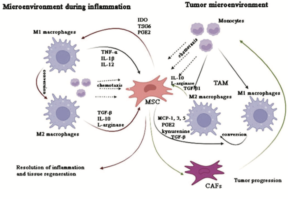
In the inflammatory microenvironment, through cytokine production, MSCs influence monocyte chemotaxis, favoring M2 phenotypes, which consequently leads to immunosuppression and resolution of inflammation. In the tumor microenvironment, MSCs influence the formation of tumor-associated macrophages (TAMs), which are predominantly M2 phenotypes, and under their influence, MSCs may differentiate into carcinoma-associated fibroblasts (CAFs), further supporting tumor progression.
Interaction between mesenchymal stem cells and macrophages in inflammatory conditions – diabetes mellitus and psoriasis as disease model systems
In chronic inflammatory conditions that lead to chronic diseases, such as diabetes mellitus (a chronic metabolic disorder characterized by low-grade inflammation) and psoriasis (an inflammatory immune skin disease), there is a disrupted balance between M1-like and M2-like macrophage types, which can result in permanent tissue damage [16],[17]. It has been shown that insulin resistance, which frequently occurs before the onset of type 2 diabetes (diabetes mellitus type II – DMT2), is fundamentally driven by a chronic, low-grade inflammatory state [18]. Specifically, TNF-α and IL-1β produced by adipose tissue macrophages, which exhibit characteristics of the M1 macrophage type, have been identified as key factors in the development of insulin resistance [19]. In patients with type 1 diabetes caused by an immune response, studies have shown increased M1-like macrophages. At the same time, it has been demonstrated that diabetic mice have increased CD169+ cells with stem cell retaining activity. It has also been shown that diabetes affects the release of stem cells via the stromal cell-derived factor-1 (SDF-1) secreted by M1- like macrophages, which prevents the release of MSCs from the bone marrow, resulting in a vicious cycle. Due to this process, the potential of MSCs to regenerate blood vessels and other organs is further impaired [20]. Therefore, it is urgent and important to utilize the potential of targeted therapies in controlling the balance between macrophages and MSCs and reducing comorbidities caused by the underlying disease.
Recent studies have shown that, in patients with psoriasis, MSCs exhibit abnormalities. These cells have a reduced ability to regulate inflammation and regeneration. Orciani et al. demonstrated that MSCs isolated from psoriatic skin lesions exhibit increased expression of vascular endothelial growth factor (VEGF) and inducible nitric oxide synthase (iNOS) [21]. Jiao et al. demonstrated that MSCs from skin lesions of psoriasis patients decreased the levels of TGF-β and its receptors, thus increasing the Th17/Treg ratio, which resulted in an immune imbalance in these patients [22]. MSCs isolated from the bone marrow of psoriasis patients also exhibited abnormalities. This aligns with research suggesting that psoriasis is associated with aberrant hematopoiesis [23].
It has also been noted that chronic psychological stress is one of the main factors in the development of psoriasis and that it simultaneously leads to the development of stress erythropoiesis with subsequent changes within the bone marrow microenvironment [24],[25],[26]. MSCs and their interactions with immune system cells may be the key players in the pathogenesis of this disease.
On the other hand, psoriatic lesions predominantly feature M1-like macrophages, which have been shown to secrete TNF-α and IL-23 [16]. However, in a mouse model of psoriasis, it has been demonstrated that the M2 phenotype plays a regulatory role [26]. Some studies suggest that macrophage depletion reduces inflammation [27]. In contrast, other studies on mice have demonstrated that the stimulation of glucocorticoid receptors in macrophages leads to M1-like macrophage polarization via phosphorylation of signal transducer and activator of transcription 1 (STAT1), whereas blocking this signaling pathway reduces inflammation, further highlighting the importance of stress in the development of psoriasis [28]. There is a limited body of research emphasizing the significance of MSC-macrophage interactions in the pathogenesis of psoriasis. Nonetheless, studies suggest a therapeutic potential of MSCs based on their ability to modulate the immune response by regulating macrophage polarization.
Interaction between mesenchymal stem cells and macrophages in the tumor microenvironment
Some hypotheses state that carcinogenesis and tumor growth are based on chronic inflammation, which disrupts the balance between proinflammatory and anti-inflammatory responses. Such an imbalance impairs an adequate immune response in eliminating tumor cells, thereby creating favorable conditions for tumor growth [29]. Many recent studies have highlighted the significant role of MSCs, macrophages, and their interactions in tumor development, due to their immunomodulatory capabilities. In addition to interacting with T and B lymphocytes during carcinogenesis, MSCs also establish interactions with macrophages, modulating their activity. Unlike the inflammation process, where the transition from M1-like to M2-like macrophage types leads to the resolution of the inflammatory process in the tumor microenvironment, M2-like macrophages contribute to cancer progression (Figure 1).
A special type of macrophage is present within the tumor microenvironment – tumor-associated macrophages (TAMs). These macrophages may exhibit phenotypic characteristics of both M1-like and M2-like macrophages and are a significant component of the immune system and the tumor microenvironment [30]. Research has shown that M1-like macrophages can eliminate tumor cells through phagocytosis or by activating the T-cell response [31].
In contrast, M2-like macrophages play a crucial role in tumor progression by secreting matrix metalloproteinases, facilitating the migration of tumor cells and tumor stromal cells. They also promote angiogenesis by releasing angiogenic molecules and expressing a range of enzymes involved in regulating angiogenesis [30]. M2-like macrophages are characterized by an increased expression of L-arginase, IL-10, and programmed cell death ligand 1 (PD-L1), which leads to immunosuppression and suppression of T-cell functions, ultimately promoting tumor growth [32]. Macrophages influence mesenchymal stem cells (MSCs) to acquire the carcinoma-associated fibroblast (CAF)-like phenotype, thereby promoting cancer progression.
MSCs, in combination with altered conditions within the tumor microenvironment, contribute to the formation of TAMs. MSCs, via PGE2 and kynurenine, with MIP-1α and MIP-2α produced by inflammatory macrophages, contribute to the conversion of inflammatory M1-like macrophages into alternatively activated, immunosuppressive M2-like macrophages [34]. In this way, MSCs and altered conditions within the tumor microenvironment modulate macrophage activity.
Studies on B16 F0 melanoma and EL4 lymphoma models have demonstrated that exosomes derived from tumor cells influence MSCs to enhance macrophage infiltration [35]. After macrophage infiltration within the tumor microenvironment, they polarize into the M2 tumor phenotypes under the influence of MCP1, MCP-3, and MCP-5, which are products of MSCs [36]. On the other hand, in breast cancer, MSC products – TGF-β, C1q, and semaphorins, contribute to the M2 polarization of macrophages [32]. As has been shown, chronic stress is one of the factors contributing to cancer development [37].
Yang et al. demonstrated that stress influences macrophage polarization, impacting cancer development [38]. On the other hand, in vivo studies have shown that stress mediators affect the intercellular interaction between MSCs and macrophages [39].
THERAPEUTIC POTENTIAL
In recent years, there has been a significant focus on the therapeutic potential of MSCs. Due to their strong immunomodulatory effects, MSCs and their derivatives show great potential in treating various inflammatory diseases. After application, MSCs migrate to sites of inflammation, which highlights their potential for treating such conditions [40]. However, MSCs obtained from different tissues express different cytokines that affect their immunomodulatory potential [41].
In addition to MSCs, numerous studies have been conducted on the therapeutic potential of MSC exosomes. MSC exosomes are considered to be potential carriers of various cytokines, immunomodulators, or even gene therapy. Studies have shown the therapeutic potential of MSCs in certain inflammatory diseases by regulating macrophage activity. It has been shown that PGE2, produced by MSCs influences macrophages by stimulating IL-10 production and inhibiting monocyte differentiation into dendritic cells [42]. The generation of PGE2 by MSCs is dependent upon TNF-α and iNOS signaling from monocytes/macrophages, indicating the complexity of their relationship [43].
In vivo and in vitro studies investigating the therapeutic efficacy of MSCs in wound healing have demonstrated that MSCs exert their immunomodulatory effects primarily through the stimulation of M2 macrophage polarization [44]. Many studies have also substantiated the therapeutic potential of MSCs in treating psoriasis. The administration of MSCs or MSC exosomes, intravenously or subcutaneously in psoriasis mouse models has resulted in significant immunomodulation and anti-inflammatory effects [45]. This manifested in the reduction of psoriasis symptoms, the normalization of epidermal differentiation, a reduction in angiogenesis and hyperproliferation, and a decrease in immune cell infiltration within the skin [42]. It has been shown that MSCs prevent keratinocyte proliferation by inhibiting TNF-α production by monocytes and macrophages [46].
Xie et al. have demonstrated that MSCs isolated from the umbilical cord reduce insulin resistance by reprogramming macrophages from the M1-like to the M2-like types in rats with type 2 diabetes mellitus [47]. Specifically, MSCs stimulate the M1-like macrophage phenotypes to increase IL-6 expression, which then leads to an increase in IL-4 receptor expression, resulting in the phosphorylation of signal transducer and activator of transcription 6 (STAT6) in macrophages and the subsequent transformation of macrophages into the M2-like phenotypes [47]. On the other hand, it has been shown that MSC treatment reduces macrophage infiltration by decreasing MCP-1 expression [48]. In diabetic nephropathy, bone marrow MSCs, umbilical cord MSCs, and their derived exosomes and microRNAs have been shown to enhance macrophage M2 polarization, leading to a pro-inflammatory response and reducing kidney damage caused by diabetes [49]. MSCs therapy has also proven beneficial in regulating mitochondrial dysfunction by stimulating arginase 1, a marker of M2-like macrophages [49].
Macrophage manipulation, in terms of activation and depletion, has shown potential therapeutic effects in inflammation and cancer. Adaptive macrophage therapy has proven effective in treating lymphoma and breast cancer. Ren et al. (2012) demonstrated that cancer-associated MSCs, which typically promote tumor growth, lose their function after specific eradication of macrophages and that in C-C chemokine receptor type 2 (CCR2)-deficient mice, the antitumorigenic effect of tumor-associated MSCs was reduced [36]. It has been shown that there is an increased production of MCP-1 after radiation therapy, which results in enhanced ability of tumors to attract MSCs [50].
CONCLUSION
Undoubtedly, the interactions between macrophages and MSCs are crucial in regulating the inflammatory microenvironment, significantly influencing disease onset and progression, as well as tissue repair and remodeling. However, their relationship can vary depending on the microenvironment. Studies suggest that modulating these interactions holds therapeutic potential, and a targeted approach tailored to specific microenvironment conditions could yield more effective results.
Informations

-
Keywords:
-
Received:
-
Revised:
-
Accepted:
-
Online first:
-
DOI:
-
Cite this article:

Sanja Momčilović
Institute for Medical Research, University of Belgrade
4 Dr Subotića Street, POB 39, 11129 Belgrade 102, Serbia
E-mail:
1. Costela-Ruiz VJ, Melguizo-Rodríguez L, Bellotti C, Illescas-Montes R, Stanco D, Arciola CR, et al. Different Sources of Mesenchymal Stem Cells for Tissue Regeneration: A Guide to Identifying the Most Favorable One in Orthopedics and Dentistry Applications. Int J Mol Sci. 2022 Jun; 23(11):6356. doi: 10.3390/ ijms23116356. [CROSSREF]
2. Song N, Scholtemeijer M, Shah K. Mesenchymal Stem Cell Immunomodulation: Mechanisms and Therapeutic Potential. Trends Pharmacol Sci. 2020 Sep;41(9):653-64. doi: 10.1016/j.tips.2020.06.009. [CROSSREF]
3. Wang LT, Liu KJ, Sytwu HK, Yen ML, Yen BL. Advances in Mesenchymal Stem Cell Therapy for Immune and Inflammatory Diseases: Use of CellFree Products and Human Pluripotent Stem Cell-Derived Mesenchymal Stem Cells. Stem Cells Transl Med. 2021 Sep;10(9):1288-303. doi: 10.1002/ sctm.21-002. [CROSSREF]
4. Li H, Dai H, Li J. Immunomodulatory properties of mesenchymal stromal/stem cells: The link with metabolism. J Adv Res. 2022 May;45:15-29. doi: 10.1016/j.jare.2022.05.012. [CROSSREF]
5. Jiang W, Xu J. Immune modulation by mesenchymal stem cells. Cell Prolif. 2020 Jan;53(1):e12712. doi:10.1111/cpr.12712. [CROSSREF]
6. Choi H, Lee RH, Bazhanov N, Oh JY, Prockop DJ. Anti-inflammatory protein TSG-6 secreted by activated MSCs attenuates zymosan-induced mouse peritonitis by decreasing TLR2/NF-κB signaling in resident macrophages. Blood. 2011 Jul;118(2):330-8. doi: 10.1182/blood-2010-12-327353. [CROSSREF]
7. Lendeckel U, Venz S, Wolke C. Macrophages: shapes and functions. ChemTexts. 2022 Apr;8(2):12. doi: 10.1007/s40828-022-00163-4. [CROSSREF]
8. Yu Y, Yue Z, Xu M, Zhang M, Shen X, Ma Z, et al. Macrophages play a key role in tissue repair and regeneration. PeerJ. 2022 Sep;10:e14053. doi: 10.7717/ peerj.14053. [CROSSREF]
9. Shapouri‐Moghaddam A, Mohammadian S, Vazini H, Taghadosi M, Esmaeili S, Mardani F, et al. Macrophage plasticity, polarization, and function in health and disease. J Cell Physiol. 2018 Sep;233(9):6425-40. doi: 10.1002/ jcp.26429. [CROSSREF]
10. Chen S, Saeed AFUH, Liu Q, Jiang Q, Xu H, Xiao GG, et al. Macrophages in immunoregulation and therapeutics. Sig Transduct Target Ther. 2023 May;8(1):207. doi: 10.1038/s41392-023-01452-1. [CROSSREF]
11. Kim J, Hematti P. Mesenchymal stem cell–educated macrophages: A novel type of alternatively activated macrophages. Exp Hematol. 2009 Dec;37(12):1445-53. doi: 10.1016/j.exphem.2009.09.004. [CROSSREF]
12. Luz-Crawford P, Djouad F, Toupet K, Bony C, Franquesa M, Hoogduijn MJ, et al. Mesenchymal Stem Cell-Derived Interleukin 1 Receptor Antagonist Promotes Macrophage Polarization and Inhibits B Cell Differentiation. Stem Cells. 2016 Feb;34(2):483-92. doi: 10.1002/stem.2254. [CROSSREF]
13. Carty F, Mahon BP, English K. The influence of macrophages on mesenchymal stromal cell therapy: passive or aggressive agents? Clin Exp Immunol. 2017 Apr;188(1):1-11. doi: 10.1111/cei.12929. [CROSSREF]
14. Jackson MV, Morrison TJ, Doherty DF, McAuley DF, Matthay MA, Kissenpfennig A, et al. Mitochondrial Transfer via Tunneling Nanotubes is an Important Mechanism by Which Mesenchymal Stem Cells Enhance Macrophage Phagocytosis in the In Vitro and In Vivo Models of ARDS. Stem Cells. 2016 Aug;34(8):2210-23. doi: 10.1002/stem.2372. [CROSSREF]
15. Chen L, Tredget EE, Wu PYG, Wu Y. Paracrine Factors of Mesenchymal Stem Cells Recruit Macrophages and Endothelial Lineage Cells and Enhance Wound Healing. PLoS ONE. 2008 Apr;3(4):e1886. doi: 10.1371/journal.pone.0001886. [CROSSREF]
16. Xia T, Fu S, Yang R, Yang K, Lei W, Yang Y, et al. Advances in the study of macrophage polarization in inflammatory immune skin diseases. J Inflamm (Lond). 2023 Oct;20(1):33. doi: 10.1186/s12950-023-00360-z. [CROSSREF]
17. Kraakman MJ, Murphy AJ, Jandeleit-Dahm K, Kammoun HL. Macrophage Polarization in Obesity and Type 2 Diabetes: Weighing Down Our Understanding of Macrophage Function? Front Immunol. 2014 Sep;5: 470. doi: 10.3389/ fimmu.2014.00470. [CROSSREF]
18. Olefsky JM, Glass CK. Macrophages, Inflammation, and Insulin Resistance. Annu Rev Physiol. 2010 Mar;72(1):219-46. doi: 10.1146/annurev-physiol021909-135846. [CROSSREF]
19. Fujisaka S, Usui I, Bukhari A, Ikutani M, Oya T, Kanatani Y, et al. Regulatory Mechanisms for Adipose Tissue M1 and M2 Macrophages in Diet-Induced Obese Mice. Diabetes. 2009 Nov;58(11):2574-82. doi: 10.2337/db08-1475. [CROSSREF]
20. Albiero M, Poncina N, Ciciliot S, Cappellari R, Menegazzo L, Ferraro F, et al. Bone Marrow Macrophages Contribute to Diabetic Stem Cell Mobilopathy by Producing Oncostatin M. Diabetes. 2015 Aug;64(8):2957-68. doi: 10.2337/ db14-1473. [CROSSREF]
21. Orciani M, Campanati A, Salvolini E, Lucarini G, Di Benedetto G, Offidani A, et al. The mesenchymal stem cell profile in psoriasis: The mesenchymal stem cell profile in psoriasis. Br J Dermatol. 2011 Sep;165(3):585-92. doi: 10.1111/j.1365-2133.2011.10438.x. [CROSSREF]
22. Jiao J, Zhao X, Wang Y, Liang N, Li J, Yang X, et al. Normal mesenchymal stem cells can improve the abnormal function of T cells in psoriasis via upregulating transforming growth factor-b receptor. J. Dermatol. 2022 Oct;9(10):F988-97. doi: 10.1111/1346-8138.16490. [CROSSREF]
23. Zhang Z, Yu Z, Tian P, Hou S, Han S, Tan X, et al. Differentially-expressed genes identified by suppression subtractive hybridization in the bone marrow hematopoietic stem cells of patients with psoriasis. Mol Med Rep. 2014 May;10(1):479-85. doi: 10.3892/mmr.2014.2203. [CROSSREF]
24. Kamiya K, Kishimoto M, Sugai J, Komine M, Ohtsuki M. Risk Factors for the Development of Psoriasis. Int J Mol Sci. 2019 Sep;20(18):4347. doi: 10.3390/ ijms20184347. [CROSSREF]
25. Momčilović S, Bogdanović A, Milošević MS, Mojsilović S, Marković DC, Kočović DM, et al. Macrophages Provide Essential Support for Erythropoiesis, and Extracellular ATP Contributes to a Erythropoiesis-Supportive Microenvironment during Repeated Psychological Stress. Int J Mol Sci. 2023 Jul;24(14):11373. doi: 10.3390/ijms241411373. [CROSSREF]
26. Hagert C, Sareila O, Kelkka T, Jalkanen S, Holmdahl R. The Macrophage Mannose Receptor Regulate Mannan-Induced Psoriasis, Psoriatic Arthritis, and Rheumatoid Arthritis-Like Disease Models. Front Immunol. 2018 Feb;9:114. doi: 10.3389/fimmu.2018.00114. [CROSSREF]
27. Wang H, Peters T, Kess D, Sindrilaru A, Oreshkova T, Van Rooijen N, et al. Activated macrophages are essential in a murine model for T cell-mediated chronic psoriasiform skin inflammation. J Clin Invest. 2006 Aug;116(8):2105- 14. doi: 10.1172/JCI27180. [CROSSREF]
28. Ge H, Mao Y, Chen W, Li Z, Yu Y, Luo S, et al. Stress Aggravates Imiquimod-Induced Psoriasiform Inflammation by Promoting M1 Macrophage Polarization. Int Immunopharmacol. 2023 Nov;124(Pt A):110899. doi: 10.1016/j. intimp.2023.110899. [CROSSREF]
29. Ostrand-Rosenberg S. Immune surveillance: a balance between protumor and antitumor immunity. Curr Opin in Genet Dev. 2008 Feb;18(1):11-8. doi: 10.1016/j.gde.2007.12.007. [CROSSREF]
30. Pan Y, Yu Y, Wang X, Zhang T. Tumor-Associated Macrophages in Tumor Immunity. Front Immunol. 2020 Dec;11:583084. doi: 10.3389/fimmu.2020.583084. [CROSSREF]
31. DeNardo DG, Ruffell B. Macrophages as regulators of tumour immunity and immunotherapy. Nat Rev Immunol. 2019 Jun;19(6):369-82. doi: 10.1038/ s41577-019-0127-6. [CROSSREF]
32. Biswas S, Mandal G, Chowdhury SR, Purohit S, Payne KK, Anadon C, et al. Exosomes Produced by Mesenchymal Stem Cells Drive Differentiation of Myeloid Cells into Immunosuppressive M2-Polarized Macrophages in Breast Cancer. J Immunol. 2019 Dec;203(12):3447-60. doi: 10.4049/jimmunol.1900692. [CROSSREF]
33. Zhang Q, Chai S, Wang W, Wan C, Zhang F, Li Y, et al. Macrophages activate mesenchymal stem cells to acquire cancer-associated fibroblast-like features resulting in gastric epithelial cell lesions and malignant transformation in vitro. Oncol Lett. 2019 Jan;17(1):747-56. doi: 10.3892/ol.2018.9703. [CROSSREF]
34. Vasandan AB, Jahnavi S, Shashank C, Prasad P, Kumar A, Prasanna SJ. Human Mesenchymal stem cells program macrophage plasticity by altering their metabolic status via a PGE2-dependent mechanism. Sci Rep. 2016 Dec;6:38308. doi: 10.1038/srep38308. [CROSSREF]
35. Lin LY, Du LM, Cao K, Huang Y, Yu PF, Zhang LY, et al. Tumour cell-derived exosomes endow mesenchymal stromal cells with tumour-promotion capabilities. Oncogene. 2016 Nov;35(46):6038-42. doi: 10.1038/onc.2016.131. [CROSSREF]
36. Ren G, Zhao X, Wang Y, Zhang X, Chen X, Xu C, et al. CCR2-Dependent Recruitment of Macrophages by Tumor-Educated Mesenchymal Stromal Cells Promotes Tumor Development and Is Mimicked by TNFα. Cell Stem Cell. 2012 Dec;11(6):812-24. doi:10.1016/j.stem.2012.08.013. [CROSSREF]
37. Vignjević Petrinović S, Milošević MS, Marković D, Momčilović S. Interplay between stress and cancer-A focus on inflammation. Front Physiol. 2023 Mar 20;14:1119095. doi: 10.3389/fphys.2023.1119095. [CROSSREF]
38. Yang J, Wei W, Zhang S, Jiang W. Chronic stress influences the macrophage M1-M2 polarization balance through β-adrenergic signaling in hepatoma mice. Int Immunopharmacol. 2024 Sep;10:138:112568. doi: 10.1016/j.intimp.2024.112568. [CROSSREF]
39. Momčilović S, Bogdanovic A, Milošević M, Gašić U, Kapor S, Vignjević Petrinović S. Interactions between mesenchymal stem cells and macrophage in stress erythropoiesis: roles for nitric oxide and purinergic signalling. Hemasphere 2024; 8(Suppl 1); 4098-9; Madrid, Spain, EHA2024, 13–16 June 2024.
40. Kidd S, Spaeth E, Dembinski JL, Dietrich M, Watson K, Klopp A, et al. Direct Evidence of Mesenchymal Stem Cell Tropism for Tumor and Wounding Microenvironments Using In Vivo Bioluminescent Imaging. Stem Cells. 2009 Oct;27(10):2614-23. doi: 10.1002/stem.187. [CROSSREF]
41. Hwang JH, Shim SS, Seok OS, Lee HY, Woo SK, Kim BH, et al. Comparison of Cytokine Expression in Mesenchymal Stem Cells from Human Placenta, Cord Blood, and Bone Marrow. J Korean Med Sci. 2009 Jan;24(4):547-54. doi: 10.3346/jkms.2009.24.4.547. [CROSSREF]
42. Diotallevi F, Di Vincenzo M, Martina E, Radi G, Lariccia V, Offidani A, et al. Mesenchymal Stem Cells and Psoriasis: Systematic Review. Int J Mol Sci. 2022;23(23):15080. doi: 10.3390/ijms232315080. [CROSSREF]
43. Németh K, Leelahavanichkul A, Yuen PST, Mayer B, Parmelee A, Doi K, et al. Bone marrow stromal cells attenuate sepsis via prostaglandin E2–dependent reprogramming of host macrophages to increase their interleukin-10 production. Nat Med. 2009 Apr;15(1):42-9. [Internet] Available from: https://www.nature.com/articles/nm.1905. [HTTP]
44. Zhang QZ, Su WR, Shi SH, Wilder-Smith P, Xiang AP, Wong A, et al. Human Gingiva-Derived Mesenchymal Stem Cells Elicit Polarization of M2 Macrophages and Enhance Cutaneous Wound Healing. Stem Cells. 2010 Oct;28(10):1856- 68. doi: 10.1002/stem.503. [CROSSREF]
45. Dairov A, Sekenova A, Alimbek S, Nurkina A, Shakhatbayev M, Kumasheva V, et al. Psoriasis: The Versatility of Mesenchymal Stem Cell and Exosome Therapies. Biomolecules. 2024 Oct; 14(11):1351. doi: 10.3390/biom14111351. [CROSSREF]
46. Ren X, Zhong W, Li W, Tang M, Zhang K, Zhou F, et al. Human umbilical cord-derived mesenchymal stem cells alleviate psoriasis through TNF-α/NFκB/MMP13 pathway. Inflammation. 2023 Jun;46(3):987-1001. doi: 10.1007/ s10753-023-01785-7. [CROSSREF]
47. Xie Z, Hao H, Tong C, Cheng Y, Liu J, Pang Y, et al. Human umbilical cord-derived mesenchymal stem cells elicit macrophages into an anti-inflammatory phenotype to alleviate insulin resistance in type 2 diabetic rats. Stem Cells. 2016 Mar;34(3):627-39. doi: 10.1002/stem.2238. [CROSSREF]
48. Lv S, Cheng J, Sun A, Li J, Wang W, Guan G, et al. Mesenchymal stem cells transplantation ameliorates glomerular injury in streptozotocin-induced diabetic nephropathy in rats via inhibiting oxidative stress. Diabetes Res Clin Pract. 2014 Apr;104(1):143-54. doi: 10.1016/j.diabres.2014.01.011. [CROSSREF]
49. Yuan Y, Li L, Zhu L, Liu F, Tang X, Liao G, et al. Mesenchymal stem cells elicit macrophages into M2 phenotype via improving transcription factor EB-mediated autophagy to alleviate diabetic nephropathy. Stem Cells. 2020 Jan;38(5):639-52. 639-52. doi: 10.1002/stem.3144. [CROSSREF]
50. Zielske SP, Livant DL, Lawrence TS. Radiation Increases Invasion of Gene-Modified Mesenchymal Stem Cells into Tumors. Int J Radiat Oncol Biol Phys. 2009 Nov;75(3):843-53. doi: 10.1016/j.ijrobp.2008.06.1953. [CROSSREF]