


Spontaneous resorption of large thoracic disc herniation: a case report
- Clinical Center of Serbia, Neurosurgery Clinic, Belgrade, Serbia
- “Zemun” Medical Center, Neurosurgery Department, Belgrade Serbia
ABSTRACT
Introduction: Thoracic disc herniation is a rare condition, accounting for 0.1 - 3.0% of all disc herniations, and may require complex surgical treatment.
Case Report: We are presenting a 40-year-old patient with Th9-Th10 level disc herniation, who showed clinical and radiological improvement after conservative treatment.
Conclusion: Our case demonstrates that, in patients with thoracic disc herniation, without myelopathy and with mild complaints, surgery should be delayed as much as possible due to the possibility of spontaneous herniated disc resorption.
INTRODUCTION
Thoracic disc herniation is a rare condition, accounting for 0.1 – 3.0% of all disc herniations, and may demand complex surgical treatment [1],[2]. The incidence of symptomatic thoracic disc herniation is estimated to be between 1 in 1,000 and 1 in 1,000,000 per year. Approximately 75% of all thoracic disc herniations are found below the level of Th8, with Th11–Th12 being the most common level, due to the higher amount of spinal mobility and weakness of the posterior longitudinal ligament. Spontaneous regression of the herniated disc was first described in 1945 by Key, most commonly involving the lumbar region with only a few cases of described spontaneous thoracic disc regression [1],[2],[3].
We report the case of a patient presenting with Th9–Th10 level disc herniation, who showed impressive clinical and radiological improvement after conservative treatment only.
CASE REPORT
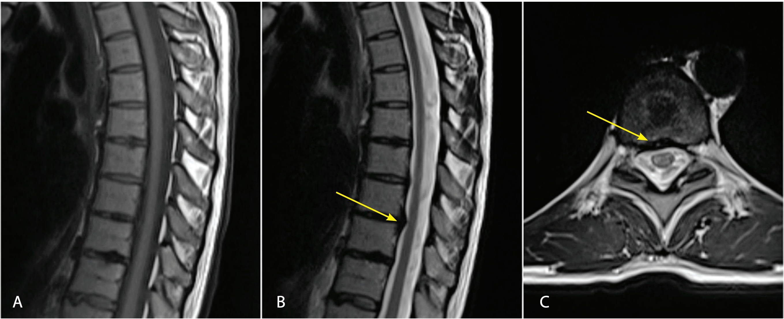
A 40-year old woman was examined in a different health care institution, complaining of pain in the right side of the thorax, difficulty breathing (during inhalation), and a feeling of heaviness in the lower extremities (more pronounced in the right leg), which had been ongoing for several months. A neurological exam showed paravertebral muscle spasm in the thoracic region, no motor weakness of the lower extremities, and absence of sphincter dysfunction. Lazarević’s sign was bilaterally negative. Pathological reflexes and sensory deficits were not present. Magnetic resonance imaging (MRI) of the cervical, thoracic, and lumbar regions was performed. A large Th9–Th10 disc herniation in the right dorsomedial region was observed, with compression of the Th9 spinal root and the anterior part of the spinal cord, without myelopathy (Figure 1). Surgery was suggested, but the patient declined. Therefore, physical therapy was performed.
A follow-up MRI was performed after two months, showing the unaltered lesion, but the clinical symptoms had improved (Figure 2). After another examination at the aforementioned institution, surgery was suggested once more, with a differential diagnosis of schwannoma established after post-contrast rim enhancement. The patient was then referred to our institution. Following an MRI re-examination, we found that the lesion had the characteristic appearance of disc herniation, and, since symptoms were minimal and already improved, we suggested non-surgical conservative treatment, including initial bedrest for several days, followed by physical therapy. The follow-up MRI of the thoracic spine, performed nine months after the first examination, revealed significant disc resorption (Figure 3). The total follow-up of the patient was 24 months, during which time no complication or neurological deterioration occurred.


DISCUSSION
Most disc herniations in the thoracic segment are asymptomatic. Clinical presentation varies depending on the level and the size of the lesion, most commonly presenting with pain in the thoracic region, with a radicular distribution, unilateral or bilateral sensory symptoms and muscle weakness, depending on the level and sphincter dysfunction [2].
Surgery is indicated in patients with motor weakness and sphincter dysfunction. Two major surgical approaches are: the posterolateral and anterior, which include either the transthoracic approach (trans-sternal, transpleural, retropleural) or thoracoscopy [3]. Due to a significant rate of complications such as: neurological deterioration, dural fistula formation, and pneumonia, special care should be taken when considering surgery in patients with mild symptoms [3],[4]. In our patient, despite the discal mass occupying a significant portion of the spinal canal, with an apparent compression effect on the spinal cord and spinal root, we decided to proceed with conservative treatment, due to the absence of myelopathy signs and lack of serious neurological symptoms.
Spontaneous resorption of a herniated disc is well documented at the cervical and lumbar levels [2],[5],[6],[7],[8]. Spontaneous resorption of thoracic disc herniation has been reported, but no major consensus has been achieved concerning the parameters that could definitely predict such an event [5]. It is believed that exposure of the disc to the epidural space, and consequently vascularization, play a major role in spontaneous herniating disc resorption [1],[9]. Neovascularization with macrophage-induced expression of matrix metalloproteinase-3 (MMP-3) and MMP-7 (which predominately degrade aggrecan and collagen proteins) facilitates the resorption process [1],[10],[11],[12]. Additionally, the tumor necrosis factor-alpha (TNF-α) induces vascular endothelial growth factor receptor (VEGFR) expression and neoangiogenesis, documented on the gadolinium-enhanced MRI as rim enhancement surrounding the herniated disc [1],[6],[13],[14],[15],[16],[17]. This rim enhancement, which was also present in our patient, can lead to misdiagnosis of intradural tumor. According to Autio et al., the thickness of the herniating disc’s rim enhancement on the MRI, the percentage of dislocation of the nucleus pulposus, and the age group of 41- 50 years, are strongly correlated with the occurrence of spontaneous resorption [1],[18]. Sakai reported a decrease of symptoms in 75% of cases where marked rim enhancement was evident [19]. Haro et al. suggested that patients experiencing no sphincter dysfunction and no progressive paresis, and showing apparent rim enhancement on the MRI, should undergo physical therapy before considering surgery [12]. Carreon et al. described the reaction of a herniating discus containing an end plate, emphasizing that the presence of endplate tissue diminishes neovascularization and further spontaneous resorption [6],[15],[16]. In addition to enzymatic degradation, two more mechanisms can be found in literature: dehydration and retraction into the intervertebral space [6],[8],[17],[18],[19]. Due to higher water content, the regression mechanism may include both dehydration and inflammation, especially in younger people. Age has been suggested as a factor influencing spontaneous resorption, probably since herniated discs in older people have less nucleus pulposus tissue and more cartilaginous annulus fibrosus tissue, which most likely inhibits the inflammatory cascade, resulting in a weaker immune response and neoangiogenesis in older patients [20]. Orief et al. suggested that, when the MRI determines high water content in the discs, conservative therapy should be carried out. The reduction of herniating mass seen on the MRI correlates well with symptom improvement. The same authors also reported a radicular pain recovery period of 3 - 6 weeks, while disc resorption visible on the MRI took 4 - 9 months to occur [6]. The same pattern was present in our patient, with clinical improvement preceding radiological proof of resolution.
The possible predictive signs of the positive evolution of thoracic disc herniation have not been fully elucidated. A study by Splendiani et al. demonstrated that MRI offers prognostic information about disk herniation evolution. Among 64 patients with 72 lumbar disk herniations, spontaneous MRI confirmed regression of disk herniation was seen in 34.72% of patients. Among these patients, free disc fragments (sequester) regressed in all cases, herniations with high signal intensity on T2W MRI sequences regressed in 85.18% of the cases, and herniations with peripheral contrast ring enhancement regressed in 83% of patients [21]. In our patient, MRI characteristics, especially high signal intensity on the T2W sequence, as well as peripheral post-contrast ring enhancement, were predictive of possible spontaneous resorption. The results of the Splendiani et al. study could probably be extrapolated to thoracic disk herniations, especially since they also showed that disc herniation evolution did not demonstrate any association with level, location, or size [20]. However, new prospective studies examining the evolution of discus hernias at the thoracic level are needed.
A recent study by Turk et al. showed that despite radiological improvement, indication for surgery still existed in a high number of patients with lumbar disc herniation, a substantial share of which developed permanent motor deficits [22]. However, the situation is more complex in the thoracic region. We believe that clinical follow-up, as well as radiological monitoring, is crucial for decision making, especially in patients with thoracic disc herniation initially presenting without neurological deficit and without clinical and MRI signs of myelopathy, which was the case with our patient.
CONCLUSION
In patients with thoracic disc herniation, without clinical or radiological signs of myelopathy, and with mild complaints, surgery should be delayed due to the possibility of spontaneous herniated disc resorption. Further studies identifying clinical and radiological signs predictive of possible thoracic disc resorption are needed. Also, studies of the physiology of spontaneous disk resorption are necessary to define therapeutic protocols, in order to avoid unnecessary surgery and surgical morbidity in patients with a high probability of spontaneous resolution.
Informations

-
Keywords:
-
Received:
-
Revised:
-
Accepted:
-
Online first:
-
DOI:
-
Cite this article:

Vuk Aleksić
Zemun Medical Center, Neurosurgery Department
9 Vukova Street, 11000 Belgrade, Serbia
E-mail:
1. Arce CA, Dohrmann GJ. Herniated thoracic disks. Neurol Clin. 1985; 3(2):383–92. [CROSSREF]
2. El-Kalliny M, Tew JM Jr, van Loveren H, Dunsker S. Surgical approaches to thoracic disc herniations. Acta Neurochir (Wien). 1991; 111(1-2):22–32. [CROSSREF]
3. Yoshihara H. Surgical treatment for thoracic disc herniation: an update. Spine (Phila Pa 1976). 2014; 39(6):E406–12 [CROSSREF]
4. Arts MP, Bartels RH. Anterior or posterior approach of thoracic disc herniation? A comparative cohort of mini-transthoracic versus transpedicular discectomies. Spine J. 2014; 14(8):1654–62. [CROSSREF]
5. Brauge D, Madkouri R, Clément R, Reina V, Brauge T, Gaillard S. What are the possibilities of spontaneous resorption of a thoracic disc herniation occupying more than 20% of the spinal canal in the asymptomatic subject? Comparative study. J Clin Neurosci. 2017; 44:269–73. [CROSSREF]
6. Orief T, Orz Y, Attia W, Almusrea K. Spontaneous resorption of sequestrated intervertebral disc herniation. World Neurosurg. 2012; 77(1):146–52. [CROSSREF]
7. Saal JA, Saal JS, Herzog RJ. The natural history of lumbar intervertebral disc extrusions treated nonoperatively. Spine (Phila Pa 1976). 1990; 15(7):683–6. [CROSSREF]
8. Teplick JG, Haskin ME. Spontaneous regression of herniated nucleus pulposus. AJR Am J Roentgenol. 1985; 145(2):371–5 [CROSSREF]
9. Komori H, Okawa A, Haro H, Muneta T, Yamamoto H, Shinomiya K. Contrast- -enhanced magnetic resonance imaging in conservative management of lumbar disc herniation. Spine (Phila Pa 1976). 1998; 23(1):67–73. [CROSSREF]
10. Haro H, Crawford HC, Fingleton B, MacDougall JR, Shinomiya K, Spengler DM, et al. Matrix metalloproteinase-3-dependent generation of a macrophage chemoattractant in a model of herniated disc resorption. J Clin Invest. 2000; 105(2):133–41. [CROSSREF]
11. Haro H, Crawford HC, Fingleton B, Shinomiya K, Spengler DM, Matrisian LM. Matrix metalloproteinase-7-dependent release of tumor necrosis factor-alpha in a model of herniated disc resorption. J Clin Invest. 2000; 105(2):143–50. [CROSSREF]
12. Haro H, Murakami S, Komori H, Okawa A, Shinomiya K. Chemonucleolysis with human stromelysin-1. Spine (Phila Pa 1976). 1997; 22(10):1098–104. [CROSSREF]
13. Haro H, Kato T, Komori H, Osada M, Shinomiya K. Vascular endothelial growth factor (VEGF)-induced angiogenesis in herniated disc resorption. J Orthop Res. 2002; 20(3):409–15. [CROSSREF]
14. Kato T, Haro H, Komori H, Shinomiya K. Sequential dynamics of inflammatory cytokine, angiogenesis inducing factor and matrix degrading enzymes during spontaneous resorption of the herniated disc. J Orthop Res. 2004; 22(4):895–900. [CROSSREF]
15. Carreon LY, Ito T, Yamada M, Uchiyama S, Takahashi HE. Neovascularization induced by anulus and its inhibition by cartilage endplate. Its role in disc absorption. Spine (Phila Pa 1976). 1997; 22(13):1429-34; discussion 1446–7. [CROSSREF]
16. Kokubun S, Sakurai M, Tanaka Y. Cartilaginous endplate in cervical disc herniation. Spine (Phila Pa 1976). 1996; 21(2):190–5. [CROSSREF]
17. Slavin KV, Raja A, Thornton J, Wagner FC. Spontaneous regression of a large lumbar disc herniation: report of an illustrative case. Surg Neurol. 2001; 56(5):333–6; discussion 7. [CROSSREF]
18. Autio RA, Karppinen J, Niinimäki J, Ojala R, Kurunlahti M, Haapea M, et al. Determinants of spontaneous resorption of intervertebral disc herniations. Spine (Phila Pa 1976). 2006; 31(11):1247–52. [CROSSREF]
19. Sakai T, Tsuji T, Asazuma T, Yato Y, Matsubara O, Nemoto K. Spontaneous resorption in recurrent intradural lumbar disc herniation. Case report. J Neurosurg Spine. 2007; 6(6):574–8. [CROSSREF]
20. Kawaguchi K, Harimaya K, Matsumoto Y, Hayashida M, Okada S, Iida K, et al. Effect of cartilaginous endplates on extruded disc resorption in lumbar disc herniation. PLoS One. 2018; 13(4):e0195946 [CROSSREF]
21. Splendiani A, Puglielli E, De Amicis R, Barile A, Masciocchi C, Gallucci M. Spontaneous resolution of lumbar disk herniation: predictive signs for prognostic evaluation. Neuroradiology. 2004; 46(11):916–22. [CROSSREF]
22. Turk O, Antar V, Yaldiz C. Spontaneous regression of herniated nucleus pulposus: the clinical findings of 76 patients. Medicine (Baltimore). 2019; 98(8):e14667. [CROSSREF]