


Two-stage repair of the intrathoracic portion of a left subclavian artery aneurysm after previous coronary artery bypass surgery
- Clinic for Vascular and Endovascular Surgery, Clinical Center of Serbia, Belgrade, Serbia
- Faculty of Medicine, University of Belgrade, Belgrade, Serbia
ABSTRACT
Introduction: We present a case of a 70-year-old patient who underwent successful two-stage repair of an intrathoracic left subclavian artery aneurysm (SAA) and who had previously undergone CABG.
Case presentation: The patient had previously undergone three-vessel coronary artery bypass graft (CABG) repair with the use of the left internal mammary artery (LIMA) to left anterior descending artery (LAD). Percutaneous coronary intervention (PCI) was performed as a back-up option in case of potential intraoperative LIMA-LAD bypass occlusion. Owing to the impossibility of hybrid repair due to a short proximal landing zone and aortic arch anomaly (bovine type), the vascular surgery review board decided to perform two-stage open surgery. Firstly, the patient underwent left carotid-subclavian (C-S) bypass with the Dacron graft, with the application of the standard supraclavicular approach. After a few days, the second procedure, through the left posterolateral thoracotomy was performed. The left subclavian artery (SA) was ligated distally to the SAA but proximally to the origin of the LIMA. The distal aortic arch at the site of the left SAA was clamped partially and the aneurysm was excised. The defect of the aortic arch was sutured and reinforced with a felt patch. Postoperatively, the patient had a good recovery, without any signs of myocardial injury or any surgery-related issues.
Conclusion: Careful planning and two-stage open surgical treatment of the left SAA, first with a left carotid-subclavian bypass, followed by aneurysm exclusion and suture of the outer aortic arch curvature may be a treatment option for patients not amenable to hybrid treatment.
INTRODUCTION
Subclavian artery aneurysms (SAAs) are rare, compared to aneurysms involving other peripheral arteries. SAA can result in severe complications and may lead to rupture, thrombosis, distal embolization and compression of adjacent structures [1]. There is paucity of data in literature regarding the surgical approach and the use of intraoperative techniques in the treatment of SAAs, especially when the aneurysm involves the first (intrathoracic) portion of the subclavian artery (SA). The situation becomes even more complicated in case of previous coronary artery bypass surgery (CABG) where the left internal mammary artery (LIMA) had been used as a graft. We present a case of a 70-year-old patient who underwent successful two-stage repair of an intrathoracic left SAA, and who had previously undergone coronary artery bypass grafting (CABG).
CASE REPORT
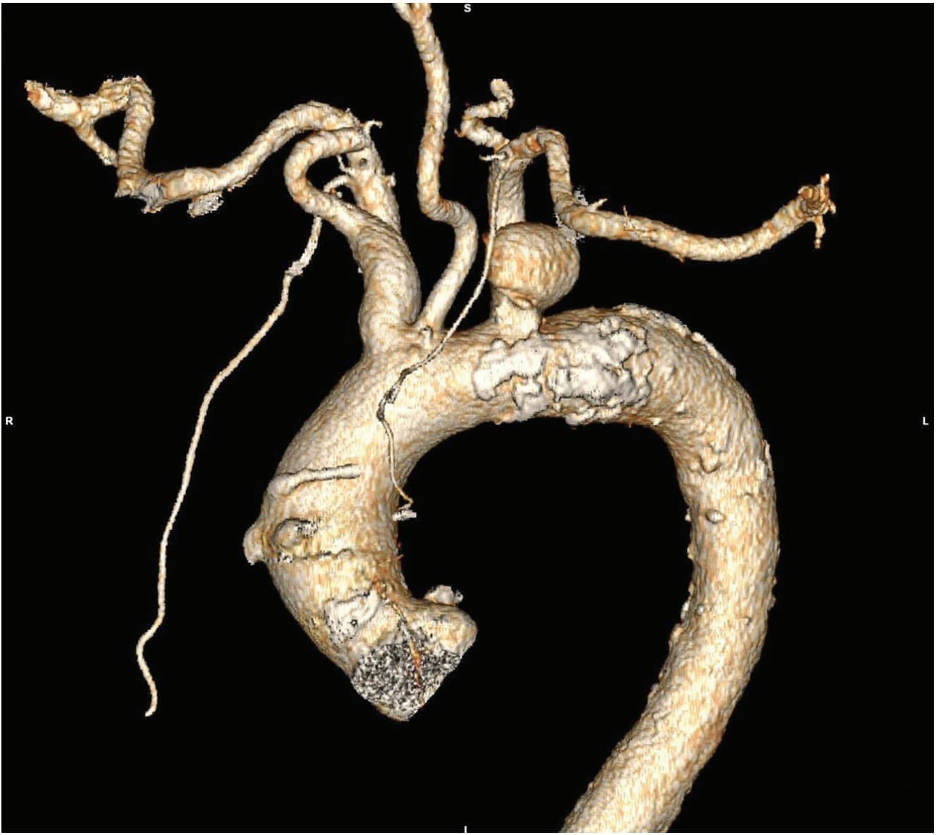
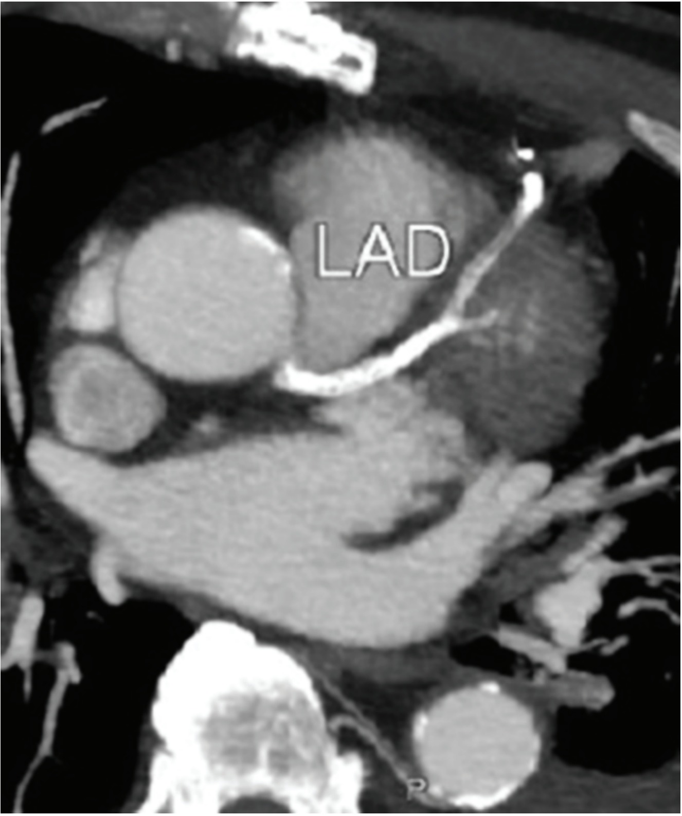
A 70-year-old patient was initially admitted to hospital due to a left saccular SAA of the first (intrathoracic) SA portion, presenting with dysphonia. Previously he had been operated on due to a large (70 mm) pararenal abdominal aortic aneurysm (AAA) and had undergone three-vessel CABG repair, performed with the use of the left internal mammary artery (LIMA) to left anterior descending artery (LAD) and two saphenous vein bypasses (one to the right coronary artery and the second to the circumflex coronary artery) (Figure 1). Coronary angiography was performed preoperatively. It was noted that both of the venous grafts as well as the LIMA-LAD bypasses were patent (Figure 2).
Due to a risk of occlusion of the LIMA-LAD during left SAA surgery, a joint cardiothoracic and vascular surgery board review meeting was convened, and it was decided, as a back-up strategy, to first perform a percutaneous coronary intervention (PCI) of the LAD, proximally to the LIMA-LAD bypass, in case of potential bypass occlusion during the vascular procedure.
The patient first underwent the planned PCI. The subsequent postintervention recovery was uneventful, and the patient was readmitted six months later for left SAA surgery. The patient had initially been considered for hybrid repair (aortic arch stent-graft implantation + extra-anatomic supra-aortic branch reconstruction). However, this was not feasible due to a short proximal landing zone associated with arch anomaly (bovine type). Due to the high risk that total aortic debranching carries (previous sternotomy with CABG), it was decided to perform two-stage open repair of the left SAA: first the left carotid-subclavian (C-S) bypass and second the excision of the left SAA through left posterolateral thoracotomy.
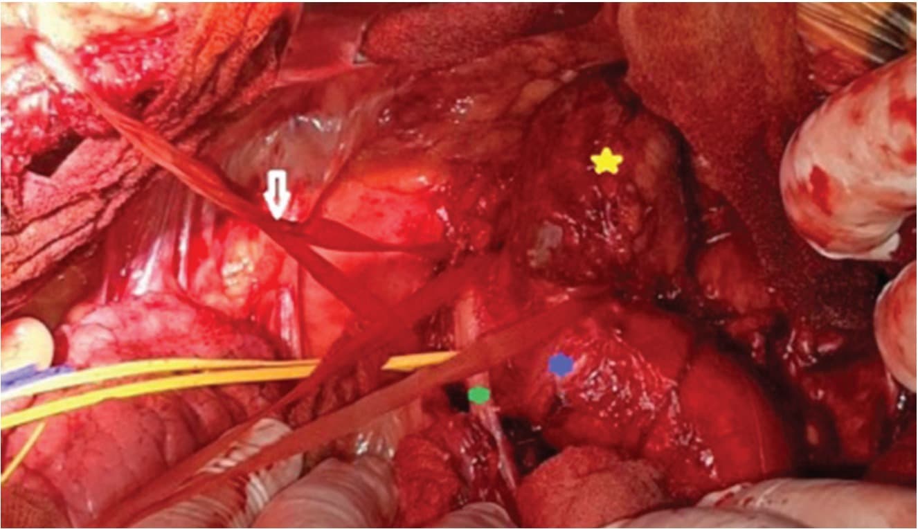
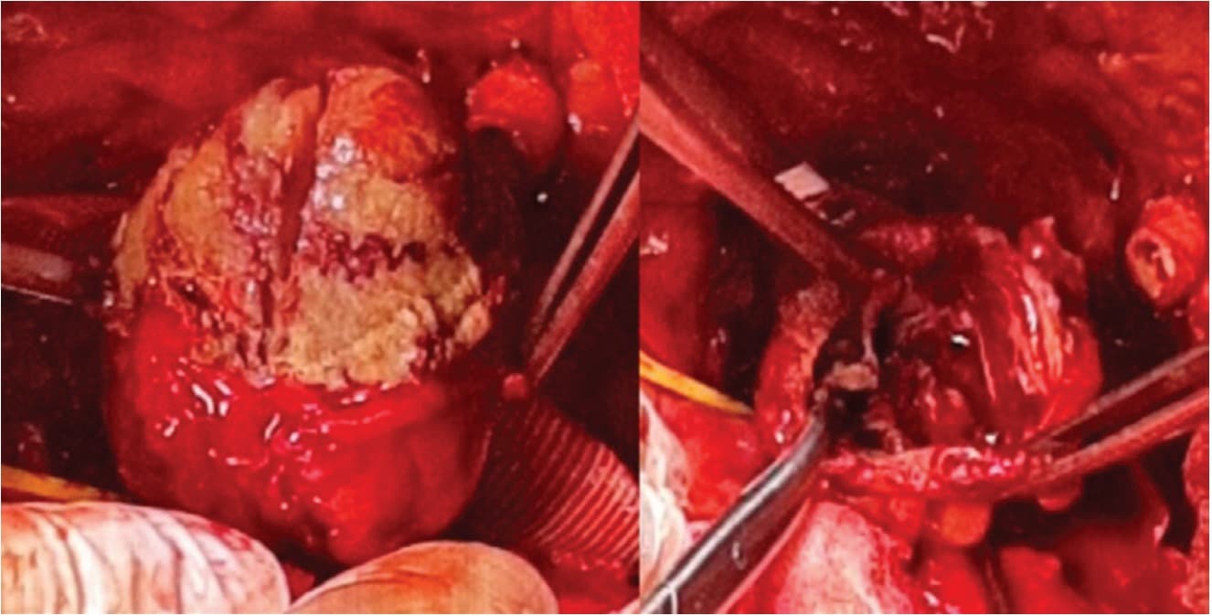
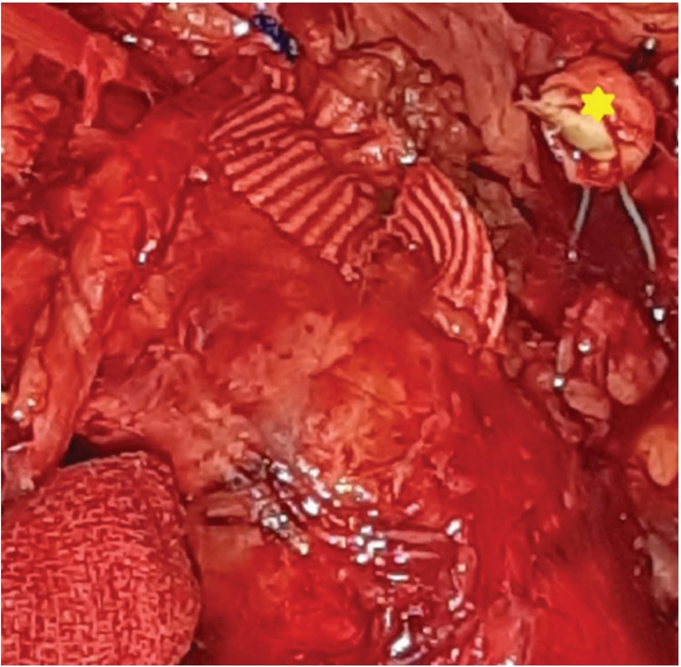
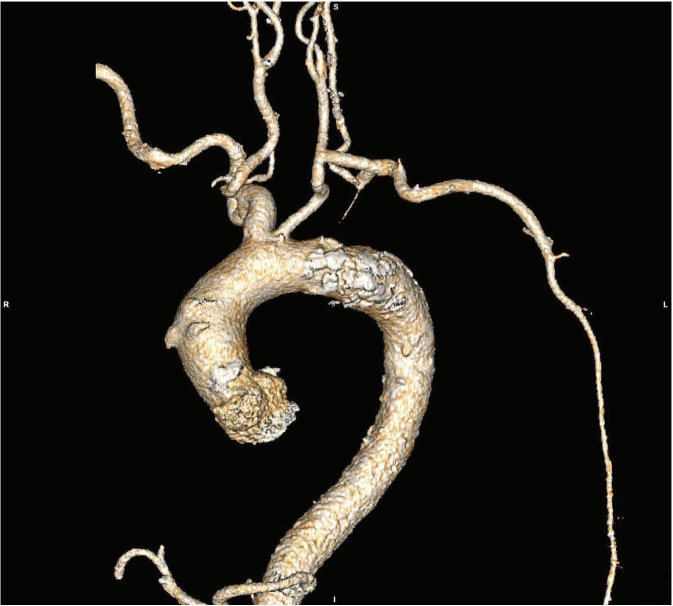
First, the patient underwent left C-S bypass with the Dacron graft with the application of the standard supraclavicular approach. The vertebral artery and LIMA could not be identified; therefore, the left SA was not proximally ligated. After a few days, the second procedure, with a definite excision of the left SAA, was performed. After endotracheal intubation with a double lumen bronchial tube and deflation of the left lung, left posterolateral thoracotomy was performed through the fifth intercostal space. All the elements of the distal aortic arch were identified (Figure 3). After systemic heparin administration (100 IU/kg), the left SA was ligated distally to the SAA and proximally to the origin of the LIMA, thus the LIMA-LAD bypass remained patent. The distal aortic arch was partially clamped at the site of the SAA and the aneurysm was opened (Figure 4). The defect of the aortic arch was sutured, and it was reinforced with a felt patch (Figure 5). After the procedure was completed, protamine-sulfate was administered and the left hemithorax was drained with the use of two drains. Postoperatively, the patient had a good recovery, without any signs of myocardial injury or any surgery-related issues. A follow-up angiography was performed before discharge, showing a patent LIMA-LAD bypass with a completely excluded left SAA and a patent left C-S bypass (Figure 6).
DISCUSSION
According to some data from literature, the SAAs represent only 1% of all repaired peripheral artery aneurysms [2]. The most common etiology of SAA is atherosclerosis. Other causes include posttraumatic dilatation, poststenotic dilatation - due to thoracic outlet syndrome [3], congenital arteriopathy (Kommerell diverticulum – aneurysm of the initial segment of the aberrant left SA) [4], and genetic arteriopathies (such as Marfan, Turner, Loeys-Dietz and vascular Ehlers-Danlos syndromes) [5],[6].
The aneurysm in this patient had an atherosclerotic etiology. The experience from the previous twelve years at our clinic, which includes 25 patients, shows that only 20% of all SAAs had an atherosclerotic etiology and that the same percentage had an intrathoracic localization [1]. In this series of patients with intrathoracic SAAs, there were 3 left SAAs and 2 right SAAs. The first three were treated with hybrid surgery (stent-graft implantation + extra-anatomic supra-aortic branch reconstruction) and the second two were treated with open surgery, with the application of the sternotomy approach.
The localization of the SAA dictates the surgical approach. The gold standard for extrathoracic SAA is open surgery, especially in genetic arteriopathies or in cases of thoracic outlet syndrome, where a simultaneous rib/muscle resection/embolectomy is often necessary. However, when it comes to intrathoracic SAAs, the surgical approach depends on the side where the aneurysm is located (right/left). On the right side, the preferred option is open surgery by means of median sternotomy. However, hybrid treatment with stentgraft implantation in the right SA with coverage of the common carotid artery and right subclavian-carotid bypass may be an option in high-risk patients.
The preferred treatment option for intrathoracic left SAA is the hybrid approach (stent-graft implantation + extra-anatomic supra-aortic branch reconstruction) due to a lower associated perioperative risk. Open surgery usually necessitates the use of partial extracorporeal circulation, temporary clamping of the left SA and reconstruction of the distal aortic arch with graft interposition. Unfortunately, in this patient, hybrid treatment was not possible due to a short and inadequate landing zone and a bovine aortic arch anomaly. The customary length of the landing zone that is considered sufficient is 15-20 mm, without any sharp angles in the lesser curvature of the aortic arch - this being necessary to provide an adequate proximal sealing zone and prevent type IA endoleak, which is considered to be procedure failure and needs to be corrected in order to prevent pulsatile flow into the aneurysm sac [7].
Consequently, a less invasive two-stage approach was chosen as a treatment option. Bearing in mind possible occlusion of the LIMA-LAD bypass during left SAA surgery, we performed PCI with recanalization of the LAD, fearing that the occlusion of the LIMA-LAD may produce severe and potentially fatal myocardial infarction.
The surgical procedure was performed in two stages. In the first stage we enabled adequate perfusion of the left arm and the left ventricle through a LIMA-LAD bypass with the help of a C-S conduit, while in the second one we performed the exclusion of the aneurysm with preservation of the flow through the LIMA-LAD with meticulous suture of the outer curvature of the distal aortic arch. There was no need for extracorporeal circulation and higher doses of intravenous heparin, whereby potential complications such as pulmonary (pulmonary hemorrhage, acute respiratory distress syndrome), coagulopathy, cooling of the patient, etc., were avoided [8].
Our case demonstrates the importance of selecting the right treatment option for each patient, especially in the presence of previous operations and cardiovascular comorbidity.
CONCLUSION
Careful planning and two-stage open surgical treatment of the left SAA, first with left carotid-subclavian bypass, followed by aneurysm exclusion and suture of the outer curvature of the aortic arch, in the second stage, may be a treatment option for patients not amenable to hybrid treatment.
Informations

-
Keywords:
-
Received:
-
Revised:
-
Accepted:
-
Online first:
-
DOI:
-
Cite this article:

Petar Zlatanović
Clinic for Vascular and Endovascular Surgery, Clinical Center of Serbia
8 Koste Todorovića Street, 11000 Belgrade, Serbia
E-mail:
1. Davidovic LB, Zlatanovic P, Ducic S, Koncar I, Cvetic V, Kuzmanovic I. Single center experience in the management of a case series of subclavian artery aneurysms. Asian J Surg. 2020 Jan; 43(1):139-47.[CROSSREF]
2. Lawrence PF, Gazak C, Bhirangi L, Jones B, Bhirangi K, Oderich G, et al. The epidemiology of surgically repaired aneurysms in the United States. J Vasc Surg. 1999 Oct; 30(4):632-40.[CROSSREF]
3. Davidović LB, Koncar IB, Pejkić SD, Kuzmanović IB. Arterial complications of thoracic outlet syndrome. Am Surg. 2009 Mar; 75(3):235-9.[CROSSREF]
4. Drullinsky D, Gill H, Bayne JP, Morin JF, Obrand D. Hybrid management of a ruptured right subclavian artery aneurysm dissection. J Vasc Surg Cases Innov Tech. 2017 Oct 9; 3(4):198-200.[CROSSREF]
5. Morisaki K, Kobayashi M, Miyachi H, Maekawa T, Tamai H, Takahashi N, et al. Subclavian artery aneurysm in Marfan syndrome. Ann Vasc Surg. 2012 Jul; 26(5):731.e1-4.[CROSSREF]
6. Beaulieu RJ, Lue J, Ehlert BA, Grimm JC, Hicks CW, Black JH 3rd. Surgical Management of Peripheral Vascular Manifestations of Loeys-Dietz Syndrome. Ann Vasc Surg. 2017 Jan; 38:10-16.[CROSSREF]
7. Riambau V, Böckler D, Brunkwall J, Cao P, Chiesa R, Coppi G, et al. Editor’s Choice - Management of Descending Thoracic Aorta Diseases: Clinical Practice Guidelines of the European Society for Vascular Surgery (ESVS). Eur J Vasc Endovasc Surg. 2017 Jan; 53(1):4-52.[CROSSREF]
8. Davidovic LB, Ilic N, Koncar I, Dragas M, Markovic M, Sindjelic R, et al. Some technical considerations of open thoracoabdominal aortic aneurysm repair in a transition country. Vascular. 2011 Dec; 19(6):333-7.[CROSSREF]
- Figure 1. Preoperative scan showing the saccular aneurysm of the intrathoracic part of the left subclavian artery with the patent LIMA-LAD bypass
- Figure 2. Preoperative scan showing the saccular aneurysm of the intrathoracic part of the left subclavian artery with the patent LIMA-LAD bypass
- Figure 3. Intraoperative photo showing all the anatomical elements of the distal aortic arch
- Figure 4. Intraoperative photo after the opening of the aneurysm sac with its thrombus - on the left; the aneurysm sac after thrombus removal - on the right
- Figure 5. Intraopearative photo after completed reconstruction
- Figure 6. Postoperative scan showing a patent left carotid-subclavian bypass, the absence of the left subclavian artery aneurysm, after its excision, and aortic arch suture