


Characteristics of patients with secondary erythrocytosis in relation to patients with polycythemia vera
- Clinic for Eye Diseases, Clinical Center of Serbia, Belgrade, Serbia
- Clinic of Hematology, Clinical Center of Serbia, Belgrade, Serbia
- Faculty of Medicine, University of Belgrade, Belgrade, Serbia
ABSTRACT
Introduction: Erythrocytosis represents elevated hemoglobin and hematocrit levels above the range of normal values. Primary erythrocytosis - polycythemia vera, is characterized by increased erythrocyte production, due to a disorder at the level of the multipotent stem cell in the bone marrow. On the other hand, secondary erythrocytosis (SE) is the result of bone marrow stimulation by an external factor.
Aim: The aim of our study was to determine parameters which are significant in differentiating SE from primary erythrocytosis - polycythemia vera (PV).
Materials and methods: This is a retrospective study involving 108 patients with SE and 111 patients with PV, who were diagnosed and treated at the Clinic of Hematology of the Clinical Center of Serbia (CCS), in the period: December 2005 – November 2018.From the patient records, the following data were extracted: demographic characteristics, laboratory parameters, spleen size, total red cell mass, serum erythropoietin (EPO) level, and spontaneous growth of the BFU-E colony.
Results: Patients with SE were younger, with a predominance of the male gender and with significantly higher serum EPO values than patients with PV. Patients with PV had significantly higher values of BFU-E, leukocyte and platelet count, spleen size, and LDH level than patients with SE. Total red cell mass analysis did not show a differential diagnostic significance.
Conclusion: Findings of normal spleen size, normal leukocyte and platelet count, normal serum LDH level, and elevated EPO, in patients, refer to the diagnosis of secondary erythrocytosis, while the findings of splenomegaly, leukocytosis, thrombocytosis, elevated serum LDH level, decreased EPO, and the presence of spontaneous BFU-E colony speak in favor of the diagnosis of polycythemia vera.
INTRODUCTION
Erythrocytosis represents elevated hemoglobin and hematocrit levels above the range of normal values. The regulation of erythropoiesis is a complex process which includes bone marrow sensitivity to oxygen and erythropoietin (EPO). The indicators of erythrocytosis are the following: the number of erythrocytes in peripheral blood, the hemoglobin (Hb) level, and the volume ratio – hematocrit (Hct).
Absolute erythrocytosis is defined as the state where the total red blood cell mass is above 125%, as compared to the reference range defined for sex and body mass, as analyzed by a highly specialized nuclear test [1]. Analysis of the total red blood cell mass enables differentiation between absolute and relative erythrocytosis, which is characterized by normal red blood cell mass, but a decreased plasma volume [2]. Erythrocytosis can be primary and secondary. Primary erythrocytosis is characterized by increased erythrocyte production, due to a disorder at the level of the multipotent stem cell in the bone marrow [3]. This is when spontaneous growth of the erythropoiesis precursor occurs, in the absence of erythropoietin (EPO) [3]. Primary erythrocytosis, also known as polycythemia vera – PV, is the result of the occurrence of an acquired mutation on the JAK2 gene [4],[5]. The JAK2-V617F mutation is described in 95% of patients, while the JAK2 exon 12 mutation is present in 3% of the patients with PV[4],[5]. These mutations lead to the production of constitutively active tyrosine kinase, which activates the erythropoietin receptor and the JAK-STAT signaling pathway, which, in turn, increases the production of erythrocytes, and often the accompanying production of leucocytes and thrombocytes.
On the other hand, secondary erythrocytosis (SE) happens as the result of the stimulation of bone marrow by an external factor. The most common states leading to secondary erythrocytosis are the following: hypoxemia caused by smoking, respiratory or cardiovascular diseases, as well as polycystic kidney disease or ectopic production of erythropoietin. Marked obesity is stated as a risk factor for secondary erythrocytosis, in case of the development of sleep apnea, i.e., sleep hypoxemia. When no cause can be defined, such a state is called idiopathic erythrocytosis.
The goal of our research was to determine the parameters relevant for the differentiation of secondary erythrocytosis (SE) from primary erythrocytosis – polycythemia vera (PV).
MATERIALS AND METHODS
This is a retrospective study involving 108 patients with the diagnosis of secondary erythrocytosis (SE) and 111 patients with the diagnosis of polycythemia vera (PV), who were diagnosed, monitored and treated at the Clinic of Hematology of the Clinical Center of Serbia, in the period between December 2005 and November 2018. In patients with PV, diagnosis was established on the basis of the criteria of the World Health Organization (WHO) from 2016 [7], (Table 1).

The following data was collected from medical records: 1) patient demographic characteristics; 2) complete blood count and white blood cell count parameters; 3) lactate dehydrogenase laboratory analyses; 4) spleen and liver size (determined with abdominal ultrasound – US); 5) erythrocyte volume; 6) presence of the erythropoiesis precursor in bone marrow or peripheral blood; 6) serum level of erythropoietin (EPO); 7) JAK2V617F mutation status.
The median follow-up for patients with SE was 32 months (range: 1 – 151), while in patients with PV it was 17 months (range: 1 – 62).
In statistical data processing, descriptive statistics methods were employed, and, depending on data distribution, parametric (Student’s t-test and analysis of variance – ANOVA) and nonparametric (the chi-squared test, the Mann-Whiteny test, the Kruskal-Wallis test) methods were employed for assessing the significance of the difference among the analyzed data. The univariate and multivariate Cox regression models were used to identify the predictors of secondary erythrocytosis in relation to polycythemia vera.
RESULTS
Patients with secondary erythrocytosis (SE)
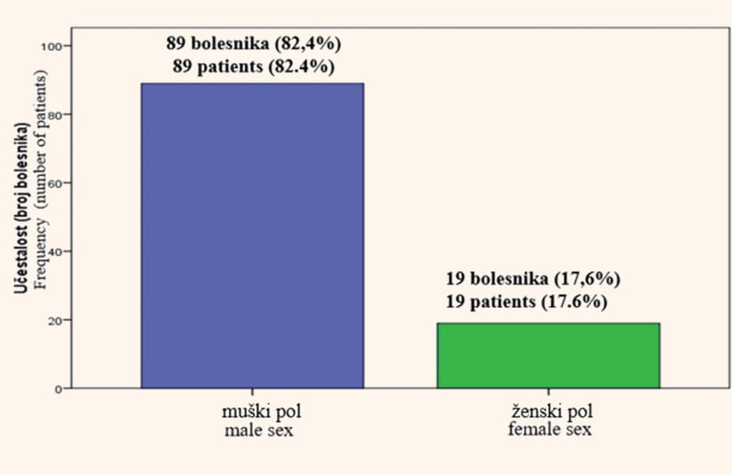
Among the 108 analyzed patients with SE, 89 were male subjects (82.4%), and 19 were female subjects (17.6%), (M:F = 4.7:1), (Figure 1). In the entire group of analyzed patients, the median age at the moment of diagnosis was 55 years (range: 19 – 88).
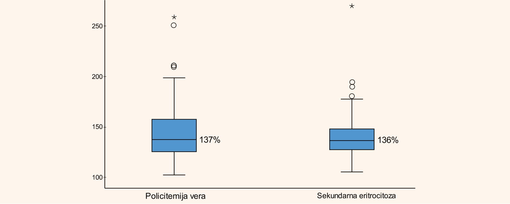
The median dimeter of the liver was 135 mm (range: 130 – 174), while the median diameter of the spleen was 100 mm (range: 90 – 150). The total erythrocyte mass was 136% (range: 105 – 269), (Figure 3). An elevated total erythrocyte mass of over 125%, which is characteristic of polycythemia vera, was present in 90 patients (83%). The median value of the oxygen saturation level (SO2) was 96% (range: 89.4 – 99). Below-normal SO2 (<92%) was noted in 5 patients. The median value of serum EPO was 9.3 IU/mL (range: 3.3 – 32.4). Among the 108 SE patients, analysis of the JAK2V617F mutation was carried out in 42 patients (38.8%), mostly during follow-up, and it was negative in all of the analyzed patients.
Patients with polycythemia vera (PV)
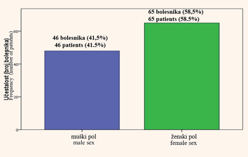
Among the 111 patients with polycythemia vera (PV) included in the study, 65 were female (58.5%) and 46 were male subjects (41.5%), (F:M = 1.4:1), (Figure2). In the entire group of analyzed patients, the median age at the moment of diagnosis was 62 years (range: 24 – 85). The median diameter of the liver was 135 mm (range 120 – 180), while the median diameter of the spleen was 121 mm (range: 90 – 255). A total of 15 patients (13.3%) were found to have hepatomegaly, while 52 patients (42%) had splenomegaly.
The median value of serum EPO was below-normal and amounted to 2 mU/mL (range: 0.06 – 31.8). Of the 101 analyzed patients, the JAK2V617F mutation was detected in 95% of the subjects, while 5% of the patients did not have this mutation. The total erythrocyte mass indicated the presence of absolute erythrocytosis amounting to 137% (range: 102 – 258), (Figure 3). The presence of absolute erythrocytosis, typical for polycythemia vera, was confirmed in 88 patients (79.2%).
As compared to SE patients, patients with PV were significantly older (p = 0.001), they had a larger spleen diameter (p < 0.001), a higher leukocyte count (12 ± 5.2x10⁹ /L vs. 7.7 ± 2.8 x10⁹ /L, p < 0.001), a higher platelet count (718 ± 354x109 /L vs. 221 ± 61x109 /L, p < 0.001), elevated serum LDH (559 ± 183 U/L vs. 360 ± 64 U/L, p < 0,001), lower serum erythropoietin (EPO) (2.5 ± 0.4 ml/mL vs. 13.8 ± 1.3 ml/mL, p < 0.001) and the presence of spontaneous BFU-E colonies (45±12 vs. 0, p = 0.011).
DISCUSSION
The present study analyzed demographic, clinical and laboratory characteristics of patients with SE and PV. The results have shown that patients with PV have a significantly higher white blood cell count, higher platelet count, higher LDH values, larger spleen size, and the presence of spontaneous BFU-E colonies, as compared to patients with SE. In patients with erythrocytosis, with a normal white blood cell count and platelet count, with a normal LDH level and a normally sized spleen, it is advised that the causes of secondary erythrocytosis should first be excluded.
In our study, the total erythrocyte mass did not demonstrate a statistically significant difference between SE patients and PV patients, and the significance of the total erythrocyte mass is reflected, most probably, in the differentiation between absolute and relative erythrocytosis [1],[3]. Serum EPO values in patients with SE are either normal or above-normal, while in patients with PV these values are typically below-normal, which indicates the significance of EPO analysis in the differentiation between secondary erythrocytosis and polycythemia vera. Below-normal serum EPO value is a minor criterion for diagnosing PV [6]. In patients with erythrocytosis it is recommended that secondary erythrocytosis causes should first be excluded. However, if the patient is a non-smoker without associated comorbidities, but has leukocytosis and/or thrombocytosis, as well as an above-normal LDH level, initial analysis of serum EPO can also be considered.
BFU-E colony values were present in patients with PV, as compared to SE patients, where these values were absent. The JAK2V617F mutation was detected in 95% of the patients with PV, which is in keeping with the above stated data from literature, while in SE patients, in whom this mutation was tested, it was not detected, which indicates that the analysis of this mutation can be significant in the differentiation between polycythemia vera and secondary erythrocytosis [4]. However, in our study, the JAK2V617F mutation was analyzed in 39% of the patients, which is a limiting factor for reaching a definitive conclusion.
CONCLUSION
Total erythrocyte mass did not demonstrate diagnostic significance in the differentiation of between primary and secondary erythrocytosis. The findings of splenomegaly, leukocytosis, thrombocytosis, above-normal serum LDH levels and below-normal serum EPO values, speak in favor of polycythemia vera. On the other hand, a normally sized spleen, a normal white blood cell count and platelet count, normal values of serum LDH and elevated EPO values, indicate secondary erythrocytosis. Therefore, in patients with erythrocytosis, who have a normal white blood cell and platelet count, a normal LDH value and a normally sized spleen, it is recommended that causes of secondary erythrocytosis should be excluded first, upon which serum EPO analysis should be performed, and the need for further hematological analysis considered, in relation to polycythemia vera.
LIST OF ABBREVIATIONS AND ACRONYMS
BFU-E – burst forming unit-erythroid
EPO – erythropoietin
Hb – hemoglobin
Hct – hematocrit
LDH – lactate dehydrogenase
PV – polycythemia vera
SE – secondary erythrocytosis
SO2 – oxygen saturation
WHO – World Health Organization
US – ultrasound
KCS – Clinical Center of Serbia
Informations

-
Keywords:
-
Received:
-
Revised:
-
Accepted:
-
Online first:
-
DOI:
-
Cite this article:

Danijela Leković
Clinic of Hematology, Clinical Center of Serbia, Belgrade, Serbia
2 Koste Todorovića Street, 11000 Belgrade, Serbia
E-mail:
1. Pearson TC, Guthrie DL, Simpson J, et al. Interpretation of measured red cell mass and plasma volume in adults: Expert Panel on Radionuclides of the International Council for Standardization in Haematology. Br J Haematol 1995; 89:748–56.[CROSSREF]
2. McMullin MF, Bareford D, Campbell P, et al. Guidelines for the diagnosis, investigation and management of polycythaemia/erythrocytosis. Br J Haematol 2005;130:174-95.[CROSSREF]
3. Prchal JF, Axelrad AA. Bone marrow responses in polycythemia vera. N Engl J Med. 1974;290(24):1382.[CROSSREF]
4. Scott LM, Tong W, Levine RL, et al. JAK2 exon 12 mutations in polycythemia vera and idiopathic erythrocytosis. N Engl J Med 2007; 356:459–68.[CROSSREF]
5. Kralovics R, Passamonti F, Buser AS, et al. A gain-of-function mutation of JAK2 in myeloproliferative disorders. N Engl J Med. 2005;352(17):779–90. [CROSSREF]
6. Gagnon DR, Zhang TJ, Brand FN. Hematocrit and the risk of cardiovascular disease—the Framingham study: a 34-year follow-up. Am Heart J 1994;127:674-82.[CROSSREF]
7. Barbui T, Thiele J, Gisslinger H, et al. The 2016 WHO classification and diagnostic criteria for myeloproliferative neoplasms: document summary and in-depth discussion. Blood Cancer J. 2018;8(2):15.[CROSSREF]
- Table 1. WHO criteria (2016) for PV diagnosis*
- Figure 1. Distribution of patients with secondary erythrocytosis by sex (male/ female)
- Figure 2. Distribution of patients with polycythemia vera by gender (male/ female)
- Figure 3. Total red cell mass in patients with polycythemia vera and in patients with secondary erythrocytosis (%)