


An interesting case of coronary-cameral fistula with angina pectoris
- University Clinical Center of Serbia, Clinic for Cardiac Surgery, Belgrade, Serbia
- University of Belgrade, Faculty of Medicine, Belgrade, Serbia
- University Clinical Center of Serbia, Center for Anesthesia, Belgrade, Serbia
ABSTRACT
Introduction: Coronary-cameral fistula (CCF) is an anomalous connection between a coronary artery and a cardiac chamber. Most CCFs are discovered incidentally during angiographic evaluation of coronary vascular disorders. We report a case of CCF with angina pectoris.
Case report: A 67-year-old woman presented with chest pain and dyspnea upon exertion. Coronary angiography showed atherosclerotic lesions in the two major coronary arteries, but also communication between three arteries and the cavity of the right ventricle (RV) through many small, diffuse fistulas. Angiography also showed a fistula between the proximal left anterior descending artery (LAD) (first septal branch) and the right ventricle, as well as between the proximal right coronary artery (RCA) (acute marginal branch) and the right ventricle. The patient qualified to undergo coronary artery bypass graft surgery (CABG) and surgical closing of the fistulas, which is why we performed, on a beating heart, double vessel revascularization by autovein graft between the ascending aorta and the RCA and between the ascending aorta and the LAD, as well as closing of the fistulas with hemoclips and polypropylene suture, with a teflon pledget.
Conclusion: Hemodynamically insignificant fistulas, which are clinically silent and not associated with other abnormal findings, most commonly do not require further treatment. Large, hemodynamically significant fistulas should be closed by ligation. However, smaller fistulas tend to get larger with age and it is recommended that early elective closure is performed in patients experiencing symptoms or in asymptomatic patients with a continuous murmur or a systolic murmur with an early diastolic component.
INTRODUCTION
Coronary-cameral fistula (CCF) is an anomalous connection between a coronary artery and a cardiac chamber. Most CCFs are discovered incidentally during angiographic evaluation of coronary vascular disorders. It was previously believed that coronary-cameral fistula, first described by Krause in 1865, is a rare anomaly of the coronary arteries [1]. We present a case of coronary-cameral fistula with angina pectoris. Selective coronary angiography revealed diffuse CCF, which included the right coronary artery (RCA) (Figure 1) and the left anterior descending artery (LAD) (Figure 2), voiding into the right ventricle (RV), as well as double coronary vessel disease.
CASE PRESENTATION
A 67-year-old woman, without known risk factors for coronary disease, presented with chest pain and dyspnea upon exertion. Her vital signs were normal. Echocardiographic examination showed preserved ejection fraction of the left ventricle (LV) – 60%, and absence of valvular heart disease, however moderate hypertrophy of the left ventricle with diastolic dysfunction was observed.
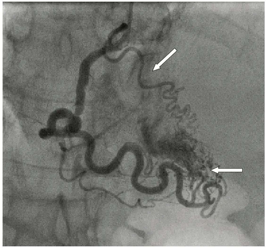
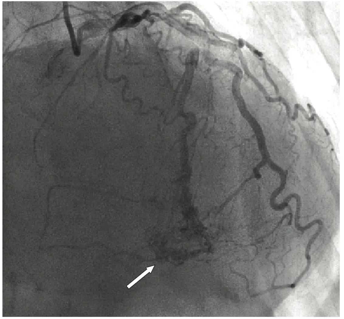
Coronary angiography showed atherosclerotic lesions in the two main coronary arteries, and it was also discovered that three coronary arteries were in communication with the right ventricular cavity through many small, diffuse fistulas (Figure 1 and Figure 2), which resulted in complete contrast opacification in the right ventricle. The coronary sinus was normal in size. Selective coronarography revealed a 90 – 99% stenosis in the distal segment of the right coronary artery (RCA) and an 80% stenosis in the short segment of the left anterior descending artery (LAD), spreading into the other, diagonal branch.
Angiography also showed a fistula between the proximal left anterior descending artery (LAD) (first septal branch) and the right ventricle, as well as between the proximal right coronary artery (RCA) (acute marginal branch) and the right ventricle.
Two-dimensional transthoracic echocardiography showed normal diameters of the cardiac ventricles. The trunk of the pulmonary artery was overflown with blood. The systolic pressure of the right ventricle was moderately elevated – up to the value of 32 mmHg. Color doppler echocardiographic examination indicated the presence of coronary artery fistula. The patient had not previously undergone any invasive cardiological diagnostic or therapeutic procedure that could be connected with CCF, and in her family anamnesis, she negated the existence of coronary-cameral fistula.
The patient’s condition indicated the need for surgical revascularization of the myocardium (coronary artery bypass graft surgery – CABG) and surgical closing of the fistula.
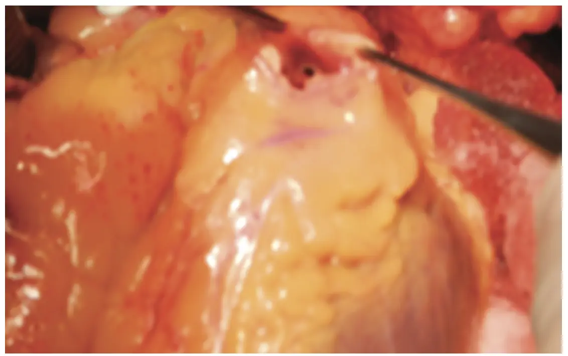
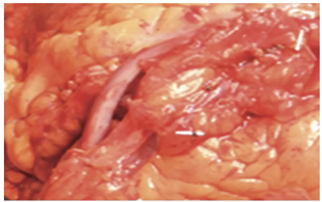
In the case of our patient, we performed, on a beating heart, double vessel revascularization by autovein graft between the ascending aorta and the RCA, and between the ascending aorta and the LAD. The fistulas were secured with two hemoclips and polypropylene suture 4-0, with a teflon pledget (Figure 3 and Figure 4). The patient was discharged from hospital in good general condition.
DISCUSSION
Coronary artery fistula is an abnormal direct connection between a coronary artery and a cardiac chamber (coronary-cameral fistula) or vein (coronary arteriovenous fistula). Coronary artery fistulas occur with an incidence of 0.1%. Rarely, they may cause myocardial ischemia, due to coronary steal syndrome, cardiac insufficiency, or spontaneous intrapericardial rupture [2]. Sixty percent of these fistulas originate from the right coronary artery, while 90% end on the right side of the heart. Coronary-cameral fistulas, from all three major coronary arteries, which end in the right ventricle, are a rare occurrence.
Coronary-cameral fistulas are most commonly congenital and may be connected to the normally occurring Thebesian veins, which drain blood from the coronary arteries after it passes through the capillaries into the cardiac ventricles [3]. They are usually clinically insignificant, and do not result in clinical symptoms and signs. Depending on the size and localization of the fistulas, in some cases, surgical ligation, epicardially or endocardially, or a percutaneous endoluminal procedure (embolization) may be performed. The procedure is difficult or impossible when the fistulas are diffuse. Therefore, despite ischemia, such a procedure was not contemplated in our patient [4].
Yamanaka and Hobbs found fistulas of small coronary arteries in 0.18% of 126,595 patients who had undergone cardiac catheterization [2]. Fistulas of this type usually develop from one coronary artery branch and drain into one of the cardiac ventricles or into one blood vessel, and are most commonly not associated with clinical signs and symptoms [6]. Nevertheless, large fistulas with a significant shunt result in signs such as the presence of a persistent murmur and symptoms such as dyspnea upon exertion, exercise intolerance, or congestive heart failure, which is when surgical treatment is necessary [5],[6],[7]. The main sites of fistula development are the right coronary artery (55%), the left coronary artery (35%), and both coronary arteries (5%). The main sites where fistulas most commonly drain are the right ventricle (40%), the right atrium (26%), pulmonary arteries (17%), less frequently the superior vena cava or the coronary sinus, and least frequently the left atrium and the left ventricle [8].
Informations

-
Keywords:
-
Received:
-
Revised:
-
Accepted:
-
Online first:
-
DOI:
-
Cite this article:

Dejan Lazović
Clinic for Cardiac Surgery, University Clinical Center of Serbia
2 Pasterova Street, 11000 Belgrade, Serbia
E-mail:
1. Krause W. Ueber den Ursprung einer akzessorischen A. coronaria aus der A. pulmonalis. Z Ratl Med 1965; 24:225-9.
2. Yamanaka O, Hobbs RE. Coronary artery anomalies in 126,595 patients undergoing coronary arteriography. Cathet Cardiovasc Diagn. 1990 Sep;21(1):28- 40. doi: 10.1002/ccd.1810210110.[CROSSREF]
3. Iadanza A, del Pasqua A, Fineschi M, Pierli C. Three-vessel left-ventricular microfistulization syndrome: a rare case of angina. Int J Cardiol. 2004 Jul;96(1):109-11. doi: 10.1016/j.ijcard.2003.04.052.[CROSSREF]
4. Iyisoy A, Arslan Z, Ozmen N, Kursaklioglu H, Amasyali B, Demirtas E. Double coronary fistulas between coronary artery and left ventricle: a case report [in Turkish]. Gulhane Tip Dergisi 2003;45:203-5.
5. Alkhulaifi AM, Horner SM, Pugsley WB, Swanton RH. Coronary artery fistulas presenting with bacterial endocarditis. Ann Thorac Surg. 1995 Jul;60(1):202-4.[CROSSREF]
6. Kaplan JD, Redberg RF. Coronary to pulmonary artery fistula detected by transthoracic echocardiography. Am Heart J. 1995 Apr;129(4):839-40. doi: 10.1016/0002-8703(95)90343-7.[CROSSREF]
7. Kidawa M, Peruga JZ, Foryś J, Krzemińska-Pakuła M, Kasprzak JD. Acute coronary syndrome or steal phenomenon - a case of right coronary to right ventricle fistula. Kardiol Pol. 2009 Mar;67(3):287-90.[HTTP]
8. Funabashi N, Komuro I. Aberrant fistula arteries from the left main branch and right coronary artery to the left pulmonary arterial sinus demonstrated by multislice computed tomography. Int J Cardiol. 2006 Jan 26;106(3):428- 30. doi: 10.1016/j.ijcard.2005.01.036.[CROSSREF]
- Figure 1. Selective coronary angiography showing a maze of fine vessels arising from the right coronary artery (proximal and distal) and communicating with the right ventricular cavity
- Figure 2. Selective coronary angiography showing multiple fistulas arising from the proximal left anterior descending coronary artery - first septal branch and communicating with the right ventricular cavity
- Figure 3. The ostium of the fistula between the RCA and the RV
- Figure 4. Ligated and sutured fistula, by pass from the aorta to the right coronary artery