


Clinical and serological features in an 11-month-old infant with paroxysmal cold hemoglobinuria
- Institute for Mother and Child Health Care of Serbia “Dr Vukan Čupić”, Belgrade, Serbia
- University of Belgrade, Faculty of Medicine, Belgrade, Serbia
ABSTRACT
Introduction: Paroxysmal cold hemoglobinuria (PCH) is an uncommon form of autoimmune hemolytic anemia (AIHA). A specific etiological factor has not been determined yet. The pathogenesis of the disease is explained by the presence of “biphasic hemolysin” or low-affinity IgG autoantibody, which in most cases binds to the erythrocyte P antigen, binding components of complement until the complete activation of the cascade and hemolysis of erythrocytes. The disease typically presents in children following an acute infection, usually involving the upper respiratory tract or gastroenteritis.
Case report: We present an 11-month-old female infant who was admitted to the Intensive Care Unit of the Institute due to vomiting, anemia, yellow discoloration of the skin and mucous membranes, dark-red-colored urine, and general weakness. Two days prior to admission, she had a nasal discharge. Initial blood tests revealed a hemoglobin level of 63 g/L, hematocrit at 17.3%, reticulocyte count of 1.76%, leukocytosis, thrombocytosis, elevated C-reactive protein at 75.7 mg/L, lactate dehydrogenase at 5365 IU/L, total/indirect bilirubin at 67.9/64.5 µmol/L, and decreased haptoglobin. No schizocytosis was evident in peripheral blood. Polyspecific direct antiglobulin test (DAT) was positive 3+ and monospecific DAT was C3d 1+. The infant was treated with intravenous immunoglobulins, a single unit of red blood cells transfusion, parenteral dual antimicrobial therapy, and a single dose of corticosteroids accompanied by intravenous hydration and correction of electrolyte disturbances. On the tenth day upon admission, blood samples were taken for the Donath-Landsteiner (DL) test, which showed the presence of “biphasic hemolysin” and confirmed the diagnosis of PCH.
Conclusion: Clinical recognition of PCH, the transient nature of “biphasic hemolysin” and the possibility of performing the DL test depend on the timely confirmation diagnosis of this rare form of AIHA.
INTRODUCTION
Paroxysmal cold hemoglobinuria (PCH) represents a rare and frequently underrecognized type of autoimmune hemolytic anemia (AIHA), characterized by intravascular hemolysis and hemoglobinuria. A specific etiological factor has not been determined yet. The pathogenesis of the disease is attributed to the presence of “biphasic hemolysin,” a low-affinity polyclonal IgG autoantibody. This autoantibody binds exclusively to erythrocytes in the peripheral circulation at temperatures lower than body temperature. In this process, the binding of complement components occurs. Although the antibody dissociates in the central circulation, the complement remains bound, leading to the complete activation of the cascade and subsequent hemolysis of erythrocytes at temperatures around 37°C. A key element in the pathophysiology of the disease is the repeated binding and dissociation of antibodies in normal circulation. In the past, PCH was a chronic manifestation of tertiary syphilis or congenital syphilis. In 1904, Austrian doctors Julius Donath and Karl Landsteiner demonstrated the biphasic nature of autoantibodies in vitro and developed a test that is still used. For this reason, the alternative names Donath-Landsteiner antibody and Donath-Landsteiner hemolytic anemia are used with equal frequency [1],[2],[3].
Over the past 60-80 years, the epidemiology and clinical picture have changed significantly. Nowadays, the disease primarily manifests in children as acute intravascular hemolysis associated with infectious agents including viral infections (adenovirus, influenza A, cytomegalovirus, Epstein-Barr virus, measles virus, mumps virus) and bacterial infections (Hemophilus influenzae, Mycoplasma pneumoniae, Escherichia coli). In clinical practice, individual cases of PCH have been confirmed following the administration of mRNA vaccine against COVID-19, heptavalent pneumococcal conjugate vaccine, and measles vaccine. Paroxysmal cold hemoglobinuria can rarely be associated with lymphoproliferative and autoimmune diseases. In a small number of patients, hemolysis occurs without a known cause (primary form) [4],[5],[6],[7].
The disease typically presents in children following an acute infection, usually involving the upper respiratory tract or gastroenteritis. The average time interval from the onset of infection to the manifestation of PCH is two weeks (two days to five weeks). The disease has a sudden onset, with hemoglobinuria present in more than twothirds of patients, followed by fever, jaundice, and /or conjunctival icterus in more than a quarter of patients. Urine has the characteristic dark red, even brown color. Severe anemia is often present. The direct antiglobulin test (DAT) usually shows positivity only for complement [8].
The diagnosis of PCH is confirmed by Donath-Landsteiner (DL) test. The detection of pathognomonic DL antibodies poses a laboratory challenge. It requires a specific sample preparation, incubation at different temperatures, and well-educated personnel [9].
Paroxysmal cold hemoglobinuria is a transient disease. It typically resolves spontaneously within a span of a few days to a few weeks.
The aim of this paper is to present a patient in whom a rare form of AIHA, PCH, was confirmed by the DL test. In the Republic of Serbia DL test can currently be done at the Blood Transfusion Department of the Institute for Mother and Child Health Care of Serbia.
CASE PRESENTATION
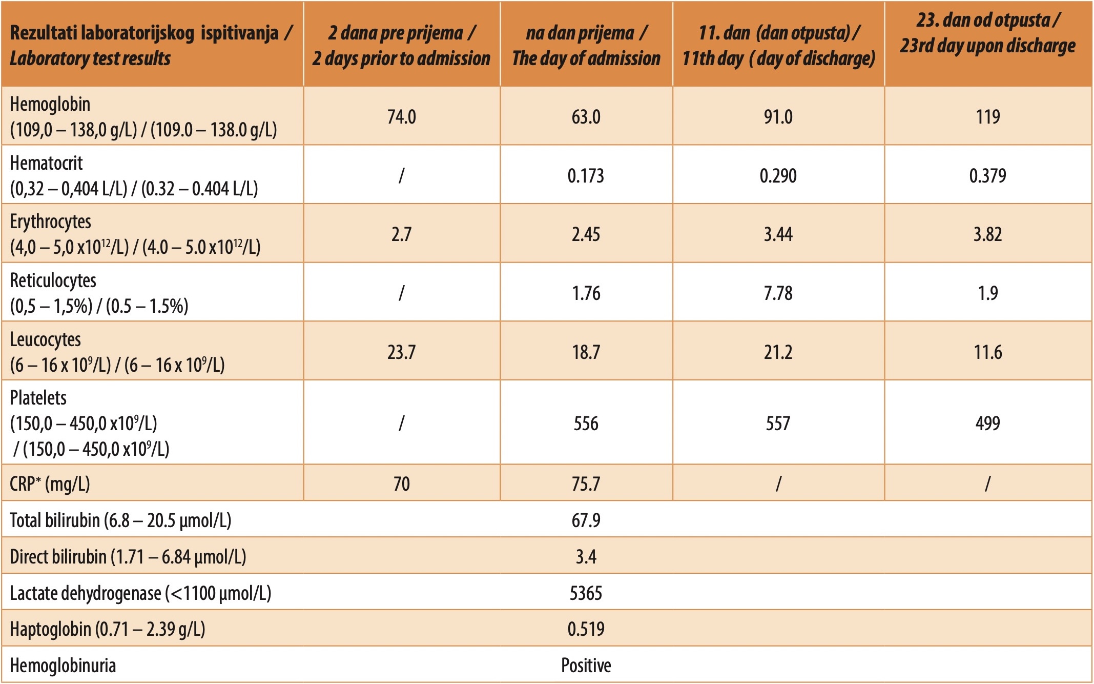
An 11-month-old female infant was transferred from a regional health center (RHC) due to vomiting, yellow discoloration of the skin and mucous membranes, and overall lethargy. She did not have a fever. Two days prior to admission, she had nasal discharge, and her mother gave her Aerius syrup. The results of the laboratory analyses done at the RHC were as follows: leucocytes 23,7 x 10⁹ /L, hemoglobin 74 g/L, erythrocytes 2,7 x 10¹² /L, CRP 70 mg/l, bilirubin 126 µmol/L. She was examined by a surgeon, who ruled out any acute surgical condition.
An infant weighing 9.670 kg and measuring 75 cm in length was admitted to the Intensive Care Unit (ICU) of the Institute. On admission to the ICU, she was found to be in a disoriented state, tachycardic with a heart rate of approximately 160 bpm, and with yellow discoloration of the skin and mucous membranes. The pharynx was slightly hyperemic, with a clear nose and no secretions present. After insertion of a urinary catheter, the obtained urine was dark red in color, with the presence of proteinuria, hematuria, and urate cylinders. Ultrasound examination of the abdomen and radiography of the abdomen and urinary tract did not reveal any pathological changes. The blood count revealed anemia, reticulocytosis and leukocytosis, thrombocytosis, elevated levels of CRP, lactate dehydrogenase and total/indirect bilirubin, and diminished haptoglobin (Table 1).
No pathological forms of erythrocytes (schizocytes) were evident in peripheral blood. The immunohematological examination revealed the following results: blood type 0, RhD positive, polyspecific DAT 3+, and monospecific DAT C3d 1+, IgG negative.
The patient was treated with intravenous immunoglobulins. Parenteral dual antimicrobial therapy (cefotaxime, metronidazole) was prescribed, followed by one dose of corticosteroid therapy and a proton pump inhibitor (pantoprazole), intravenous hydration, and correction of electrolyte disturbances. One unit of compatible erythrocytes was transfused to treat anemia. Differential diagnostic examination for suspected hemolytic uremic syndrome (HUS) was supplemented with immunological testing (ANCA, ANA, anti-dsDNA, C3, C4). A blood sample was collected and prepared for potential transfer to a center abroad for diagnosing atypical HUS. Later on, the girl was in a stable and improving general condition, with normal vital signs, and decreasing yellow discoloration of the skin.
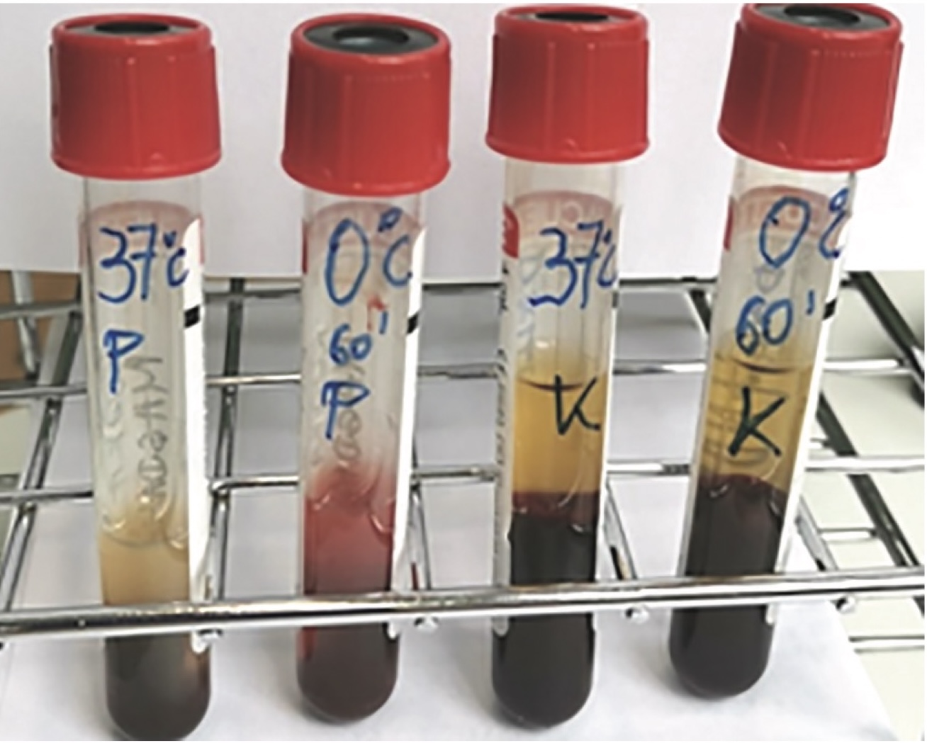
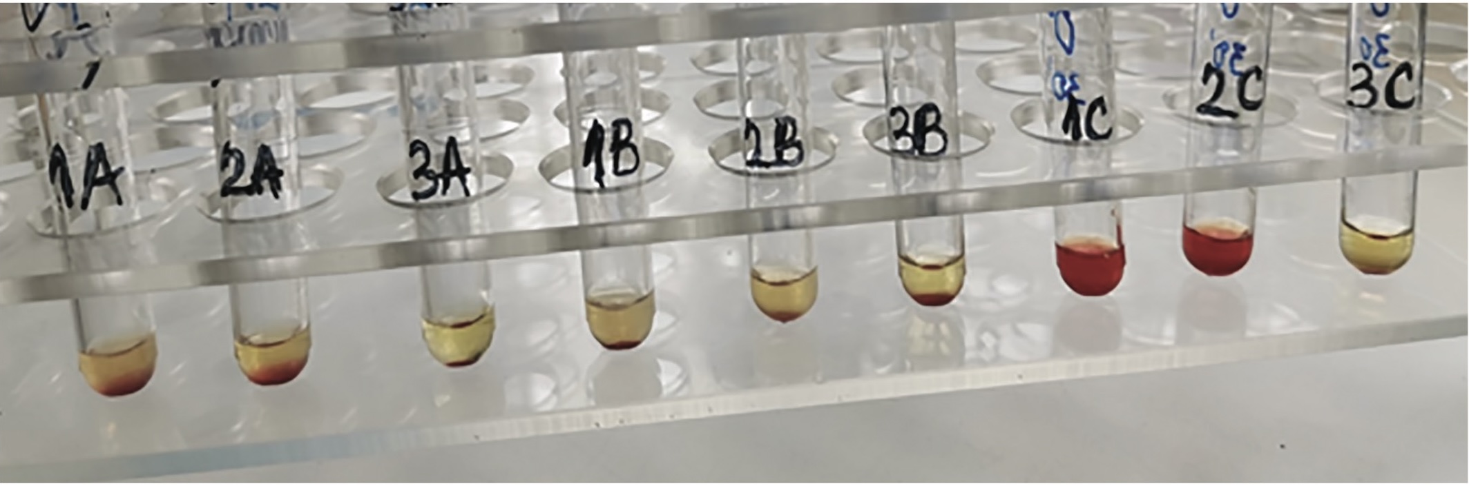
During the evaluation of the disease, PCH was suspected. On the tenth day upon admission, blood samples were taken for the direct and indirect Donath-Landsteiner (DL) test. Positive results were obtained for the presence of DL autoantibodies, i.e., hemolysis of erythrocytes was observed in patient samples incubated both with and without the addition of exogenous complement, first at 0°C and then at 37°C (Image 1 and Image 2).
On the eleventh day upon admission, the infant was discharged in a stable condition, with normal vital signs and physical examination findings. She was sent home with instructions for regular check-ups with her pediatrician.
DISCUSSION
The 11-month-old infant, exhibiting clinical and laboratory signs of erythrocyte hemolysis, presents a complex diagnostic challenge.
Autoimmune hemolytic anemia is rare in the pediatric population and its estimated incidence is 0.81 cases per 100,000 in those under 18 years of age [10]. While the initial treatment of severe anemia involves hemodynamic stabilization and transfusion of compatible red blood cells, subsequent treatment and prognosis vary depending on the etiology. An acute or severe episode of AIHA, as seen in this case, can be life-threatening. However, consensus guidelines for managing these patients are limited due to the low incidence of the disease and the challenges in recognizing atypical cases. In this case, there was clinical and laboratory evidence for intravascular hemolysis with DAT positivity only for the complement component C3d. The evaluation of the disease, considering the typical clinical presentation, also suggested PCH.
Clinicians should keep in mind that suspicion of PCH should not depend on the presence of an active infection, and that infection does not always have to precede the development of PCH [8],[11]. Intravascular hemolysis and rapidly progressing anemia may cause symptoms such as general weakness, pallor, abdominal pain, nausea, and vomiting [8].
The systematic review by Jacobs et al. included 125 publications where a total of 230 patients whose PCH diagnosis had been confirmed by DL test were presented. The gender-based frequency distribution was as follows: male/female, 124/106 (ratio 1.17:1). The median age is 5 years (ranging from 8 months to 91 years), with an interquartile range of 2.6 to 51 years. The lowest average value of hemoglobin was found in the age group 10-19 years (48 g/l; range 23-106 g/l), followed by the age group 0-9 years (53 g/l; range 20-129 g/l), and the age group 20-29 years (54 g/l; range 27 -161 g/l) [8].
The specificity of the biphasic autoantibody was confirmed in 31% (71/230), patients. IgG anti-P was confirmed in 59/71 (83,1%) patients. Other rarely present anti-erythrocyte antibodies include IgG anti-I, IgM anti-I, anti-PP1Pk (Tja), IgG anti-i, IgM anti-P, IgA anti-P, anti-Pr-like antibodies, and IgM antibody with undetermined specificity [8].
The origin of autoantibodies in PCH has not been clarified. According to one model, the P antigen is altered by an infectious agent, or this agent leads to cross-reactivity. An infectious agent can induce changes in the surface globoside (P antigen) on the erythrocyte membrane, which increases its immunogenicity. Another hypothesis posits that glycosphingolipids become immunogenic upon binding to viral antigens. A third hypothesis proposes that increased lymphocyte activation or immune dysregulation caused by a virus leads to heightened production of autoantibodies. All three mechanisms can be present in each individual case of PCH [12],[13],[14].
Direct antiglobulin test is a golden standard for AIHA. In PCH there is usually positivity only for complement, but it may be negative for both C3d and IgG.
The diagnosis of PCH is confirmed by DL test. Since this test is rarely performed, it can be done only in reference laboratories or in certain hospital blood banks [11],[15]. The test is deemed positive if the patient’s serum, either with or without added complement from a healthy individual’s serum, shows hemolysis in tubes that were pre-incubated at 0°C and subsequently at 37°C. Conversely, the test is negative if there is no erythrocyte hemolysis in any tube incubated separately at 0°C and 37°C. The test tubes containing serum from a healthy person act as a negative control, and their contents should not exhibit hemolysis [9],[11].
The biphasic autoantibody is transient and detectable only during the phase of acute hemolysis, which is why, in suspected cases, the DL test should be performed as early as possible [11]. A negative DL test result does not exclude PCH in an appropriate clinical context. The most common reason for false-negative results is a low and undetectable DL antibody titer. Other potential causes of false-negative results include autoadsorption of DL antibodies when serum separation is not carried out at 37°C or neutralization of anti-P antibodies by globoside in fresh serum, which is added as a complement source. Adding an exogenous complement is important, because it is assumed that the patient has exhausted their complement due to the hemolytic process. Apart from false negatives, false positives are also possible, particularly in individuals with monophasic IgM autoantibodies with a broad thermal range [16].
Paroxysmal cold hemoglobinuria is a transient disease. It requires adequate care of the patient that involves managing body temperature, ensuring adequate rest, hydration, and avoiding exposure to cold. In case of severe anemia, supportive treatment with erythrocyte transfusion is required. The patient should be warmed, but the use of blood warmers for RBC transfusion should be approached cautiously, as there is insufficient evidence to fully support this practice. Erythrocytes with the p or P-null phenotype are uncommon and not readily available for emergency transfusions. Nevertheless, most patients requiring transfusion achieve satisfactory posttransfusion hemoglobin despite presumed or documented P incompatibility [11]. P antigen is one of three antigens from the GLOB blood group system. This system is designated number 028 according to the classification of the Working Party for Red Cell Immunogenetics and Blood Group Terminology of the International Society of Blood Transfusion. The gene that encodes the synthesis of the antigen P, B3GALNT1, was cloned in 1998. It is located on the long arm of chromosome 3 (3q26.1) [17]. Individuals lacking the P antigen expression are very uncommon. The estimated frequency of phenotype p is 5.8/1,000,000 European inhabitants. A higher frequency of 141/1,000,000 was registered in Västerbotten county in the North of Sweden. Additionally, the p phenotype is more prevalent among the Japanese population and within the Amish community [18].
Based on the presentation of three patients, Harada and Sonoda noted a seasonal pattern in the disease, occurring predominantly in spring or autumn. They suggested that daily fluctuations in air temperature might be linked to the temperature-dependent DL antibody. We have similar experience with the DL test that we introduced as a standard operating procedure in 2017. In the first patient, a 23-month-old girl, hemolytic crisis related to PCH was confirmed by DL test in November, and in an 11-month-old female infant it was confirmed in March [15],[19].
Empirical treatment for AIHA typically starts with corticosteroids. Their administration can be stopped when the diagnosis of PCH is confirmed as there is no evidence to support the benefit of their use [11]. The refractory, recurrent, and chronic forms of PCH have shown successful responses to more intensive immunosuppressive therapies, including intravenous immunoglobulins, azathioprine, or rituximab. Eculizumab, a humanized anti-C5 monoclonal antibody, has been utilized in three patients with PCH so far [20],[21],[22],[23].
The clinical recognition of PCH, the transient nature of DL antibodies, and the feasibility of conducting the DL test all depend on promptly confirming the diagnosis of this rare form of AIHA.
ABBREVIATIONS
ANA – antinuclear antibody
ANCA – antineutrophil cytoplasmic antibody
Anti-dsDNA – anti-double-stranded DNA
AIHA – autoimmune hemolytic anemia
CRP – C-reactive protein
DAT – direct antiglobulin test
DL – Donath-Landsteiner
GLOB – the name of a blood group system
HUS – hemolytic uremic syndrome
IgG – immunoglobulin
G IgM – immunoglobulin M
ICU – Intensive Care Unit
PCH – paroxysmal cold hemoglobinuria
RHC – regional health center
Informations

-
Keywords:
-
Received:
-
Revised:
-
Accepted:
-
Online first:
-
DOI:
-
Cite this article:

Zorica Radonjić
Institute for Mother and Child Health Care of Serbia “Dr Vukan Čupić”,
6-8 Radoja Dakića Street, 11070 Belgrade, Serbia
E-mail:
1. Cooling LL. Kids, colds, and complement: paroxysmal cold hemoglobinuria. Transfusion. 2017; 57:1332-5. doi: 10.1111/trf.14128. [CROSSREF]
2. Shanbhag S, Spivak J. Paroxismal cold hemoglobinuria. Hematol Oncol Clin N Am. 2015; 29(3):473-8. doi: 10.1016/j.hoc.2015.01.004. [CROSSREF]
3. Donath JL, Landsteiner K. Uber paroxysmale haemoglobinurie. Munch Med Wochenschr. 1904; 51:1590-3.
4. Eder AF. Review: acute Donath-Landsteiner hemolytic anemia. Immunohematology. 2005; 21(2):56-62. doi:10.21307/immunohematology-2019-394 [CROSSREF]
5. Bunch C, Schwartz FC, Bird GW. Paroxysmal cold haemoglobinuria following measles immunization. Arch Dis Child. 1972; 47(252):299-300. doi: 10.1136/ adc.47.252.299. [CROSSREF]
6. Hayashi H, Yasutomi M, Hayashi T, Yuasa M, Kawakita A, Hata I, et al. Paroxysmal cold hemoglobinuria caused by an IgM-classDonath-Landsteiner antibody. Pediatr Int. 2013;55(5):664-6. doi: 10.1111/ped.12110. [CROSSREF]
7. Misawa K, Yasuda H, Koyama D, Inano T, Inoguchi A, Shirasu C, et al. Adult paroxysmal cold hemoglobinuria following mRNA COVID-19 vaccination.eJHaem 2022; 3(3):992-5. doi: 10.1002/jha2.508. [CROSSREF]
8. Jacobs JW, Figueroa Villalba CA, Booth GS, Woo JS, Stephens LD, Adkins BD. Clinical and epidemiological features of paroxysmal cold hemoglobinuria: a systematic review. Blood Advance. 2023; 7(11):2520-7. doi: 10.1182/bloodadvances.2022009516. [CROSSREF]
9. Williams JD, Jayaprakash RK, Kithany H, Tighe MP. How to use Donath-Landsteiner test to diagnose paroxysmal cold hemoglobinuria (PCH). Arch Dis Child Educ Pract Ed 2022; 107:199-206. doi: 10.1136/archdischild-2020-319568. [CROSSREF]
10. Aladjidi N, Jutand MA, Beaubois C, Fernandes H, Jeanpetit J, Coureau G, et al. Reliable assessment of the incidence of childhood autoimmune hemolytic anemia. Pediatr Blood Cancer. 2017; 64(12):e26683. doi: 10.1002/pbc.26683. [CROSSREF]
11. Chou ST,Schreiber AD. Autoimmune hemolytic anaemia. In: Orkin SH, Fisher DE, Grinsburg D, Thomas Look A, Lux SE, Nathan DE, editors. Nathan and Oski’s Hematology and Oncology of Infancy and Childhood, 8th ed. Philadelphia: Elsevier Saunders; 2015. p 411-30.
12. Haukenes G. Demonstration of host antigens in the myxovirus membrane: Lysis of virus by antibody and complement. Acta Pathol Microbiol Scand. 1977;85(2):125-8. doi: 10.1111/j.1699-0463.1977.tb01685.x. [CROSSREF]
13. Nordhagen R, Stensvold K, Winsnes A, Skyberg D, Støren A. Paroxysmal cold haemoglobinuria. The most frequent acute autoimmune haemolytic anaemia in children? Acta Paediatr Scand. 1984;73(2):258-62. doi: 10.1111/j.1651- 2227.1984.tb09939.x. [CROSSREF]
14. Klenk HD, Choppin PW. Glycosphingolipids of plasma membranes of cultured cells and an enveloped virus (SV5) grown in these cells. Proc Natl Acad Sci. 1970;66(1):57-64. doi:10.1073/pnas.66.1.57. [CROSSREF]
15. Radonjić Z, Andrić B, Šerbić O, Mićić D, Kuzmanović M, Jovanović Srzentić S, et al. A rare case report of autoimmune haemolytic anemia in a female child due to Donath-Landsteiner antibody. Transfus Clin Biol. 2020; 27:83-6. doi: 10.1016/j.tracli.2020.03.001. [CROSSREF]
16. Jacobs JW, Booth GS, Woo JS, Stephens LD, Figueroa Villalba CA, Adkins BD. Challenges in recognising paroxysmal cold hemoglobinuria. Transfus Med 2024; 34 (1):71-3. doi: 10.1111/tme.13024. [CROSSREF]
17. Hellberg A, Westman JS, Olsson ML. An update on the GLOB blood group System and collection. Immunohematology 2013; 29(1):19-24. [CROSSREF]
18. Hellberg A, Westman JS, Thuresson B, Olsson ML. P1PK: the blood group systemthat changed its name and expanded. Immunohematology 2013;29:25-33. [CROSSREF]
19. Harada N, Sonoda M. Adenovirus-associated paroxysmal cold hemoglobinuria as chilly month hemolytic crisis. Pediatr Infect Dis J. 2024; 43(4):e147-8. doi: 10.1097/INF.0000000000004230. [CROSSREF]
20. Prince SD, Winestone LE, Nance SJ, Friedman DF. Recurrent Donath-Landsteiner hemolytic anemia: a pediatric case report. Transfusion. 2017;57(6):1401- 6. doi: 10.1111/trf.14032. [CROSSREF]
21. Koppel A, Lim S, Osby M, Garratty G, Goldfinger D. Rituximab as successful therapy in a patient with refractory paroxismal cold hemoglobinuria. Transfusion. 2007;47(10):1902-4. doi:10.1111/j.1537-2995.2007.01414.x. [CROSSREF]
22. Pelletier J, Ward C, Borloz M, Ickes A, Guelich S, Edwards Jr.E. A case of childhood severe paroxysmal cold hemoglobinuria with acute renal failure successfully treated with plasma exchange and eculizumab. Case Rep Pediatr. 2022; v2022. doi: 10.1155/2022/3267189. [CROSSREF]
23. Lau-Braunhut SA, Stone H, Collins G, Berentsen S, Braun BS, Zinter MS. Paroxysmal cold hemoglobinuria successfully treated with complement inhibition. Blood Adv. 2019;3(22):3575-8. doi:10.1182/bloodadvances.2019000897. [CROSSREF]
- Table 2. Multiple regression analysis
- Figure 1. The result of the direct Donath-Landsteiner test P – patient samples; K – healthy person samples (control) Hemolysis of patient erythrocytes when patient sample was incubated at 0 ºC, followed by the incubation at 37ºC. No hemolysis was observed in control tube. No hemolysis was seen when patient sample and control sample were incubated at 37ºC.
- Figure 2. The result of the indirect Donath-Landsteiner test Hemolysis of donor erythrocytes was observed when patient serum or a mixture of patient serum and normal serum (tubes 1C and 2C) were incubated at 0 ºC followed by incubation at 37ºC. No hemolysis was seen when patient serum or mixed serum were incubated at 0ºC (tubes 1A and 2A ) or 37ºC (tubes 1B and 2B). No hemolysis was observed in control tubes containing normal serum only (tubes 3A,3B, 3C)