


Correlation of cytogenetics and grade of bone marrow fibrosis in primary myelofibrosis
- University Clinical Center of Serbia, Clinic for Hematology, Belgrade, Serbia
- University Clinical Center of Serbia, Department of Pathohistology, Belgrade, Serbia
- University of Belgrade, Faculty of Medicine, Institute for Medical Statistics and Informatics, Belgrade, Serbia
- University Clinical Center of Serbia, Clinic for Hematology, University of Belgrade, Faculty of Medicine, Belgrade, Serbia
ABSTRACT
Introduction/Aim: Primary myelofibrosis is a clonal myeloproliferative neoplasm characterized by bone marrow fibrosis and extramedullary hematopoiesis. Fibrosis often complicates bone marrow aspiration and biopsy, further hindering disease diagnosis. Despite this, cytogenetic analysis is possible from bone marrow aspirate or peripheral blood. Karyotype is an independent prognostic parameter in primary myelofibrosis. The prognostic significance of bone marrow fibrosis grade in primary myelofibrosis remains debatable. This study aimed to investigate the impact of bone marrow fibrosis on overall survival in patients with primary myelofibrosis, as well as to examine the correlation between karyotype and bone marrow fibrosis grade in these patients.
Materials and Methods: The study of the correlation between karyotype and bone marrow fibrosis grade included 120 out of 136 patients with primary myelofibrosis. We used the stratification of chromosomal abnormalities according to the recommendations of prognostic scoring systems: Lille, Mayo, International, and Dynamic International. Karyotype analysis was performed using the conventional cytogenetic method. Bone marrow biopsies of patients were stained using the Gordon-Sweet method.
Results: In 136 patients with primary myelofibrosis, no statistically significant difference in survival was observed (p = 0.084) regardless of the bone marrow fibrosis grade. Additionally, no correlation was found between karyotype or chromosomal aberrations of varying risk levels and bone marrow fibrosis grade (Lille p = 0.293; Mayo p = 0.108; International Prognostic Scoring System p = 0.086; Dynamic International Prognostic Scoring System p = 0.613) in 120 patients with primary myelofibrosis.
Conclusion: Although we did not establish a correlation between karyotype and bone marrow fibrosis grade, we believe that both parameters should be incorporated into new prognostic scoring systems for primary myelofibrosis.
INTRODUCTION
Primary myelofibrosis (PMF) is a clonal myeloproliferative neoplasm (MPN) characterized by bone marrow (BM) fibrosis and extramedullary hematopoiesis [1],[2]. Patients with PMF present with heterogeneous clinical features. It is known that unfavorable prognostic factors include older age, the presence of constitutional symptoms, anemia, decreased or increased leukocyte count, thrombocytopenia, circulating myeloblasts, and the presence of clonal chromosomal aberrations [3],[4].
Cytogenetic data in PMF are often scarce and inconclusive. Their prognostic significance is limited by the difficulty in obtaining high-quality metaphases from bone marrow aspirates [5]. However, studies reporting successful cytogenetic analysis have shown that recurrent cytogenetic abnormalities are observed in approximately one-third of patients at diagnosis, and their frequency increases throughout the disease. The most common aberrations in PMF (+1q, 13q-, 20q-, +9, +8) occur in twothirds of patients with an abnormal karyotype [6]. Other abnormalities, such as balanced translocations, complex aberrant karyotypes, and the coexistence of two or more unrelated pathological clones, are rare. Nevertheless, cytogenetics remains one of the key prognostic parameters in current Prognostic Scoring Systems (PSS) for PMF.
Unlike cytogenetics, fibrosis is a crucial histopathological finding in BM biopsies of PMF patients and represents a primary diagnostic criterion for PMF according to the World Health Organization (WHO) classification [7]. Fibrosis is a reactive phenomenon caused by abnormal megakaryocyte maturation, leading to the release of increased amounts of inflammatory and fibrogenic cytokines. These cytokines stimulate the deposition of reticulin and collagen fibers, the formation of new blood vessels, and osteosclerosis [8].
According to the European consensus, BM fibrosis is assessed in hematopoietic areas using a semi-quantitative scale with four levels: grade 0 – absent; grade 1 – a loose network of reticulin with many intersections, especially in perivascular areas; grade 2 – diffuse and dense increase reticulin with extensive intersections, occasionally with focal bundles of collagen and/or focal osteosclerosis; grade 3 – diffuse and dense increase reticulin with extensive intersections and coarse collagen bundles, often associated with osteosclerosis [9]. These criteria were reinforced and revised by the new WHO diagnostic criteria for PMF introduced in 2016 [10]. There is still no clear consensus on BM fibrosis as a prognostic parameter in PMF.
The aim of our study, whose results are presented in this paper, was to investigate whether there is a correlation between cytogenetic data and the degree of bone marrow (BM) fibrosis in biopsied samples of patients with a confirmed diagnosis of primary myelofibrosis (PMF). We tested this correlation using PSS (Lille, Mayo, IPSS, and DIPS), which are the most commonly and extensively used scoring systems in clinical practice for PMF prognosis. Additionally, these PSS clearly define chromosomal aberrations as an independent prognostic parameter for PMF patients. Each applied PSS stratifies the karyotype, whether regular or aberrant, as well as chromosomal aberrations, into prognostic groups of varying risk levels (favorable, unfavorable, intermediate-1, intermediate-2), which facilitated the examination of the correlation between karyotype, or chromosomal aberrations of different risk levels, with the degree of BM fibrosis on a scale of 0-3 according to the European consensus (MF-0, MF-1, MF-2, MF-3) [9].
PATIENTS AND METHODS
Patients
From January 2004 to December 2010, a total of 144 patients with PMF were diagnosed at the Clinic for Hematology of the University Clinical Center of Serbia in Belgrade. All patients met the WHO diagnostic criteria for PMF [11],[12]. Clinical, laboratory, and pathohistological data, along with cytogenetic results, were collected retrospectively until 2013. Patients with post-polycythemia vera myelofibrosis (post-PV-MF) and post-essential thrombocythemia myelofibrosis (post-ET-MF) were excluded from the study.
Out of the 144 newly diagnosed PMF patients, pathohistological reports were available for 136 patients (136/144). Cytogenetic analysis was not possible in 16 of the 136 patients (16/136). The core focus of this study was the clinical and diagnostic parameters of 120 patients in whom the correlation between karyotype and BM fibrosis grade could be assessed. Their data were available in the clinical archives.
A total of 24 out of 144 PMF patients (24/144) diagnosed and treated at the clinic during the study period were excluded from the study because they lacked either a pathohistological or cytogenetic report. At the time of data collection for this study, 78 patients (65%) were alive, while 42 patients (35%) had not survived.
Data on BM aspirates and biopsies used for the research were obtained from the archive with the consent of the patients or their families. The Ethics Committee of the University Clinical Center of Serbia in Belgrade approved the study.
METHODS
Cytogenetic analysis
Conventional cytogenetic analysis was performed on metaphases obtained from unstimulated cells in bone marrow aspirates or peripheral blood cultures using a previously published technique [13]. Karyotypes were interpreted according to the International System for Human Cytogenetic Nomenclature [14].
Pathohistological analysis
BM biopsies were stained using the Gordon-Sweet method. Fibrosis was assessed using the Bauermeister system from 1971 [15] (scale 0–4) and the consensus of the revised European system (scale 0–3).
Statistics
Statistical analysis of the correlation between cytogenetics and BM fibrosis was performed using several PSS. Survival was measured from diagnosis to the last contact or death. Data were presented as median (min-max) or n (%). Differences between Lille, IPSS, Mayo, and DIPSS categories by BMF were analyzed using the Mann-Whitney U test (for two groups) and the Kruskal-Wallis’s test (for three or four groups). Survival was estimated using the Kaplan-Meier curve and the Log-rank test for group comparisons. All analyses were conducted using SPSS 20.0 (IBM Corp.). Probability values (p) ≤ 0.05 were considered statistically significant.
RESULTS
Given that BM fibrosis is one of the key parameters for diagnosing PMF and one of the two main parameters examined in our study, we also analyzed overall survival in 136 PMF patients based on the degree of BM fibrosis. The statistical analysis of overall survival revealed that survival time did not differ significantly according to BM fibrosis grade (p = 0.084). However, there were only three patients with grade 0 fibrosis (MF-0) in the study group. For this reason, we excluded the MF-0 group from the statistical analysis, after which we found a statistically significant difference (p = 0.043) in survival among patients with fibrosis grades MF-1, MF-2, and MF-3.
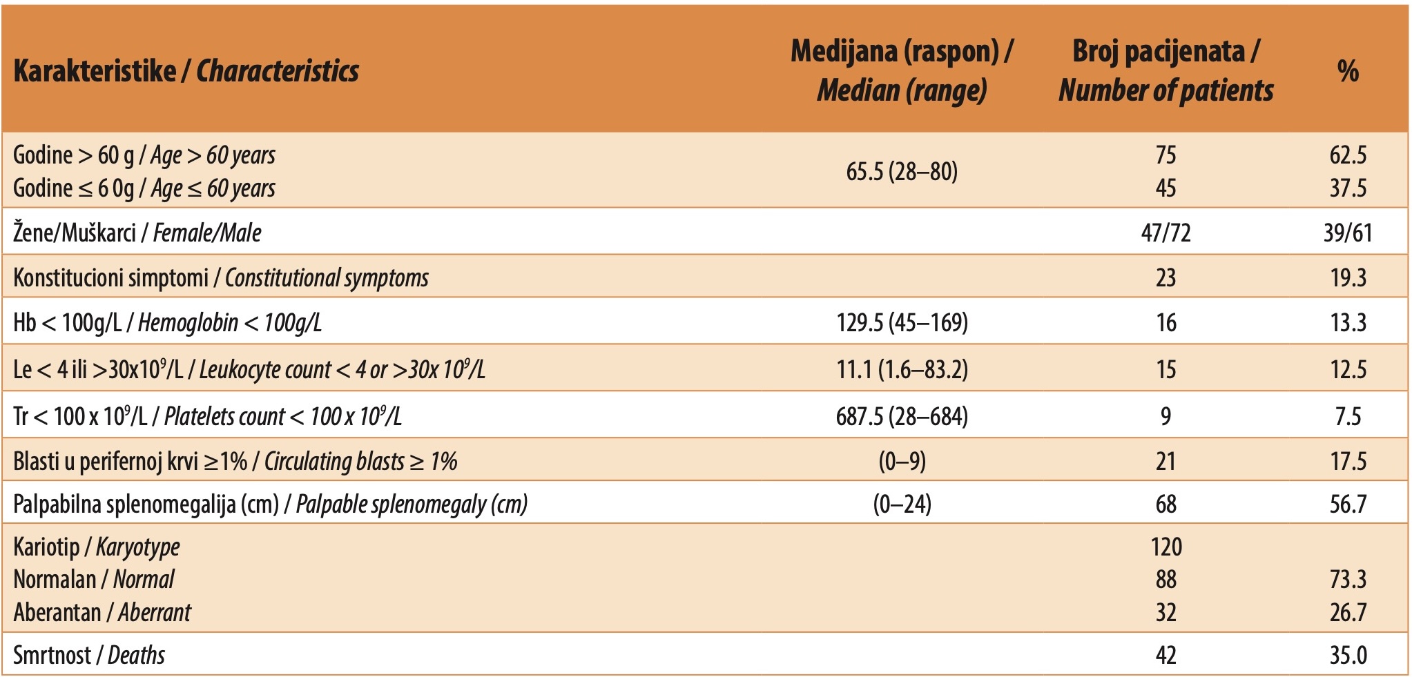
From this PMF patient group, we selected 120 patients with complete clinical, pathohistological, and cytogenetic data. Table 1 presents their characteristics at the time of diagnosis. The median age was 65.5 years (range 28–80), with 45 patients (37.5%) being younger than 60 years. The female-to-male ratio was 47/72. The median follow-up duration in 2013 was 83 months, with 42 deaths (35.0%) recorded. Constitutional symptoms were observed in 23 patients (19.3%), hemoglobin < 100 g/L in 16 (13.3%), leukocyte count < 4 or > 30 × 10⁹/L in 15 (12.5%), platelet count < 100 × 10⁹/L in 9 (7.5%), circulating blasts ≥ 1% in 21 (17.5%), and splenomegaly in 68 (56.7%) patients. A normal karyotype was present in 88 patients (88/120 or 73.3%), while 32 patients (32/120 or 26.7%) had an aberrant karyotype (Table 1).
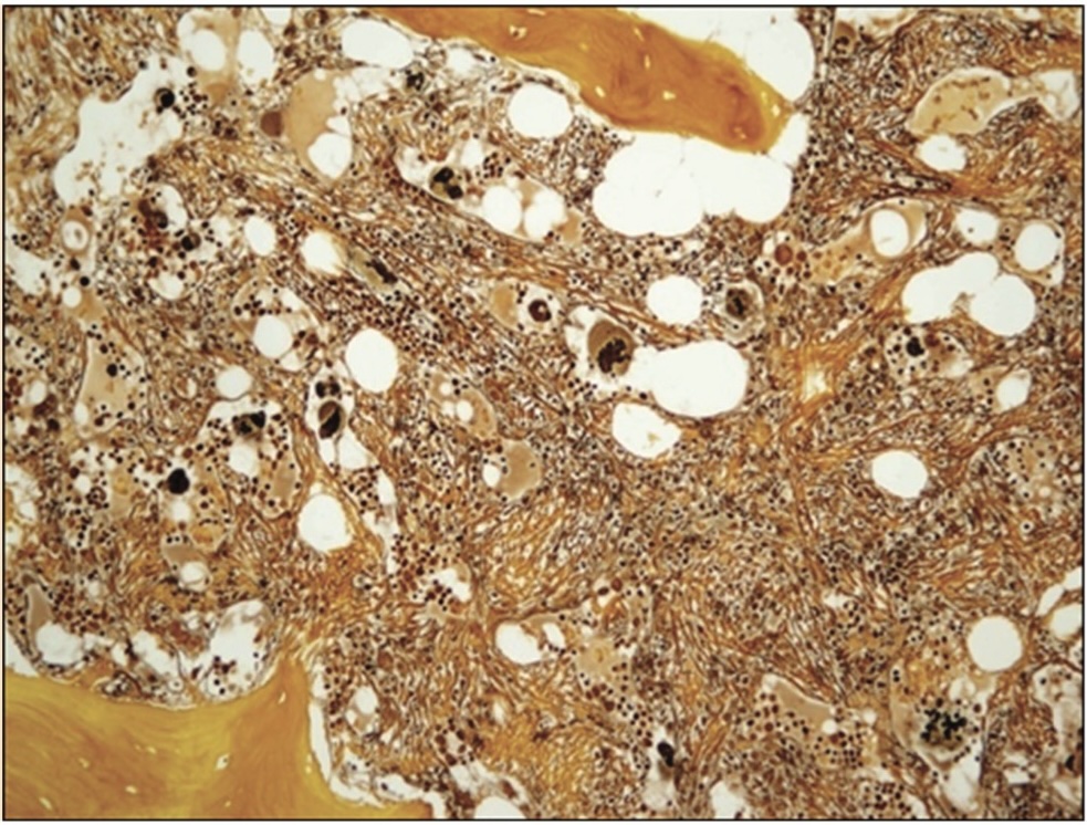
All patients were classified according to the European consensus on BM fibrosis grading [9]. Examples of the morphological characteristics of PMF and BM fibrosis grades are shown in Figure 1. The stratification of patients based on fibrosis grade and cytogenetic data is presented in Table 2. The rows of Table 2 display the PSS classifications and the stratification of chromosomal aberrations into prognostic risk groups of different levels, while the columns present the stratified BM fibrosis grades of patients on a scale from 0 to 3. The distribution of patients with an adequate karyotype (normal or aberrant) or chromosomal aberrations of a specific risk level, along with the corresponding BM fibrosis grade, is illustrated in the table.

An indicator of the accuracy of the distribution of patients by number and percentage is provided in the penultimate column, which displays the total number of patients examined. For each PSS, the total number of patients must be 120 (n = 120), meaning that the sum of the percentage representation of patients with a corresponding karyotype or chromosomal aberrations and a specific bone marrow (BM) fibrosis grade must equal 100%.
The distribution of BM fibrosis among our patients (n = 120) revealed that the highest number (n = 55, 45.8%) had fibrosis grade MF-2, followed by patients with fibrosis grade MF-3 (n = 42, 35%). Fewer patients had fibrosis grade MF-1 (n = 20 or 16.7%), and as mentioned, the lowest number had fibrosis grade MF-0 (n = 3 or 2.5%) (Table 2).
Statistical analysis of the correlation between BM fibrosis and karyotype, using the Lille PSS [3] for cytogenetics, did not reveal a statistically significant difference (p = 0.293) (Table 2). There was no correlation between a regular or aberrant karyotype and the degree of bone marrow (BM) fibrosis in patients. Among patients with a normal karyotype (favorable), the majority had fibrosis MF-2 (50.0%), followed by MF-3 (30.7%) and MF-1 (19.3%). Among patients with an aberrant karyotype (unfavorable), the highest proportion had a fibrosis grade of MF-3 (46.9%), followed by MF-2 (34.4%), and those with fibrosis grades of MF-1 and MF-0 (9.4%) (Table 2).
Using the cytogenetic stratification of the Mayo PSS [16] to examine the association between karyotype and BM fibrosis grade, no statistically significant difference was confirmed in the frequency of BM fibrosis grades among the observed patient groups with different karyotypes (p = 0.108) (Table 2). The highest number of patients with fibrosis grade MF-2 (50.0%) was found in those with a normal karyotype (favorable). In the group of patients with 13q- or 20q- aberrations (intermediate), most had MF-3 (80.0%), while one patient (20.0%) had MF-2. There were no patients with BM fibrosis grades MF-1 or MF-0.
Among patients with other chromosomal aberrations in their karyotype, the majority had BM fibrosis grade MF-3 (40.7%), followed by MF-2 (37.0%), while MF-1 and MF-0 were present at the same frequency (9.4%).
Examining the association between BM fibrosis and karyotype using IPSS [17] also showed no significant statistical difference in the frequency of BM fibrosis grades (p = 0.086) (Table 2). There was no correlation between karyotype and chromosomal aberrations of different prognostic significance (favorable, intermediate-1, intermediate-2, unfavorable) with BM fibrosis grade.
Among patients with favorable chromosomal aberrations (+9 or 13q- or 20q-), the majority had a fibrosis grade of MF-3 (62.5%), followed by MF-2 (25.0%), while one patient had a grade of MF-0 (12.5%). There were no patients with MF-1 fibrosis. The distribution of BM fibrosis grades in patients with a normal karyotype (intermediate-1) was the same as in the Lille and Mayo PSS classifications.
In the group of patients with other chromosomal abnormalities (intermediate-2), most had MF-2 (42.9%), followed by MF-3 (33.3%), while MF-1 fibrosis was present in 14.3% of cases. The lowest number of patients had MF-0 fibrosis (9.5%). In the group of patients with on chromosome 8 and a complex karyotype (unfavorable), all patients had MF-3 fibrosis (100.0%).
The application of DIPSS [18] also did not reveal a statistically significant association (p = 0.613) between cytogenetic results and the documented degree of bone marrow fibrosis (KS). According to DIPSS recommendations, patients were classified into only two prognostic categories based on karyotype findings. The favorable category includes chromosomal aberrations that are considered suitable prognostic parameters, as in the previously mentioned PSSs, along with some other aberrations (t/dup(1q), other solo and double favorable aberrations), as well as a normal karyotype. The unfavorable category comprises poor prognostic chromosomal aberrations, such as those found in IPSS (+8, complex karyotype), along with others, including (-5/5q-, -7/7q-, i(17q), inv(3), 12p-, and 11q23).
Among patients with favorable karyotypic characteristics, the majority had fibrosis grade MF-2 (46.8%), followed by those with fibrosis grade MF-3 (33.9%), then those with MF-1 (17.4%), while the smallest number of patients had MF-0 fibrosis (1.9%). Among patients with unfavorable karyotypic alterations, fibrosis grade MF-3 was the most common (45.4%), followed by patients with MF-2 fibrosis (36.4%). Patients with MF-1 and MF-0 fibrosis grades were represented with the same frequency (9.1%).
DISCUSSION
Overall survival in PMF depends on the disease’s clinical, laboratory, cytogenetic, and molecular characteristics. It is influenced by patient age, physical condition, and comorbidities. To address this, numerous PSSs have been developed, integrating these factors to categorize PMF patients into different risk levels. The imperative of risk stratification in PMF is to select the most favorable, personalized therapy that ensures a better quality of life and prolonged survival.
In the history of PSS application in PMF, cytogenetics has been recognized as an independent prognostic parameter. Early survival studies based on cytogenetics indicated that a normal karyotype was a favorable prognostic factor, while an aberrant karyotype was considered unfavorable [3]. Later, it was found that certain chromosomal aberrations (+9, 13q-, 20q-) were associated with more prolonged survival compared to a normal karyotype. These aberrations were defined as good prognostic markers [16]. Conversely, other karyotypic rearrangements (+8 or complex karyotype, -5/5q-, -7/7q-, 11q23, i(17q), inv(3), 12p-) were linked to shorter survival and were classified as poor prognostic markers in other PSSs [17],[18]. Overall, cytogenetic analysis and karyotyping play a significant role in the diagnosis of PMF but an even greater role in its prognosis.
The situation is different when it comes to BM fibrosis in PMF and MPN in general. BM fibrosis of varying degrees can be associated with the three main MPN entities-polycythemia vera (PV), essential thrombocythemia (ET), and primary myelofibrosis (PMF)-either at presentation, as in PMF, or following disease progression in PV and ET. The updated 2008 WHO classification adopted the European consensus on BM fibrosis grading [9], which introduced a four-tiered system ranging from grade 0 to 3 [7]. The progression of BM fibrosis follows a gradual evolution from absent or minimal reticulin fibrosis to extensive reticulin and/or collagen fibrosis, often accompanied by osteosclerosis. Precise assessment of fibrosis grade is crucial for the initial classification of MPNs as well as for identifying disease progression [19]. Although BM fibrosis is a key diagnostic criterion for PMF, its grading has not been incorporated into conventional PSSs. Recently, it has been emphasized that an accurate assessment of BM fibrosis grade is a critical factor in predicting PMF prognosis [8].
By analyzing the overall survival of all 136 PMF patients based on the degree of BM fibrosis, we found no statistically significant difference in survival (p = 0.084). This could lead to the controversial conclusion that the degree of fibrosis is not an important factor in survival for PMF patients, despite being one of the key diagnostic criteria. However, when we repeated the analysis excluding the three patients with MF-0 fibrosis, statistical significance in survival emerged (p = 0.043). By removing only three MF-0 patients from the cohort of 136, it was confirmed that fibrosis indeed influences overall survival in PMF. This also suggests that all diagnostic parameters should be carefully considered before definitively diagnosing PMF or determining whether the disease represents a secondary transformation of PV or ET into myelofibrosis.
The majority of our patients had BM fibrosis grades MF-2 and MF-3 (46% and 35%). Nearly one-third had MF-3, which is particularly interesting given that our retrospective study is based on patient data collected during initial specialist examinations and at the time of primary diagnosis.
According to some literature data [20], in the profibrotic phase of PMF (MF-0 fibrosis), an aberrant karyotype is present in 18% of patients, while an unfavorable aberrant karyotype (≥ 3 aberrations, +8, -5/5q-, -7/7q-, i(17q), inv(3), 12p-, 11q23, complex karyotype) is found in 4–8% of cases. Based on these data, we can conclude that lower degrees of BM fibrosis (MF-0 and MF-1) correlate with favorable chromosomal aberrations and a normal karyotype, whereas higher fibrosis grades (MF-2 and MF-3) are associated with unfavorable cytogenetics.
We conducted a similar analysis in our patient group. We employed simple karyotype categorizations (normal and aberrant) based on the Lille PSS recommendations [3]. Additionally, we applied advanced karyotype categorizations that included specific chromosomal aberrations (+9, 13q-, 20q-, +8, complex karyotype) with different levels of unfavorable prognosis, as classified by Mayo PSS [16] and IPSS [17]. The DIPSS model [18], which proposes only two karyotype categories (favorable and unfavorable chromosomal aberrations), was also applied. In none of these PSS models did we find a statistically significant correlation between karyotype and BM fibrosis grade (Lille p = 0.293; Mayo p = 0.108; IPSS p = 0.086; DIPSS p = 0.613) (Table 2).
Our primary aim was to test the association between cytogenetic and histopathological findings in PMF patients, assuming that a correlation exists between these two diagnostic and prognostic parameters. The fact is that we used PSS models in which normal or aberrant karyotypes and chromosomal aberrations were stratified into prognostic categories based on their impact on overall survival in PMF patients. On the other hand, BM fibrosis is considered an unfavorable diagnostic finding for PMF patients, with higher fibrosis grades indicating a more advanced disease stage. Based on these facts, we expected a positive association or direct correlation between cytogenetic and histopathological findings-that is, that aberrant karyotypes or “unfavorable” chromosomal aberrations would be more frequently observed in patients with MF-2 and MF-3 fibrosis, as confirmed by statistical analysis. However, our results were contrary to this expectation, as we found no direct correlation between prognostically “unfavorable” chromosomal aberrations and higher degrees of BM fibrosis (MF-2 and MF-3) or between a normal karyotype and “favorable” chromosomal aberrations with lower fibrosis grades (MF-0 and MF-1) in PMF.
CONCLUSION
To the best of our knowledge, our study is the first attempt to investigate the correlation between cytogenetics and histopathology-two crucial diagnostic and prognostic parameters for PMF. The study includes 136 PMF patients, which represents a relatively large number of cases at the time of diagnosis for a single clinical and diagnostic center in the Western Balkans. In 120 patients, we analyzed the correlation between cytogenetic findings and histopathological data on BM fibrosis grade using relevant PSS models.
Although we expected a positive correlation between karyotype (normal, aberrant) and chromosomal aberrations of different prognostic significance (favorable, unfavorable) with BM fibrosis grade, following the recommendations of the European consensus, our results showed the opposite. There was no correlation between karyotype type or chromosomal aberrations of different prognostic significance and BM fibrosis grade in patients with PMF.
However, we intend to continue investigating the correlation between BM fibrosis grade, cytogenetics, and “driver” mutations in PMF through a future prospective study.
ABBREVIATIONS AND ACRONYMS
PMF – primary myelofibrosis
MPN – myeloproliferative neoplasm
BM – bone marrow
PSS – prognostic Scoring System
WHO – world health organization
PV – polycythemia vera
ET – essential thrombocythemia
Appendix:
This paper presents some of the results from the scientific research study conducted by Dr. Vesna Đorđević as part of her doctoral thesis entitled “Type and Frequency of Chromosomal Aberrations and the Significance of the JAK2V617F Molecular Marker in Primary Myelofibrosis.”
Informations

-
Keywords:
-
Received:
-
Revised:
-
Accepted:
-
Online first:
-
DOI:
-
Cite this article:

Vesna Đorđević
Clinic for Hematology, University Clinical Center of Serbia
2 Dr Koste Todorovića Street, 11000 Belgrade, Serbia
E-mail:
1. Pizzi M, Croci GA, Ruggeri M, Tabano S, Dei Tos AP, Sabattini E, et al. The classification of myeloproliferative neoplasms: rationale, historical background and future perspectives with focus on unclassifiable cases. Cancers (Basel). 2021 Nov 12;13(22):5666. doi: 10.3390/cancers13225666. [CROSSREF]
2. Arber DA, Orazi A, Hasserjian RP, Borowitz MJ, Calvo KR, Kvasnicka HM, et al. International consensus classification of myeloid neoplasms and acute leukemias: integrating morphologic, clinical, and genomic data. Blood 2022 Sep 15;140(11):1200-1228.doi: 10.1182/blood.2022015850. [CROSSREF]
3. Dupriez B, Morel P, Demory JL, Lai JL, Simon M, Plantier I, et al. Prognostic factors in agnogenic myeloid metaplasia: a report on 195 cases with a new scoring system. Blood. 1996 Aug 1;88(3):1013-8. doi: 10.1182/blood. V88.3.1013.1013. [CROSSREF]
4. Tefferi A, Gangat N, Animesh Pardanani A, Crispino JD. Myelofibrosis: genetic characteristics and the emerging therapeutic landscape. Cancer Res. 2022 Mar 3;82(5):749–763. doi: 10.1158/0008-5472.CAN-21-2930. [CROSSREF]
5. Djordjevic V, Dencic-Fekete M, Jovanovic J, Bizic S, Jankovic G, Bogdanovic A, et al. Cytogenetics of agnogenic myeloid metaplasia: a study of 61 patients. Cancer Genet Cytogenet. 2007 Feb;173(1):57-62. doi:10.1016/j.cancergencyto.2006.09.021. [CROSSREF]
6. Đorđević V, Denčić-Fekete M, Jovanović J, Soldatović I, Janković G, Bogdanović A. Significance of cytogenetic-risk categories and refined international prognostic scoring system for overall survival in primary myelofibrosis: a single-center experience. Vojnosanit Pregl. 2020;77(5):516-24. doi: 10.2298/ VSP171129117D. [CROSSREF]
7. Thiele J, Kvasnicka HM, Tefferi A, et al. Primary myelofibrosis. In: Swerdlow SH, Campo E, Harris NL, Jaffe ES, Pileri SA, Stein H, Thiele J, Vardiman JW, editors. WHO classification of tumors of haematopoietic and lymphoid tissues, 4th ed. Lyon, France: IARC Press; 2008. p. 44-7.
8. Guglielmelli P, Rotunno G, Pacilli A, Rumi E, Rosti V, Delaini F, et al. Prognostic impact of bone marrow fibrosis in primary myelofibrosis. A study of the AGIMM group on 490 patients. Am J Hematol. 2016 Sep;91(9):918-22. doi: 10.1002/ajh.24442. [CROSSREF]
9. Thiele J, Kvasnicka HM, Facchetti F, Franco V, Walt JVD, Orazi A. European consensus on grading bone marrow fibrosis and assessment of cellularity. Haematologica. 2005 Aug 90(8):1128-32. Available from: https://haematologica.org/article/view/3634. [HTTP]
10. Arber DA, Orazi A, Hasserjian R, Tkiele J, Borowitz MJ, Le Beau MM, et al. The 2016 revision to the World Health Organization classification of myeloid neoplasms and acute leukemia. Blood. 2016 May 19;127(20):2391-405. doi: 10.1182/blood-2016-03-643544. Erratum in: Blood. 2016 Jul 21;128(3):462-3. doi:10.1182/blood-2016-06-721662. [CROSSREF]
11. Vardiman JW, Harris NL, Brunning RD. The World Health Organization (WHO) classification of the myeloid neoplasms. Blood. 2002 Oct 1;100(7): 2292-302. doi: 10.1182/blood-2002-04-1199. [CROSSREF]
12. Vardiman JW, Thiele J, Arber DA, Brunning RD, Borowitz MJ, Porwit A, et al. The 2008 revision of the World Health Organization (WHO) classification of myeloid neoplasms and acute leukemia: rationale and important changes. Blood. 2009 Jul 30;114(5):937-51. doi: 10.1182/blood-2009-03-209262. [CROSSREF]
13. Novak A, Kruskic M, Ludoski M, Jurukovski V. Rapid method for obtaining high-quality chromosome banding in the study of hematopoietic neoplasia. Cancer Genet Cytogenet. 1994 Jun;74(2):109-14. doi: 10.1016/0165- 4608(94)90007-8. [CROSSREF]
14. Hastings RJ, Moore S, Chia N, editors. ISCN 2024 – An International System for Human Cytogenomic Nomenclature (2024). Cytogenet Genome Res. 2024;164(Suppl 1):1-224. doi: 10.1159/000538512. [CROSSREF]
15. Bauermeister DE. Quantitation of Bone Marrow Reticulin-A Normal Range. Am J Clin Pathol. 1971 Jul;56(1):24-31. doi: 10.1093/ajcp/56.1.24. [CROSSREF]
16. Tefferi A, Dingli D, Li CY, Dewald GW. Prognostic diversity among cytogenetic abnormalities in myelofibrosis with myeloid metaplasia. Cancer. 2005 Oct 15;104(8):1656-60. doi: 10.1002/cncr.21358. [CROSSREF]
17. Hussein K, Pardanani AD, Van Dyke DL, Hanson CA, Tefferi A. International Prognostic Scoring System – independent cytogenetic risk categorization in primary myelofibrosis. Blood. 2010 Jan 21;115(3):496-9. doi: 10.1182/blood-2009-08-240135. [CROSSREF]
18. Caramazza D, Begna KH, Gangat N, Vaidya R, Siragusa S, Van Dyke DL, et al. Refined cytogenetic-risk categorization for overall and leukemia-free survival in primary myelofibrosis: a single center study of 433 patients. Leukemia. 2011 Jan;25(1):82-8. doi: 10.1038/leu.2010.234. [CROSSREF]
19. Pozdnyakova O, Wu K, Patki A, Rodig SJ, Thiele J, Hasserjian RP. High concordance in grading reticulin fibrosis and cellularity in patients with myeloproliferative neoplasms. Mod Pathol. 2014 Nov;27(11):1447-54. doi: 10.1038/ modpathol.2014.69. [CROSSREF]
20. Gianelli U, Thiele J, Orazi A, Gangat N, Vannucchi AM, Tefferi A, et. International Consensus Classification of myeloid and lymphoid neoplasms: myeloproliferative neoplasms. Virchows Arch. 2023 Jan;482(1):53-68. doi: 10.1007/ s00428-022-03480-8. [CROSSREF]
- Table 1. Baseline characteristics of the 120 enrolled patients
- Picture 1. MF-3, Gordon Sweet x200, Dense reticulin fibers, and bundles of collagen
- Table 2. Stratification of 120 patients with primary myelofibrosis according to the cytogenetic data from current prognostic scoring systems and the European consensus on grading of bone marrow fibrosis