


Melanocytoma of the optic nerve head coexisting with juxtapapillary choroidal nevus: Case report and literature review
- University Clinical Center of Serbia, Eye Hospital, Belgrade, Serbia
- Mitrović Eye Clinic, Belgrade, Serbia
- University of Belgrade, Faculty of Medicine, Belgrade, Serbia
ABSTRACT
Introduction: Melanocytoma of the optic nerve head (melanocytoma papillae) is a rare, benign pigmented lesion originating from melanocytes within the optic nerve head. Despite its benign nature, melanocytoma can present diagnostic challenges due to its potential mimicry of melanomas and associated complications, including visual field defects and, in rare cases, malignant transformation. Accurate diagnosis relies on multimodal imaging, including fluorescein angiography, optical coherence tomography (OCT), and enhanced depth imaging-OCT (EDI-OCT).
Case report: A 40-year-old female presented with a pigmented lesion on the left optic nerve head, which had been monitored at another institution for the past 10 years. Ophthalmologic examination revealed a sharply demarcated, elevated, gray-black lesion with clinical features typical of melanocytoma. Adjacent to the melanocytoma, a juxtapapillary choroidal nevus was observed. The diagnostic workup, including fluorescein angiography and B-scan ultrasonography, confirmed the benign nature of the lesion. The patient was advised to undergo regular follow-up to monitor for potential progression or malignant transformation.
Conclusion: This case underscores the importance of auxiliary diagnostic methods and long-term surveillance in managing melanocytoma of the optic nerve head. The coexistence of a juxtapapillary choroidal nevus further complicates the diagnostic process, emphasizing the need for regular follow-up to enable the timely detection of potential complications.
INTRODUCTION
Melanocytoma of the optic nerve head (melanocytoma papillae) is a rare, benign melanocytic lesion originating from melanocytes within the optic nerve head. Initially described by Limas and Ulrich in 1962, melanocytoma is histologically distinct from melanomas but shares a similar dark pigmentation due to heavily pigmented, round, or polygonal cells containing abundant melanin granules. While typically asymptomatic, its clinical significance lies in its potential to cause visual field defects, optic disc swelling, and, in rare cases, malignant transformation [1].
This lesion frequently coexists with juxtapapillary choroidal nevi, which are pigmented, flat lesions located near the optic disc. Studies suggest that up to 50% of melanocytoma cases are associated with such nevi, further complicating clinical differentiation from more aggressive pathologies such as melanoma [2],[3].
Advancements in imaging techniques, such as fluorescein angiography, optical coherence tomography (OCT), enhanced depth imaging-OCT (EDI-OCT), and B-scan ultrasonography have significantly improved the ability to distinguish melanocytoma from malignant lesions [4].
Although typically benign, melanocytomas can exhibit locally aggressive behavior, potentially leading to optic nerve compression or involvement of adjacent structures, such as the retina and choroid. They are most commonly diagnosed in middle-aged individuals with no significant gender predilection [5],[6]. Long-term follow-up is crucial to monitor for complications, such as visual impairment, optic neuropathy, or the rare but serious risk of malignant transformation [7].
This report presents a rare case of melanocytoma of the optic nerve head with a coexisting juxtapapillary choroidal nevus, emphasizing the diagnostic and management challenges associated with this condition. A comprehensive literature review provides additional insights into its clinical, imaging, and histopathological features, highlighting the importance of multidisciplinary care in ensuring optimal outcomes for affected patients [8],[9],[10].
CASE REPORT
A 40-year-old female patient presented to our clinic with a known history of a pigmented lesion on the left optic nerve, first identified over 10 years ago during a routine ophthalmologic examination. The patient reported no visual symptoms; her medical history was unremarkable, with no systemic illnesses or family history of ocular diseases. Visual acuity was 20/20 bilaterally without correction.
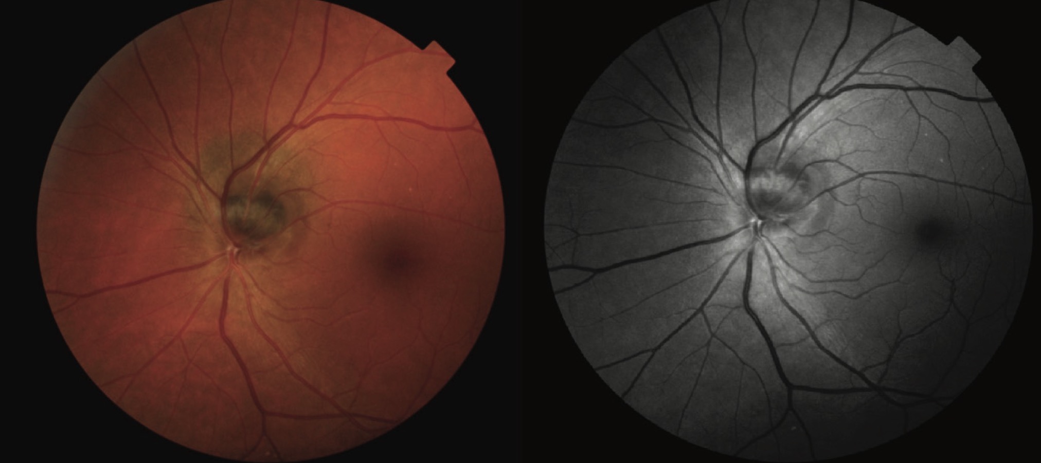
The ophthalmoscopic evaluation revealed a sharply demarcated, elevated, gray-black lesion on the optic nerve head of the left eye. Adjacent to the melanocytoma, a juxtapapillary choroidal nevus was noted, presenting as a flat, greenish-brown lesion with distinct borders. There were no signs of optic disc edema, and retinal vessels appeared normal (Figure 1). Automated perimetry revealed a small, non-progressive central scotoma.
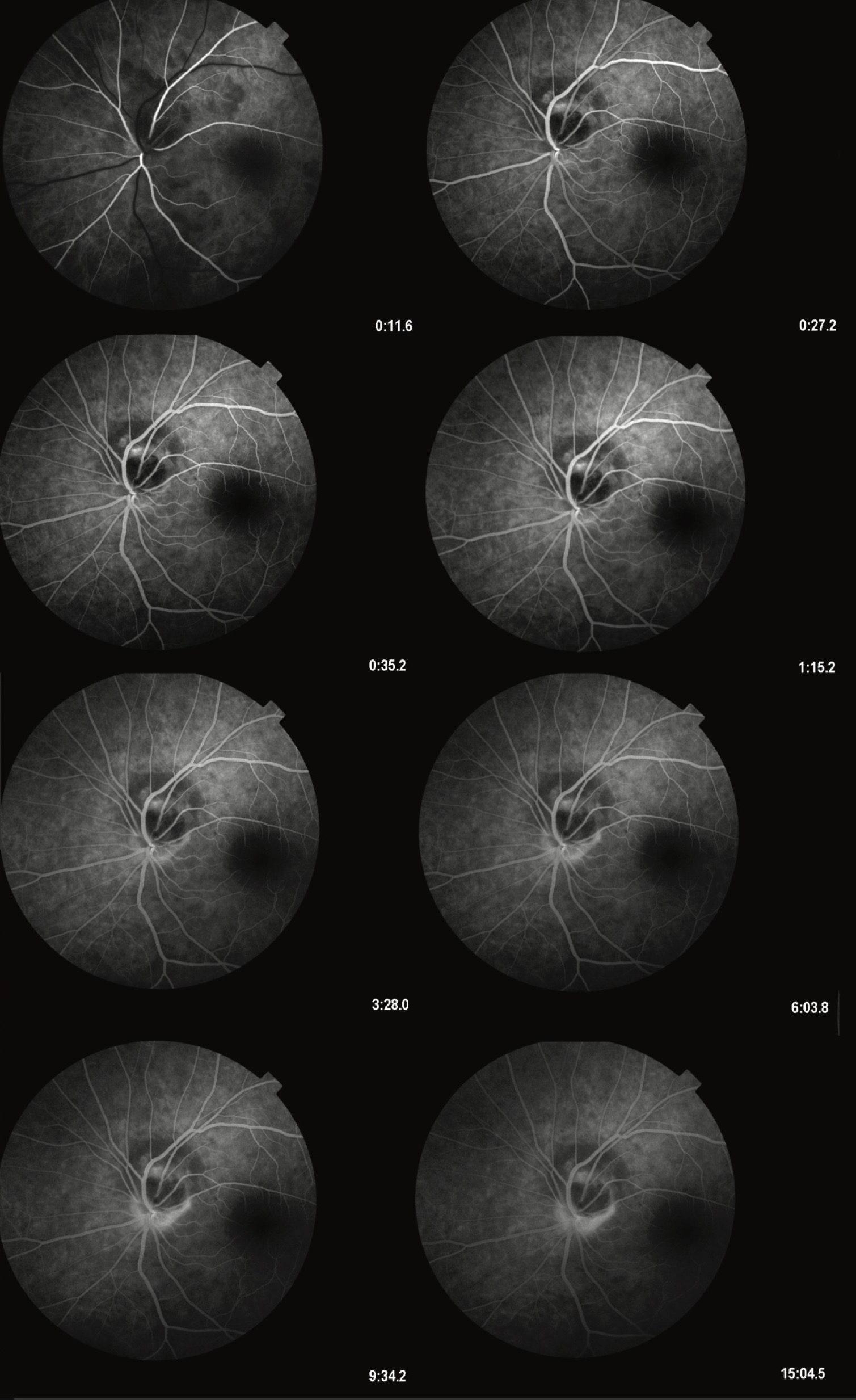
Fluorescein angiography demonstrated a complete blockage of fluorescence corresponding to the melanocytoma, with no evidence of leakage or neovascularization (Figure 2). The juxtapapillary nevus exhibited persistent hypofluorescence throughout all angiographic phases. Optical coherence tomography confirmed the presence of an elevated lesion with shadowing effects but without disruption of the retinal layers. B-scan ultrasonography revealed a solid lesion with moderate internal reflectivity.
Given the benign nature of the lesion and the absence of visual symptoms or complications, the patient was advised to undergo regular follow-up examinations every six months to monitor for potential progression, such as enlargement, visual field loss, or signs of malignant transformation. The patient was educated about the symptoms of possible complications, including sudden vision changes, which would necessitate immediate medical attention. Surgical intervention was deemed unnecessary at this stage.
DISCUSSION
Melanocytoma of the optic nerve head is an uncommon, benign lesion that can mimic more aggressive conditions such as melanoma, highlighting the importance of accurate diagnosis. Shields et al. reported that melanocytoma occurs in approximately 1% of the general population, with a higher prevalence among individuals of darker pigmentation [1],[4]. These lesions typically remain stable, although growth and malignant transformation have been documented in rare cases, with an estimated transformation rate of 1% [7].
Several case reports in the literature emphasize the clinical variability and diagnostic challenges of melanocytoma. Burgos-Blasco et al. presented a case of a patient with optic nerve melanocytoma, highlighting the use of multimodal imaging, including OCT and OCT angiography, to confirm the diagnosis and monitor lesion stability over time [5]. Another case by Kita et al. detailed a 38-year-old patient who experienced visual field defects due to optic nerve compression; follow-up revealed lesion stability over five years without intervention [9].
Mannat et al. described a rare instance of melanocytoma extending into the peripapillary retina, causing subretinal fluid accumulation and mild visual disturbances. The lesion remained stable with no signs of malignant transformation during a three-year follow-up [6]. Additionally, Garza-Garza et al. reported a series of cases where advanced imaging techniques, such as EDI-OCT, provided invaluable insights into lesion morphology and depth, aiding in differentiating benign from malignant lesions [8]. As seen in this case, the coexistence of juxtapapillary choroidal nevi complicates the clinical picture.
According to Gass [2], the presence of such nevi increases the diagnostic challenge, necessitating the use of multimodal imaging for differentiation. Fluorescein angiography and OCT are pivotal in demonstrating characteristic blockage of fluorescence and confirming the lesion’s benign nature [3],[5]. EDI-OCT further enhances diagnostic accuracy by providing high-resolution images of the optic nerve head and surrounding structures [6].
Histopathological studies reveal densely packed melanocytes with abundant melanin granules and low mitotic activity, consistent with a benign course [10]. However, factors such as lesion growth, optic nerve compression, or visual field defects warrant closer surveillance and potential intervention. Recent studies have emphasized the importance of genetic and molecular profiling to differentiate melanocytoma from melanoma in ambiguous cases [8].
Management of melanocytoma primarily involves regular monitoring to detect changes in lesion morphology or function. Shields et al. recommend follow-up intervals of six months for the first year and annual evaluations thereafter if the lesion remains stable [10]. Patient education regarding optic neuropathy or malignant transformation symptoms is crucial for timely intervention. In cases of significant growth, progressive visual field loss, or diagnostic uncertainty, surgical excision, and histopathological examination may be necessary.
CONCLUSION
Melanocytoma of the optic nerve head is a rare and typically benign lesion, yet its potential for local aggressiveness and rare malignant transformation necessitates careful and continuous monitoring. This case underscores the importance of a multidisciplinary approach, incorporating advanced imaging techniques and regular follow-up, to optimize patient outcomes and prevent complications. A thorough understanding of the lesion’s clinical and histopathological features, supported by a literature review, is essential for effective management.
Informations

-
Keywords:
-
Received:
-
Revised:
-
Accepted:
-
Online first:
-
DOI:
-
Cite this article:

Dolika D. Vasović
Eye Hospital, University Clinical Centre of Serbia
2 Pasterova Street, 11000 Belgrade, Serbia
E-mail:
1. Shields JA, Demirci H, Mashayekhi A, Shields CL. Melanocytoma of optic disc in 115 cases: the 2004 Samuel Johnson Memorial Lecture, part 1. Ophthalmology. 2004 Sep;111(9):1739-46. doi: 10.1016/j.ophtha.2004.02.016. [CROSSREF]
2. Gass JD. Stereoscopic atlas of macular disease: diagnosis and treatment. 4th ed. St. Louis: Mosby; 1997. 1061 p.
3. Miller NR. Primary tumours of the optic nerve and its sheath. Eye (Lond). 2004 Nov;18(11):1026-37. doi: 10.1038/sj.eye.6701592. [CROSSREF]
4. Raval V, Reddy R, Kaliki S, Das T, Singh AD. Optic nerve head melanocytoma: Optical coherence tomography/angiography features. Indian J Ophthalmol. 2021 Feb;69(2):332-6. doi: 10.4103/ijo.IJO_710_20. [CROSSREF]
5. Burgos-Blasco B, Ventura-Abreu N, Jimenez-Santos M, Narvaez-Palazon C, Saenz-Francés F, Santos-Bueso E. Multimodal imaging in optic nerve melanocytoma: Optical coherence tomography angiography and other findings. J Fr Ophtalmol. 2020 Dec;43(10):1039-46. doi: 10.1016/j.jfo.2020.01.032. [CROSSREF]
6. Mannat G, Meenakshi S, Navya NK, Subina N. Optic disc melanocytoma: a case report. Rom J Ophthalmol. 2021 Jan-Mar;65(1):89-92. doi: 10.22336/ rjo.2021.18. [CROSSREF]
7. Zhou N, Xu X, Wei W. Optical coherence tomography angiography characteristics of optic disc melanocytoma. BMC Ophthalmol. 2020 Oct 27;20(1):429. doi: 10.1186/s12886-020-01676-7. [CROSSREF]
8. Garza-Garza LA, Ruiz-Lozano RE, Ancona-Lezama D, González-Godinez S, Garza-León M. Multimodal imaging assessment of a “micro” optic disk melanocytoma: a case report. Arch Soc Esp Oftalmol (Engl Ed). 2021 Dec;96(12):663-7. doi: 10.1016/j.oftale.2020.10.004. [CROSSREF]
9. Kita Y, Hollό G, Murai A, Kita R, Hirakata A. Optical coherence tomography angiography findings of an optic disc melanocytoma in a glaucoma eye. Int Ophthalmol. 2019 Mar;39(3):677-82. doi: 10.1007/s10792-018-0839-9. [CROSSREF]
10. Shields JA, Demirci H, Mashayekhi A, Eagle RC Jr, Shields CL. Melanocytoma of the optic disk: a review. Indian J Ophthalmol. 2019 Dec;67(12):1949-58. doi: 10.4103/ijo.IJO_2039_19. [CROSSREF]
- Figure 1. The image presents a fundus photograph of a melanocytoma located on the optic nerve head, accompanied by a juxtapapillary choroidal nevus. The melanocytoma is visible as a sharply demarcated, elevated, dark gray-black lesion on the optic disc, consistent with its characteristic pigmentation. Adjacent to the melanocytoma, the choroidal nevus appears as a flat, greenish-brown area with distinct borders
- Figure 2. This image depicts a series of frames from fluorescein angiography (FA) of a melanocytoma of the optic nerve head, accompanied by a juxtapapillary choroidal nevus. The angiographic sequence illustrates the absence of leakage or staining over the melanocytoma, consistent with its benign nature. The lesion appears as a dark, sharply demarcated area on the optic disc, causing blockage of fluorescence. Adjacent to the melanocytoma, the choroidal nevus is visible as a hypofluorescent area