


The use of rotating hinge knee prosthesis in revision total knee arthroplasty
- Institute for Orthopedic Surgery “Banjica”, Belgrade, Serbia
- University of Belgrade, Faculty of Medicine, Belgrade, Serbia
- University of Banja Luka, Faculty of Medicine, Banja Luka, Bosnia and Herzegovina
ABSTRACT
Introduction: The number of total knee arthroplasty (TKA) procedures shows a growing trend in orthopedic hospitals worldwide. Although TKA has an excellent long-term survival rate, it is not surprising that, given the increased life expectancy and aging population with a more significant number of comorbidities, the frequency of revision TKA will also rise. Common indications for revision TKA represent aseptic loosening and periprosthetic fractures.
Case reports: A 60-year-old female patient presented to our hospital with complaints of pain and instability of her right knee, accompanied by decreased range of motion. Primary TKA was performed in another institution nine years ago. Radiographic evaluation demonstrated aseptic loosening of TKA components, with subsidence of the tibial component into varus malposition. A 73-year-old female patient was admitted to our Hospital because of an injury to her left knee resulting from a fall from a standing height that happened five days before the presentation. Primary TKA was performed 14 years ago in our Hospital. A trauma X-ray series of the injured knee revealed a Lewis type IIB periprosthetic fracture of the tibial plateau. In both patients, revision TKA was performed using a rotating hinge implant with augmentation of the tibial defect caused by loosening in the first case and fracture in the second case. Postoperative recovery was successful in both cases, with radiographic imaging showing adequate position of the implant at 2-year follow-up.
Conclusion: The use of rotating hinge knee prostheses in revision total knee arthroplasty demonstrates promising outcomes, particularly in cases involving significant bone loss and instability.
INTRODUCTION
The number of total knee arthroplasty (TKA) procedures shows a growing trend in orthopedic hospitals worldwide. It is expected that in the United States, the number of patients undergoing TKA will increase sevenfold from 2004 to 2030 [1]. Although TKA has a great to excellent long-term survival rate, with some studies demonstrating this rate to be 89–96% in 15-year interval, it is not surprising that, given the increased life expectancy and aging population with a more significant number of comorbidities, the frequency of revision TKA will also rise [2],[3]. Several studies have examined the epidemiological aspects of revision TKA. They discovered that the most common indications are aseptic loosening, periprosthetic joint infection (PJI), instability, patellofemoral complications, and periprosthetic knee fractures [4],[5].
Alongside PJI and instability, aseptic loosening is one of the most common reasons for revision of knee arthroplasties. It is multifactorial in origin, and presumed risk factors that have a crucial role in pathogenetic pathways of prosthesis component loosening include failure of cement fixation (at the cement-bone interface in particular), osteolysis due to the response of histiocytic cells to polyethylene wear, component malalignment, use of constrained prosthetic designs, and improper soft tissue and ligament tension [6].
Unlike aseptic loosening, periprosthetic fractures represent a rare indication for revision TKA. Their prevalence ranges from 0.3% to 5.5%, with distal femur fractures being more common than proximal tibia fractures [7]. However, some studies demonstrate an equal occurrence of fractures on the femoral and tibial sides, with rates ranging from 0.3% to 2.5% on the femoral side and from 0.4% to 1.7% on the tibial side, respectively [8]. Felix et al. developed the most frequently used classification system for proximal tibial periprosthetic fractures, which has excellent clinical value because it guides and facilitates the choice of treatment plan [9].
Revision TKA represents a great challenge for orthopedic surgeons. The most critical issues that should be addressed during surgery include component alignment, joint line level, extension-flexion gap mismatch, and the biomechanics of the patellofemoral joint and extensor apparatus. Extensive bone loss and instability in both the coronal and sagittal planes often necessitate the use of a constrained, hinged implant design with metal augments and press-fit or cemented stem extensions [10].
This case series demonstrates two cases in which revision TKA was indicated due to aseptic loosening and periprosthetic tibia fracture, in which the revision procedure was performed using NexGen® Rotating Hinge Knee (Zimmer Biomet, Warsaw, Indiana, United States).
CASE REPORTS
Case 1
A 60-year-old female patient presented to our hospital with complaints of pain and instability of her right knee, accompanied by decreased range of motion and difficulty walking. Primary TKA was performed in another institution nine years ago because of primary knee osteoarthritis. The first symptoms appeared three years before presentation, and they worsened significantly throughout the last couple of months, interfering significantly with the performance of activities of daily living (ADL). The patient did not report any other notable medical history related to the current knee problem.
During the physical examination, we initially observed an antalgic and unstable gait pattern, characterized by varus thrust and recurvatum deformity. Ambulation was impossible without assistive devices (crutches) in both arms. Upon inspection, with the patient comfortably positioned on the examination bed, we found minor swelling in the right knee, which was in varus alignment. There was an old, healed surgical scar on the anterior aspect of the right knee, with no signs of inflammation. The active range of motion (ROM) was 0–90°, and the passive ROM was -10–100°, with 10° of hyperextension. The finding of knee instability was confirmed while performing a varus stress test, which demonstrated a significant opening on the lateral side. The vascular and neurologic status of the right leg was intact. We obtained clinical and functional American Knee Society Scores (KSS) at the end of the physical exam, which were 35 and 27, respectively.
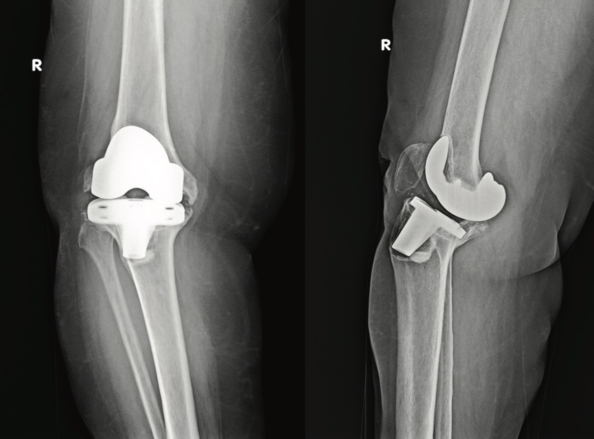
The radiologic assessment included a standard X-ray series of the right knee (Figure 1). AP and lateral views demonstrated severe component loosening, with more than 5 mm of radiolucent zones surrounding both the tibial and femoral components. The tibial component subsided into a varus position, with the tip of the tibial keel nearly protruding through the lateral plateau. The lateral view also showed a femoral component in the extended position and a significant increase in posterior tibial slope.
Given the clinical findings (severe, bidirectional instability) and the expected bone loss evident on knee radiographs, we decided to perform a revision TKA with a rotating hinge knee implant. We obtained informed consent after discussing treatment options, possible complications, and postoperative functional expectations with the patient. The patient was admitted to the Hospital one day before the planned surgery. Thromboembolic prophylactic therapy (low molecular weight heparin, LMWH) was started one day before the surgery and continued for five weeks. Given the higher incidence of periprosthetic joint infection after revision TKA, antibiotic treatment (cephazolin) was started preoperatively and continued for five days after surgery.
After standard preoperative preparation of the surgical field, under the conditions of general anesthesia, a longitudinal incision was made in the anterior aspect of the right knee following the old surgical scar. Given the expected prolonged surgical time, we decided to perform a revision TKA without inflating the tourniquet. After soft tissue dissection, standard medial parapatellar arthrotomy was done, and the joint space was revealed. Initially, we removed the fibrotic scar tissue and adhesions from the suprapatellar pouch and medial and lateral gutters. Joint aspiration fluid and tissue samples were sent to the microbiology department for extempore biopsy to check for the possible presence of pathogenic bacteria. After soft tissue release, Homan retractors were placed posteriorly, allowing for the luxation of the knee prosthesis. We then began by extracting the loosened prosthesis components. The polyethylene insert was removed using a special extractor. The femoral component was then detached at the component-cement interface using narrow osteotomes and a reciprocating saw, with extreme care being taken to avoid any iatrogenic bone loss. Extraction of the femoral component was completed with the use of a slap hammer. The tibial component was removed in a similar fashion using a reciprocating saw.
After assessing bone loss and ligamentous stability, we proceeded with preparing the medullary canal of the tibia using reamers of progressively increasing diameter. A tibial recut was made to flatten the surface of the tibial plateau and allow the accommodation of a trabecular whole block (size 10 mm) and a half block augment (size 5 mm), which was placed medially. After selecting the appropriate size of the tibial platform and determining its position, we prepared the distal femur. We began by reaming the medullary canal, following the same procedure as for the tibia. We then assessed the size and rotation of the femoral component, taking into account the mediolateral diameter calculated from the trans-epicondylar distance. We then placed the trial femoral component and performed balancing of flexion and extension gaps.
Given the preoperative recurvatum deformity, one of the imperatives was to put the femoral component more distally, which necessitated the use of distal augments. The resultant flexion-extension gap mismatch was solved with posterior femoral augments. Besides proper alignment and stability in extension and flexion, the joint line level was assessed using available anatomic landmarks, such as the medial and lateral epicondyles and the patella.
After trial reduction, knee kinematics and stability were verified through a full range of motion. The tourniquet was then inflated, and bone surfaces were cleaned with a pulsatile lavage system. Cement was applied into the medullary canals of the tibia and femur, and final components were placed. Once the cement was cured, the final polyethylene insert was inserted, and the hinge post mechanism was assembled. The wound was then thoroughly irrigated and sutured following the anatomic layers.
Rehabilitation began on the first postoperative day, following the hospital’s protocols. Early mobilization included passive and active range-of-motion exercises to promote flexibility and reduce joint stiffness. Full weight-bearing was permitted as tolerated, with patients using assistive devices such as crutches or a walker to ensure stability during ambulation. Achieving an adequate range of knee flexion was a priority, reinforced through the use of a continuous passive motion (CPM) apparatus, typically set at a predetermined speed and range and adjusted gradually based on patient tolerance and progress.
Gradual progression of strengthening exercises targeting the quadriceps, hamstrings, and surrounding musculature was initiated to support joint stability and function. Additionally, patient education emphasized activities of daily living and joint protection strategies to optimize functional recovery. The rehabilitation plan was tailored to meet the individual needs of each patient, with close clinical monitoring to address any complications and facilitate optimal outcomes.
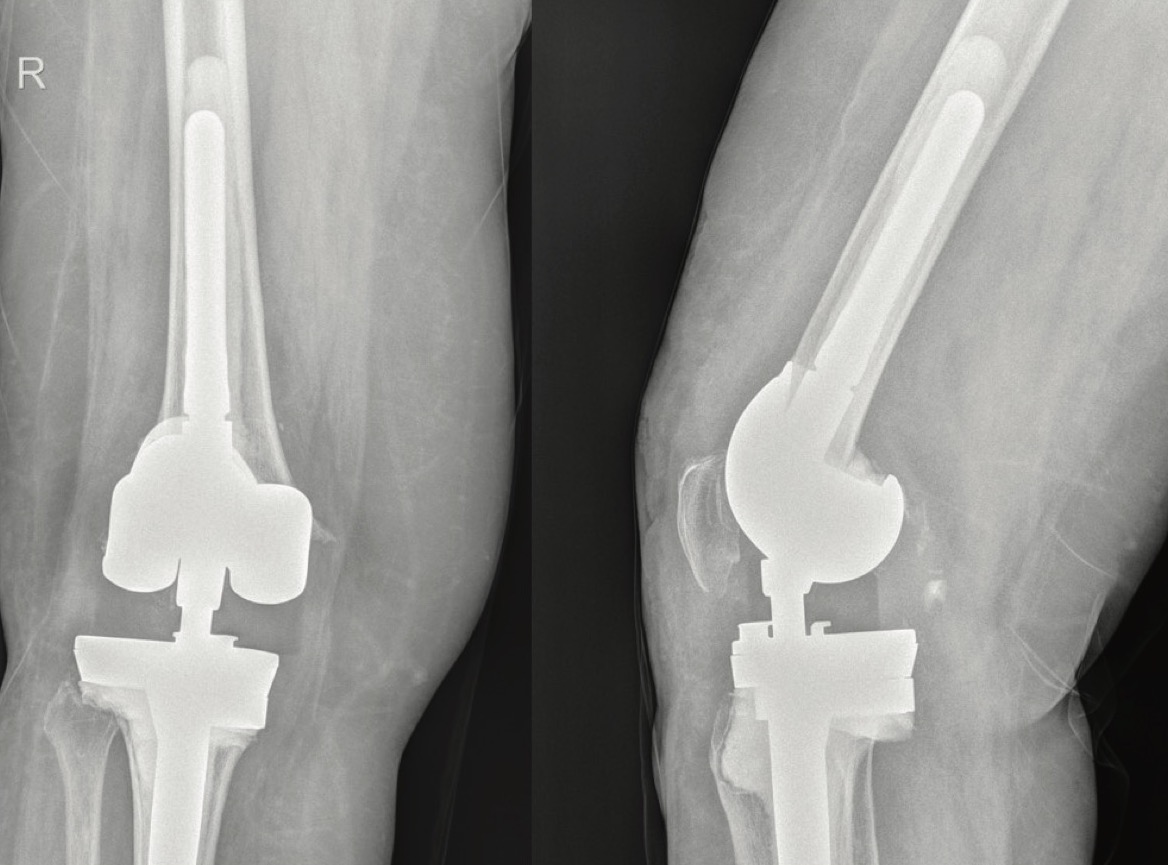
The postoperative course was uneventful, and the patient was discharged from the Hospital on the 5th postoperative day. The first postoperative control was scheduled one month after surgery, and the patient presented with a significantly improved gait pattern and a notable increase in right knee range of motion. Follow-up continued for two years, and at the last visit, the patient returned entirely to normal functional status and was free of any symptoms. This improvement was further confirmed with clinical and functional KSS, which measured 88 and 95, respectively. Control radiographs confirmed well-positioned TKA components without signs of loosening (Figure 2).
Case 2
A 73-year-old female patient was admitted to our hospital because of an injury to her left knee that resulted from a fall from a standing height that happened five days before the presentation. Her chief complaint is pain in the left knee in an attempt to bear weight. Primary TKA was performed 14 years ago in our Hospital. A focused physical examination at the Emergency Unit revealed significant swelling and deformity of the left knee, with movement of the left knee inhibited by pain. An old surgical scar was inspected on the front aspect of the left knee. There were no skin lesions or disturbance of local vascular and neurologic status.
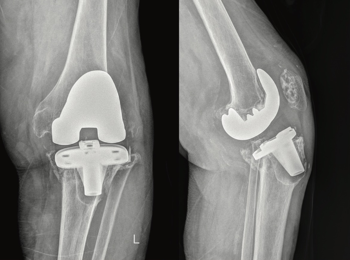
A trauma X-ray series of the injured knee was obtained. They demonstrated a periprosthetic fracture of the tibial plateau, type IIB, according to the classification system proposed by Lewis et al. (fracture spanning across the entire proximal tibia with tibial component loosening) (Figure 3). The tibial component subsided into a varus position, with its tip protruding through the anterolateral cortex of the proximal tibial metaphysis. The lateral view also demonstrated a drastically increased tibial slope.
Given the failure of the tibial component caused by a periprosthetic fracture, we decided to perform a revision TKA again using an RHK implant. We obtained informed consent and proceeded with the preoperative preparation. The same protocol for thromboembolic and antibiotic prophylactic therapy was followed, given the complexity of surgical treatment and the high risk of complications in this age group. The procedure was performed under general anesthesia. After consultation with the attending anesthesiologist, we decided to use a Cell Saver machine to collect and process the aspirated blood and then return it to the patient due to the expected blood loss during the surgery. The surgical technique did not differ in any aspect from the first case. Special care was taken during the extraction of the tibial component to preserve as much bone as possible. Given the fracture pattern and poor bone quality, we had to resect part of the proximal tibia to reach good-quality bone that would allow for proper seating of the tibial component. To resolve the problem with the joint line level and the expected loose extension and flexion gap, we again decided to augment the tibia with the 10 mm whole block trabecular augment. A revision of the femoral component was also required to accommodate the large box necessary to seat the hinge mechanism. Femoral preparation also necessitated the use of distal and posterior augments. The patellar button was removed due to evidence of wear, and we did not want to pursue implanting another button, given the poor quality of the remaining bone.
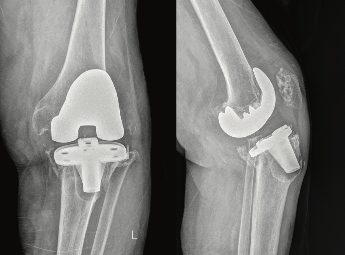
The identical rehabilitation protocol was followed in this case, except that full weight-bearing was not allowed for the first three postoperative weeks. The patient was discharged from the Hospital after seven days and returned for regular control one month after the surgery with satisfactory results. The control X-ray of the left knee, taken 2 years after surgery, showed well-positioned components with no signs of implant failure (Figure 4). Informed consent was obtained from the patients for the publication of their cases, in accordance with ethical standards.
DISCUSSION
The management of complex revisions in TKA remains a formidable challenge in orthopedic surgery, particularly when addressing issues such as significant bone loss, ligamentous instability, or multiple prior surgeries. In these scenarios, traditional fixed-bearing or semi-constrained knee prostheses may not provide the necessary functional stability and range of motion. This case series highlights the application of rotating hinge knee prostheses as a viable solution in revision TKA for patients experiencing these complications.
Rotating hinge prostheses offer several advantages over conventional designs, primarily due to their inherent stability and adaptability to anatomical conditions that are compromised. The design features a rotating mechanism that allows for an enhanced range of motion, reduces impingement, and facilitates better flexion-extension dynamics. This can be particularly beneficial in revision cases where the integrity of the soft tissue envelope is compromised, allowing for more predictable outcomes and improved patient satisfaction [11],[12]. Our findings are consistent with previous literature, which demonstrates improved functional outcomes and implant stability in similar patient populations [13],[14].
Our case series demonstrated satisfactory clinical outcomes, including pain reduction and improved functional scores, aligning with the existing literature advocating for the use of rotating hinge prostheses in similar revision scenarios [15]. Notably, the incorporation of patient-reported outcome measures (PROMs) underscored the importance of assessing not only clinical metrics but also the perceived quality of life among patients post-operatively. Comparing our results with prior research emphasizes the clinical significance of rotating hinge designs in achieving a balance between stability and mobility in complex revision scenarios.
However, the use of rotating hinge knee prostheses is not without its challenges. The increased complexity of surgical technique, potential for higher rates of complications such as infections and component loosening, and the need for meticulous preoperative planning and assessment must be emphasized. Risks include mechanical failure (e.g., hinge dislocation or fracture), aseptic loosening, and infection – all of which can adversely impact outcomes and may require further surgical intervention. Strategies to mitigate these risks include the careful selection of candidates, thorough evaluation of host bone quality, and interdisciplinary collaboration for optimal perioperative management [15]. Recognizing these potential tations align with findings from the literature, which note that long-term durability remains an area for ongoing investigation.
Ultimately, while the adoption of rotating hinge knee prostheses in revision TKA presents unique advantages, it necessitates a comprehensive understanding of the specific indications and contraindications, as well as an open dialogue about patient expectations. This case series serves as a reminder of the importance of tailored surgical solutions in the context of revision arthroplasty, pushing forward the boundaries of what can be achieved in complex knee reconstructions. Further large-scale studies with extended follow-up are warranted to validate these findings and establish long-term outcomes associated with this approach [16].
CONCLUSION
The use of rotating hinge knee prostheses in revision total knee arthroplasty demonstrates promising outcomes, particularly in cases involving significant bone loss and instability. This case series underscores the importance of individualized surgical planning and thorough preoperative assessment in achieving optimal patient outcomes. Further research is necessary to validate these findings and establish long-term efficacy.
Informations

-
Keywords:
-
Received:
-
Revised:
-
Accepted:
-
Online first:
-
DOI:
-
Cite this article:

Nikola Žarković
Institute for Orthopedic Surgery “Banjica”
28 Mihaila Avramovića Street, 11000 Belgrade, Serbia
E-mail:
1. Kurtz SM, Lau E, Ong K, Zhao K, Kelly M, Bozic KJ. Future young patient demand for primary and revision joint replacement: national projections from 2010 to 2030. Clin Orthop Relat Res. 2009 Oct;467(10):2606-12. doi: 10.1007/ s11999-009-0834-6. [CROSSREF]
2. Gioe TJ, Novak C, Sinner P, Ma W, Mehle S. Knee arthroplasty in the young patient: survival in a community registry. Clin Orthop Relat Res. 2007 Nov;464:83-7. doi: 10.1097/BLO.0b013e31812f79a9. [CROSSREF]
3. Long WJ, Bryce CD, Hollenbeak CS, Benner RW, Scott WN. Total knee replacement in young, active patients: long-term follow-up and functional outcome: a concise follow-up of a previous report. J Bone Joint Surg Am. 2014 Sep 17;96(18):e159. doi: 10.2106/JBJS.M.01259. [CROSSREF]
4. Neuhaus HJ, Maier K. Rotating hinge prosthesis for primary and revision knee arthroplasty: comparison and indications. Biomed Res Int. 2022 Jan 30;2022:9930675. doi: 10.1155/2022/9930675. [CROSSREF]
5. Ucpunar H, Camurcu Y, Buyuk AF, Aydin A, Tas SK, Sahin V. Comparison of contemporary surgical indications for revision total knee arthroplasty between 2005-2011 and 2012-2017 in a Referral Center. Arch Bone Jt Surg. 2022 Jan;10(1):32-7. doi: 10.22038/ABJS.2021.47457.2326.
6. Tarazi JM, Chen Z, Scuderi GR, Mont MA. The epidemiology of revision total knee arthroplasty. J Knee Surg. 2021 Nov;34(13):1396-401. doi: 10.1055/s0041-1735282. [CROSSREF]
7. Smolle MA, Hörlesberger N, Maurer-Ertl W, Puchwein P, Seibert FJ, Leithner A. Periprosthetic fractures of hip and knee – a morbidity and mortality analysis. Injury. 2021 Nov;52(11):3483-8. doi: 10.1016/j.injury.2021.01.015. [CROSSREF]
8. Canton G, Ratti C, Fattori R, Hoxhaj B, Murena L. Periprosthetic knee fractures. A review of epidemiology, risk factors, diagnosis, management and outcome. Acta Biomed. 2017 Jun 7;88(2S):118-28. doi: 10.23750/abm. v88i2-S.6522.
9. Felix NA, Stuart MJ, Hanssen AD. Periprosthetic fractures of the tibia associated with total knee arthroplasty. Clin Orthop Relat Res. 1997 Dec;(345):113- 24.
10. von Hintze J, Niemeläinen M, Sintonen H, Nieminen J, Eskelinen A. Outcomes of the rotating hinge knee in revision total knee arthroplasty with a median follow-up of 6.2years. BMC Musculoskelet Disord. 2021 Apr 7;22(1):336. doi: 10.1186/s12891-021-04205-9. [CROSSREF]
11. Lim JBT, Pang HN, Tay KJD, Chia SL, Lo NN, Yeo SJ. Increased constraint of rotating hinge knee prosthesis is associated with poorer clinical outcomes as compared to constrained condylar knee prosthesis in total knee arthroplasty. Eur J Orthop Surg Traumatol. 2020 Apr;30(3):529-35. doi: 10.1007/ s00590-019-02598-x. [CROSSREF]
12. Malcolm TL, Bederman SS, Schwarzkopf R. Outcomes of varus valgus constrained versus rotating-hinge implants in total knee arthroplasty. Orthopedics. 2016 Jan-Feb;39(1):e140-8. doi: 10.3928/01477447-20151228-07. [CROSSREF]
13. Kouk S, Rathod PA, Maheshwari AV, Deshmukh AJ. Rotating hinge prosthesis for complex revision total knee arthroplasty: a review of the literature. J Clin Orthop Trauma. 2018 Jan-Mar;9(1):29-33. doi: 10.1016/j.jcot.2017.11.020. [CROSSREF]
14. Spranz DM, Koch KA, Reiner T, Hetto P, Gotterbarm T, Merle C. Mid-term results of complex primary total knee arthroplasty using a rotating-hinge implant. Knee. 2022 Jan; 34:34-41. doi: 10.1016/j.knee.2021.10.001. [CROSSREF]
15. Efe T, Roessler PP, Heyse TJ, Hauk C, Pahrmann C, Getgood A, et al. Mid-term results after implantation of rotating-hinge knee prostheses: primary versus revision. Orthop Rev (Pavia). 2012 Dec 11;4(4):e35. doi: 10.4081/or.2012.e35. [CROSSREF]
16. Schlechter M, Theil C, Gosheger G, Moellenbeck B, Schwarze J, Puetzler J, et al. Good mid-term implant survival of a novel single-design rotating-hinge total knee arthroplasty. J Clin Med. 2023 Sep 22;12(19):6113. doi: 10.3390/ jcm12196113. [CROSSREF]
- Figure 1. Preoperative right knee AP and lateral view of 60-year old female patient suffering from aseptic loosening of total knee prosthesis, with severe malposition of tibial component into varus and femoral component into extension
- Figure 2. Postoperative AP and lateral views of the same patient at 2-year follow-up, showing no signs of component malalignement or loosening
- Figure 3. Preoperative left knee AP and lateral view of 73-year old female patient who suffered from proximal tibia periprosthetic fracture following fall from standing height
- Figure 4. Postoperative AP and lateral views of the same patient at 2-year follow-up, showing no signs of component malalignement or loosening