


Sclerochoroidal calcification mimicking choroidal osteoma: a case report
- University Clinical Centre of Serbia, Eye Hospital, Belgrade, Serbia
- University of Belgrade, Faculty of Medicine, Belgrade, Serbia
- Mitrović Eye Clinic, Belgrade, Serbia
ABSTRACT
Introduction: Sclerochoroidal calcifications are uncommon, benign ocular findings characterized by the deposition of calcium and phosphate within the sclera and adjacent choroid. These lesions are most frequently discovered incidentally during routine ophthalmologic evaluation, particularly in elderly individuals, and may resemble more serious intraocular pathologies.
Case report: We present the case of a 62-year-old male patient referred for evaluation of a subretinal lesion in the peripheral retina of the left eye, initially detected during a routine ophthalmologic examination. The patient was asymptomatic, with no visual complaints. Fundus examination revealed a yellowish, placoid lesion with indistinct borders located suprapapillary, between the superior vascular arcades. B-scan ultrasonography demonstrated high internal reflectivity with posterior acoustic shadowing. Fluorescein angiography revealed a hypofluorescent lesion without leakage or staining. The lesion remained stable over several years, and systemic workup showed no evidence of metabolic abnormalities. Based on clinical and imaging findings (B-scan ultrasonography and fluorescein angiography), the diagnosis of sclerochoroidal calcification was established.
Conclusion: Sclerochoroidal calcifications may clinically and ultrasonographically mimic more serious intraocular lesions. B-scan ultrasonography and fluorescein angiography, play a key role in distinguishing these benign findings from other conditions. In asymptomatic patients without signs of progression, annual follow-up is recommended, with no need for therapeutic intervention.
INTRODUCTION
Sclerochoroidal calcifications are rare, benign ocular findings characterized by abnormal deposition of calcium and phosphate within the sclera and adjacent choroid. These lesions are most frequently observed in elderly individuals and are often identified incidentally during routine ophthalmic examinations or imaging performed for unrelated complaints. Although typically asymptomatic, their appearance may closely resemble more serious intraocular pathologies, including choroidal osteoma, amelanotic choroidal melanoma, metastatic tumors, or inflammatory granulomas. Therefore, accurate diagnosis is essential to prevent unnecessary interventions [1],[2].
The etiology of sclerochoroidal calcifications can be idiopathic or associated with systemic abnormalities in calcium-phosphate metabolism, such as hyperparathyroidism, chronic kidney disease, or other endocrine disorders [3],[4]. Clinically, these lesions present as yellowish, placoid areas with indistinct borders, most often located between the superior vascular arcades. They are frequently bilateral and demonstrate high internal reflectivity on B-scan ultrasonography due to calcium content, typically accompanied by posterior acoustic shadowing [5].
This case report describes a male patient with a previously identified, stable subretinal lesion in the peripheral retina of the left eye. Clinical evaluation, including ophthalmoscopy, B-scan ultrasonography, and fluorescein angiography (FA), revealed a lesion with features of sclerochoroidal calcification. Considering that such lesions may ophthalmoscopically resemble, most commonly, a choroidal osteoma and, much more rarely, an amelanotic choroidal melanoma, this case emphasizes the importance of detailed clinical assessment in the differential diagnosis of calcified choroidal lesions.
CASE REPORT
A 62-year-old male patient was referred to the Clinic for Eye Diseases, University Clinical Centre of Serbia, for evaluation of a tumorous lesion on the periphery of the left ocular fundus, which had been initially detected in another institution in 2019 during a routine ophthalmological examination. The patient reported no visual complaints, including changes in visual acuity, visual field disturbances, or photopsias.
B-scan ultrasonography of the left eye (OS) showed a subretinal lesion localized in the superior fundus, with a flattened appearance, measuring 9.96 × 8.07 mm in base and 1.05–1.54 mm in thickness. The lesion demonstrated high internal reflectivity and an acoustic shadow, indicating dense calcification. Based on its echographic characteristics, the lesion was initially considered a choroidal osteoma.
The patient’s ophthalmological history included primary open-angle glaucoma diagnosed five years earlier, for which he was receiving topical antiglaucoma therapy (Xalatan once daily). There was no history of ocular trauma, inflammation, or surgery. His systemic medical history was notable for hypertension and type 2 diabetes mellitus, both well controlled under therapy. A positive family history of glaucoma was also noted.
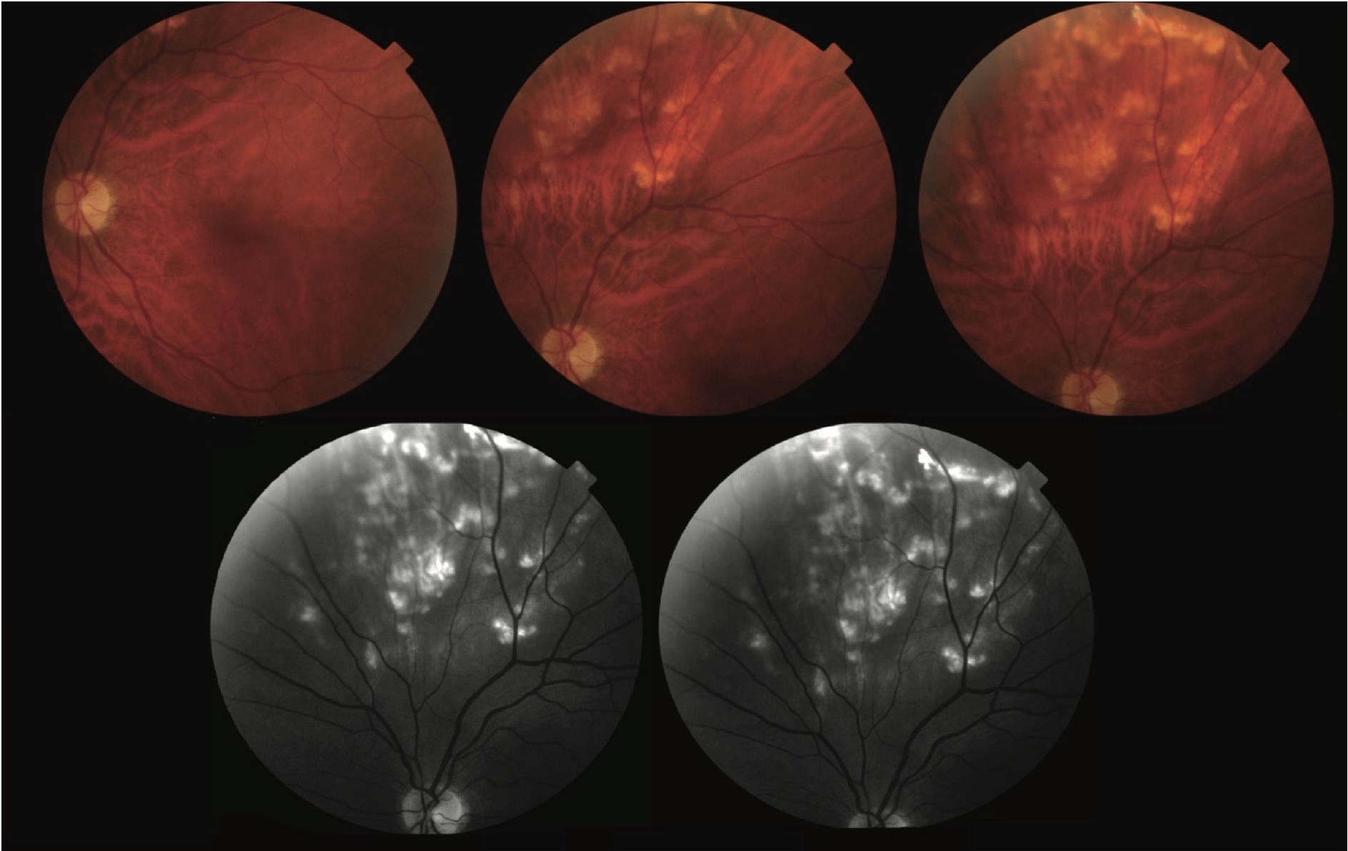
On examination, best-corrected visual acuity was 0.8 in both eyes. The anterior segments were within normal limits. Intraocular pressures were well controlled under therapy (15 mmHg in the right eye and 13 mmHg in the left eye). Fundus examination of the left eye revealed a yellowish subretinal lesion located suprapapillary (Figure 1), between the superior-nasal and superior-temporal vascular arcades. The lesion had indistinct borders and was slightly elevated, without pigmentary changes or exudation on its surface. The surrounding retina appeared normal, and the macula and optic disc were unremarkable.
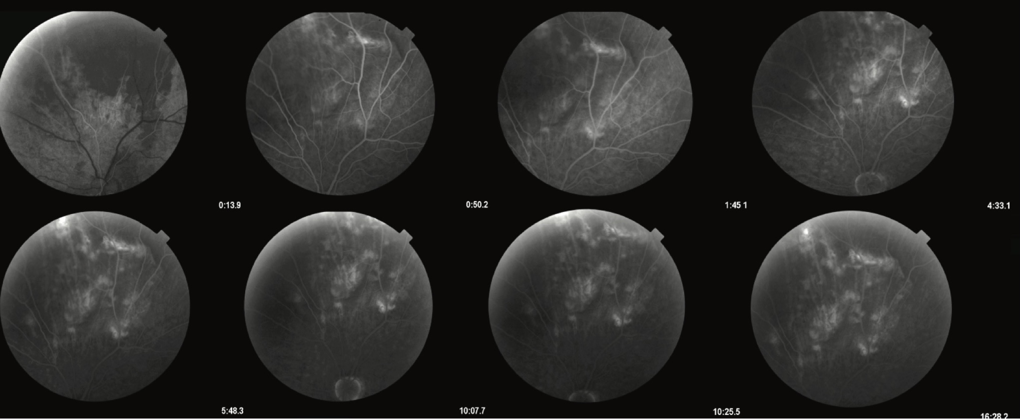
Fluorescein angiography (FA) showed a predominantly hypofluorescent lesion in the early phases, without significant leakage or staining in the later phases (Figure 2). Yellowish placoid changes were observed on the lesion surface, without subretinal fluid or choroidal neovascularization. The retinal vessels and optic disc were normal.
Based on ophthalmoscopy and additional imaging (B-scan ultrasonography and FA), the lesion was most consistent with sclerochoroidal calcification. However, the differential diagnosis included, primarily, choroidal osteoma, as well as amelanotic choroidal melanoma, choroidal hemangioma, metastatic lesions, and inflammatory pathology (focal chorioretinitis). The patient was advised to continue annual ophthalmological follow-up to monitor for potential changes in lesion size or the development of complications.
DISCUSSION
Sclerochoroidal calcifications are often discovered incidentally, but may represent a diagnostic challenge because their clinical appearance can mimic other entities such as choroidal osteoma, amelanotic choroidal melanoma, metastatic choroidal lesions, choroidal hemangiomas, or granulomatous infiltrates associated with inflammatory or hematologic diseases [1],[2]. For an accurate diagnosis, correlation of ophthalmoscopic, ultrasonographic, and angiographic findings is required.
In our patient, the lesion was localized in the peripheral fundus and remained stable over time. On ultrasonography, it showed high internal reflectivity with posterior acoustic shadowing, features characteristic of calcified intraocular lesions [3]. Fluorescein angiography demonstrated a uniformly hypofluorescent pattern without leakage or staining, indicating the non-vascular and benign nature of the lesion [4]. The absence of subretinal fluid or retinal pigment epithelial changes further reduced the likelihood of malignancy or inflammation.
Within the differential diagnosis, choroidal osteoma is of primary importance due to overlapping imaging characteristics. However, choroidal osteomas typically occur in younger patients, are most often localized in the peripapillary or macular regions, and can be associated with visual impairment, particularly when complicated by choroidal neovascularization or tumor decalcification [5]. In contrast, sclerochoroidal calcifications usually appear in older patients, are often located peripherally, and rarely affect visual acuity [6].
Sclerochoroidal calcifications may be associated with systemic disorders of calcium-phosphate metabolism, including hyperparathyroidism, chronic kidney disease, and other metabolic disturbances. Therefore, systemic evaluation is mandatory. A detailed laboratory work-up should include measurement of total and ionized calcium, phosphates, parathyroid hormone (PTH), 25(OH) vitamin D, 1,25(OH)₂ vitamin D, creatinine, urea, estimated GFR (calculated from serum creatinine), alkaline phosphatase, and basic electrolytes such as sodium, potassium, chloride, and magnesium [7],[8]. In our patient, laboratory testing revealed no abnormalities in calcium-phosphate metabolism, suggesting a primary or age-related form of the disease.
Given their benign course, sclerochoroidal calcifications rarely require therapeutic intervention. Nevertheless, long-term monitoring is recommended to detect potential morphological changes. In this case, the absence of progression and lack of visual symptoms confirmed the diagnosis of sclerochoroidal calcification rather than choroidal osteoma or any of the other aforementioned differential diagnoses.
Informations

-
Keywords:
-
Received:
-
Revised:
-
Accepted:
-
Online first:
-
DOI:
-
Cite this article:

Dolika D. Vasović
Eye Hospital, University Clinical Centre of Serbia
2 Pasterova Street, 11000 Belgrade, Serbia
E-mail:
1. Gündüz AK, Tetik D. Diagnosis and management strategies in sclerochoroidal calcification: a systematic review. Clin Ophthalmol. 2023 Sep 11;17:2665-86. doi: 10.2147/OPTH.S399058. [CROSSREF]
2. Shields CL, Hasanreisoglu M, Saktanasate J, Shields PW, Seibel I, Shields JA. Sclerochoroidal calcification: clinical features, outcomes, and relationship with hypercalcemia and parathyroid adenoma in 179 eyes. Retina. 2015 Mar;35(3):547-54. doi: 10.1097/IAE.0000000000000450. [CROSSREF]
3. Honavar SG, Shields CL, Demirci H, Shields JA. Sclerochoroidal calcification: clinical manifestations and systemic associations. Arch Ophthalmol. 2001 Jun;119(6):833-40. doi: 10.1001/archopht.119.6.833. [CROSSREF]
4. Mani X, Johnson R. A case of sclerochoroidal calcification masquerading as a retained intraocular foreign body. Radiol Case Rep. 2023 Mar 25;18(5):2034- 8. doi: 10.1016/j.radcr.2023.02.049. [CROSSREF]
5. Sood S, Friedman S. Sclerochoroidal calcification with optic nerve calcification in a patient with primary hyperparathyroidism and severe vision loss. Retin Cases Brief Rep. 2024 Jul 1;18(4):499-502. doi: 10.1097/ ICB.0000000000001418. [CROSSREF]
6. Lopez JM, Rabinovich M, Colantuono D, Souied EH. Multimodal and retro-mode imaging in sclerochoroidal calcification: a case report. Arch Soc Esp Oftalmol (Engl Ed). 2024 May;99(5):213-7. doi: 10.1016/j.oftale.2024.02.002. [CROSSREF]
7. Thomson AC, Brown GT, Dolores-Rodriguez A, Hunter AA. A Case of extensive bilateral idiopathic sclerochoroidal calcification and review of literature. Int Med Case Rep J. 2021 Oct 28;14:749-55. doi: 10.2147/IMCRJ.S336237. [CROSSREF]
8. Brahma VL, Shah SP, Chaudhry NA, Prenner JL. Bilateral idiopathic sclerochoroidal calcifications. Open Ophthalmol J. 2017 Apr 27;11:76-9. doi: 10.2174/1874364101711010076. [CROSSREF]
- Figure 1. Fundus photograph of the left eye demonstrating a yellowish, placoid subretinal lesion with indistinct borders located suprapapillary, between the superior nasal and superior temporal vascular arcades. The lesion is consistent with sclerochoroidal calcification and appears elevated, without associated subretinal fluid, hemorrhage, or pigmentary changes. The surrounding retinal tissue, macula, and optic disc appear unremarkable
- Figure 2. Fluorescein angiography of the left eye showing a hypofluorescent subretinal lesion located suprapapillary, between the superior vascular arcades. The lesion demonstrates absence of leakage or staining in both early and late phases, consistent with a benign, avascular process such as sclerochoroidal calcification