


Significance of the preventive ultrasound diagnostics in the early detection of rare and aggressive thyroid carcinomas – case report
- Obrenovac Health Centre, Radiology Department, Obrenovac, Serbia
- University Clinical Center of Serbia, Department of Pathology and Medical Cytology, Belgrade, Serbia
- University of Belgrade, Faculty of Medicine, Institute of Pathology, Belgrade, Serbia
ABSTRACT
Introduction: Medullary thyroid carcinoma (MTC) is an aggressive tumor associated with a significantly poorer prognosis compared to other types of thyroid cancer. Doppler ultrasound and familiarity with the Thyroid Imaging Reporting and Data System (TIRADS) nodule classification are essential.
Objective: This case highlights the significance of precise ultrasound imaging, crucial for early detection and subsequent management of such aggressive disease.
Case report: We describe the case of a 31-year-old woman who underwent systemic ultrasound imaging. Upon ultrasound examination, a 5 mm x 4 mm x 3 mm hypoechoic, ill-defined solid nodule with echogenic foci (TIRADS 4, score 6) was identified in the mid portion of the left thyroid lobe. Given the suspicious ultrasound features, the location of the nodule, and the elevated serum calcitonin level, the micronodule raised concerns for malignancy, particularly suggesting medullary thyroid microcarcinoma. The patient underwent thyroidectomy, and histopathological analysis confirmed the diagnosis. The histopathology report described a small white nodule (5 mm) in the left lobe. There was no remarkable necrosis, and amyloid deposits were inconspicuous. Lymphovascular invasion was not observed, and mitoses were rare. The tumor stage was T1 (pTNM), and no distant metastases were found. C-cell hyperplasia was noted around the tumor. Immunohistochemical analysis showed positive reactions for calcitonin, chromogranin, and mCEA. The proliferative index was low, approximately 4%. After surgery, serum calcitonin levels decreased. Three years after the surgery, the patient is in good general condition and is under regular monitoring. In the meantime, the patient has become a mother.
Conclusion: Early detection and treatment of micro-MTC are crucial, as these tumors have the potential for aggressive behavior. Adequate ultrasound imaging assessment could significantly improve the prognosis and outcome for these patients.
INTRODUCTION
Medullary thyroid carcinoma (MTC) is a rare tumor, accounting for 5% of thyroid malignancies. MTC is an aggressive tumor, associated with a significantly poorer prognosis compared to other types of thyroid cancer [1]. MTC is responsible for approximately 13% of deaths related to thyroid cancer, indicating its considerable clinical aggressiveness compared to follicular-derived neoplasms (FDN) [2]. Its potential to metastasize to regional lymph nodes early in its progression often results in elevated serum calcitonin levels. While the necessity of lymphadenectomy in the absence of palpable nodal disease sparks debate, studies have shown that successful normalization of serum calcitonin levels correlates with enhanced survival. A combined assessment of ultrasound and biochemical features is associated with the prognosis of MTC [1],[3].
Serum calcitonin serves as a key tumor marker and is highly specific for MTC [4]. Patients who smoke or use certain medications, such as proton-pump inhibitors (PPIs), may induce false-positive results, thereby reducing the specificity of serum calcitonin. In such cases, diagnosis is based on a continuous rise in serum calcitonin levels over time, despite discontinuation of tobacco use and PPI therapy, combined with the presence of a suspicious solitary micronodule in the central region of the upper/mid thyroid lobe [1].
The concept of micro-MTC, typically defined as MTC measuring less than 1 cm, has predominantly emerged within familial MTC contexts or as a result of calcitonin screening initiatives. This study describes the management of patients with sporadic micro-MTC in the absence of such screening programs [1],[5].
Thyroid nodules are commonly discovered incidentally during neck ultrasound (US) examinations in adults. In such instances, a comprehensive understanding of thyroid nodule characteristics on high-frequency Doppler ultrasound and familiarity with the Thyroid Imaging Reporting and Data System (TIRADS) nodule classification are essential [6],[7]. Thyroid nodules with ultrasound features suggestive of malignancy, such as marked hypoechogenicity, irregular margins, microcalcifications, a taller-than-wide shape, and high stiffness on elastography, warrant fine-needle aspiration cytology (FNAC), followed by cytological evaluation according to the 2010 Bethesda classification, regardless of nodule size. Additionally, Doppler US evaluation may reveal supplementary suspicious features, including disorganized central vasculature and a high resistance index (RI > 0.75). MTC may also exhibit distinctive US Doppler features [1],[8].
The natural history of MTC has been difficult to delineate due to its rarity. The significance of medullary microcarcinomas remains elusive despite their frequent detection, often through calcitonin screening programs. Serum calcitonin levels above 100 pg/mL are generally indicative of macro-MTC [1]. This lack of consensus extends to the clinical significance and optimal management of micro-MTC, particularly within sporadic and occult settings [1],[2].
This study highlights the significance of precise ultrasound imaging, crucial for early detection and further management, in order to prevent a poor prognosis for such an aggressive disease.
CASE REPORT
We describe a 31-year-old woman who underwent systemic ultrasound imaging. Upon ultrasound examination, a 5 mm x 4 mm x 3 mm hypoechoic ill-defined nodule with echogenic foci (TIRADS 4) was identified in the mid portion of the left thyroid lobe (Figure 1). Additionally, her serum calcitonin level was measured at 17.7 pg/mL. Serum calcitonin was repeated several times over a three-month period. Given the suspicious ultrasound features, the location of the nodule, and the elevated serum calcitonin level, the micronodule raised concerns for malignancy, particularly suggesting medullary thyroid microcarcinoma.
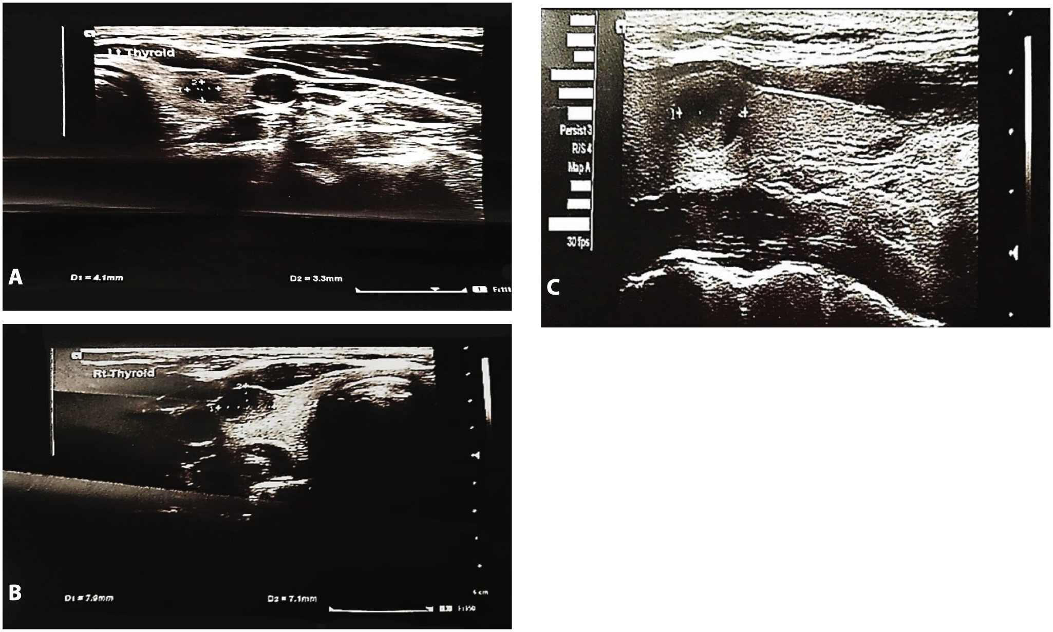
The patient was referred to a tertiary institution for further investigation of her thyroid gland. Subsequently, she underwent thyroidectomy, and histopathological analysis confirmed the diagnosis. The patient did not undergo a preoperative biopsy. The histopathology report described a small white nodule in the left lobe, with a diameter of 5 mm (Figure 2). There was no remarkable necrosis, and amyloid was inconspicuous. Lymphovascular invasion was not observed, and mitoses were rare. The tumor stage was T1 (pTNM). Lymph node status was not evaluated histopathologically, and no distant metastases were found. C-cell hyperplasia was observed around the tumor, and the rest of the thyroid tissue showed stromal changes.
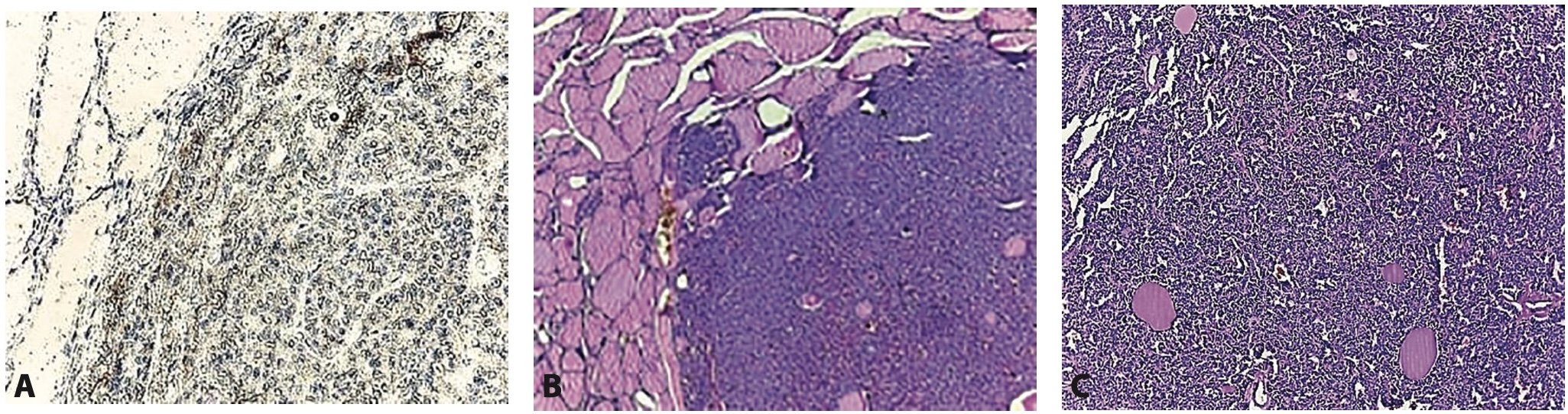
Immunohistochemical analysis showed positive reactions for calcitonin, chromogranin, and mCEA. Negative staining was observed with HBME, CD56, CK19, and galectin-3. S100 was found in rare, single cells. The proliferative index was low, approximately 4%.
The patient did not report any similar disease or genetic inheritance in her family history. After surgery, serum calcitonin levels decreased. The genetic analysis performed did not reveal any specific alterations associated with the detected MTC, suggesting a sporadic form of the disease.
The patient was referred for clinical and imaging follow-up, which includes regular serum calcitonin measurements and thorough clinical examinations. Ultrasound examinations are performed every six months.
Three years after the surgery, the patient is in good general condition and is under regular monitoring. In the meantime, the patient has become a mother.
DISCUSSION
Micro-medullary carcinomas are often detected incidentally during prophylactic thyroidectomy procedures. Advancements in ultrasound technology now enable the detection of smaller nodules before surgical decisions are made. Early resection offers a significant possibility of cure in micro-MTC cases [2].
Considering the small size of micro-MTC, no cases of tumor recurrence or distant metastases were observed during clinical follow-up. The prevailing notion in the literature indicates that micro-MTCs are associated with higher cure rates and a lower risk of metastases and mortality compared to larger tumors, with only rare cases exhibiting clinically aggressive behavior [9]. Our findings further support this notion.
Since the prognosis of MTC is strongly influenced by tumor volume and extent of spread, radiologists must be proficient in identifying specific imaging features characteristic of MTC. Early and accurate diagnosis is paramount for optimizing surgical management and long-term outcomes in patients with this aggressive tumor [1],[8].
MTC typically originates in the upper or mid portion of the central thyroid lobe, where C (parafollicular) cells are concentrated. Radiologists should be alert to the presence of hyperechoic punctuations on ultrasound imaging, which may correspond to amyloid deposits or calcifications. Such findings should prompt the radiologist to perform a fine needle aspiration cytology (FNAC) for further evaluation [10]. Given the association between MTC and multiple endocrine neoplasia (especially RET mutations) radiologists should carefully evaluate for enlarged parathyroid glands situated posterior to the thyroid lobes, medial to the cervical carotid sheaths [1].
While some cases of MTC are associated with multiple endocrine neoplasia (MEN) types IIa or IIb, the majority arise sporadically. The discovery of small (less than 1 cm) occult MTCs, known as micro-MTCs, during pathological examination of thyroid specimens resected for benign conditions or non-MTC thyroid cancers, has been reported in both patients with and without familial histories of MEN or MTC. The prognostic implications and optimal surgical management of sporadic micro-MTC remain unclear [1],[11].
The majority of MTC cases are sporadic, occurring without a familial predisposition. However, up to 25% of cases are associated with one of three familial syndromes: Multiple Endocrine Neoplasia type 2A (MEN2A), Multiple Endocrine Neoplasia type 2B (MEN2B), and Familial Medullary Thyroid Carcinoma (FMTC). These familial cases are attributed to germline gainof-function autosomal-dominant mutations in the RET proto-oncogene [2],[5]. Monoallelic germline RET K666N mutations are associated with a risk of developing familial MTC, exhibiting age-dependent expressivity. However, these variants show low penetrance for other tumors typically seen in MEN2 syndromes [12].
Sporadic MTC typically presents with a palpable thyroid nodule in individuals aged between their 5th and 6th decades of life. In contrast, familial cases tend to occur at a younger age, and their presentation varies depending on the associated syndrome. For instance, MEN2A is characterized by the presence of pheochromocytoma and hyperparathyroidism, while MEN2B is associated with features such as pheochromocytoma, mucosal neuromas, and a marfanoid habitus. Importantly, familial MTC cases often present multifocally or bilaterally and are frequently associated with C-cell hyperplasia [2],[13],[14].
Differentiating MTC from more common thyroid malignancies such as papillary thyroid carcinoma (PTC) using ultrasound examination alone can be challenging. Some studies have suggested that combining various TIRADS scoring systems improves diagnostic accuracy [15], while others have demonstrated the effectiveness of radiomics models in distinguishing between these entities [16]. Evaluation of vascularity characteristics on ultrasound can assist in differentiating MTC from benign thyroid nodules. MTC is notably characterized by a distinctive penetrating branching vascular pattern. Integrating these vascular features into ultrasound-based risk stratification systems may enhance the identification of MTC among nodules with low to intermediate suspicion, thus facilitating more precise clinical management [17]. Novel imaging methods for MTC have highlighted the significance of CCK 2 R-Targeting Compound [18].
Navigating treatment decisions for incidentally discovered sporadic micro-MTC poses challenges, especially concerning the surgical approach and management of biochemical or radiologically detected metastatic disease. In cases with a preoperative diagnosis of MTC, total thyroidectomy with central compartment lymph node dissection is routinely performed. Lateral neck dissections are reserved for cases with clinically relevant nodal involvement [5].
Previous studies have described a correlation between a less aggressive phenotype and smaller, incidentally found tumors. Notably, only tumors larger than 0.5 cm exhibited evidence of vascular invasion or extrathyroidal extension. Desmoplastic reactions were also observed, with larger tumors more likely to exhibit such reactions. However, the relationship between desmoplasia, tumor size, and the presence of nodal metastases could not be conclusively established [1],[5]. Assessment of Ki-67 expression represents a predictive histopathological parameter that can be used alongside ultrasonographic features to evaluate the aggressiveness of MTC [19].
Medullary thyroid carcinoma (MTC) poses a significant clinical challenge as it exhibits limited responsiveness to chemotherapy and radiation therapy. Consequently, the cornerstone of its management primarily revolves around surgical excision of the thyroid gland with lymph node dissection. Lymph node involvement is prevalent in a considerable proportion of cases, particularly in patients with advanced tumors (T3–T4 tumors), highlighting the aggressive nature of this malignancy. Therefore, early preoperative diagnosis is crucial to facilitate appropriate treatment planning, typically achieved through preoperative fine needle aspiration analysis in conjunction with serum calcitonin measurements [2],[20].
Routine preventive screening for MTC in the general population is primarily based on the measurement of serum calcitonin levels, rather than prophylactic ultrasound examination. Ultrasound imaging is typically considered in patients with persistently elevated calcitonin levels. Despite the low incidence of micro-medullary thyroid carcinoma and the fact that ultrasound is not a standard screening method for this disease, an adequate and precise primary radiological examination can significantly influence the clinical course, especially when the disease is detected at an early stage [10],[21].
Prophylactic thyroidectomy is generally recommended by the age of five or earlier, especially in the presence of elevated serum calcitonin levels. However, the expression and progression of the disease within high risk categories can vary significantly. Consequently, the timing of prophylactic thyroidectomy is no longer based only on genotype, but rather on individualized clinical parameters, with particular emphasis on serum calcitonin concentrations [11].
CONCLUSION
Although micro-medullary thyroid carcinoma is a rare entity and ultrasound is not a standard screening method for its detection, early diagnosis remains essential due to the tumor’s potential for aggressive behavior. Serum calcitonin measurement continues to be the primary tool for early detection; however, in patients with elevated or persistently rising calcitonin levels, targeted and precise ultrasound imaging can play a critical role. An accurate initial radiological assessment, particularly when the disease is detected at an early stage, can significantly influence the clinical course, improve prognosis, and contribute to better patient outcomes.
Informations

-
Keywords:
-
Received:
-
Revised:
-
Accepted:
-
Online first:
-
DOI:
-
Cite this article:

Ljubiša Jovanović
Department of Pathology and Medical Cytology, University Clinical Center of Serbia
26 Dr Koste Todorovića Street, 11000 Belgrade, Serbia
E-mail:
1. Pillarisetty VG, Katz SC, Ghossein RA, Tuttle RM, Shaha AR. Micromedullary thyroid cancer: how micro is truly micro? Ann Surg Oncol. 2009 Oct;16(10):2875-81. doi: 10.1245/s10434-009-0595-1. [CROSSREF]
2. Samulski TD, Livolsi VA, Montone K, Baloch Z. The variable pathologic presentations of medullary and micro-medullary thyroid carcinoma: an institutional experience. Pathol Res Pract. 2014 Mar;210(3):182-5. doi: 10.1016/j. prp.2013.12.004. [CROSSREF]
3. Zhao J, Yang F, Wei X, Mao Y, Mu J, Zhao L, et al. Ultrasound features value in the diagnosis and prognosis of medullary thyroid carcinoma. Endocrine. 2021 Jun;72(3):727-34. doi: 10.1007/s12020-020-02510-2. [CROSSREF]
4. Trimboli P, Mian C, Piccardo A, Treglia G. Diagnostic tests for medullary thyroid carcinoma: an umbrella review. Endocrine. 2023 Aug;81(2):183-93. doi: 10.1007/s12020-023-03326-6. [CROSSREF]
5. Ospina NS, Maraka S, Donegan D, Morris JC. Clinical features of a family with multiple endocrine neoplasia type 2A caused by the D631Y RET mutation. Thyroid. 2017 Oct;27(10):1332-4. doi: 10.1089/thy.2016.0536. [CROSSREF]
6. Grani G, Sponziello M, Filetti S, Durante C. Thyroid nodules: diagnosis and management. Nat Rev Endocrinol. 2024 Dec;20(12):715-28. doi: 10.1038/ s41574-024-01025-4. [CROSSREF]
7. Matrone A, Gambale C, Biagini M, Prete A, Vitti P, Elisei R. Ultrasound features and risk stratification systems to identify medullary thyroid carcinoma. Eur J Endocrinol. 2021 Jun 28;185(2):193-200. doi: 10.1530/EJE-21-0313. [CROSSREF]
8. Sipos JA. the history of thyroid ultrasound: past, present, and future directions. Endocr Pract. 2024 Dec;30(12):1220-6. doi: 10.1016/j.eprac.2024.08.010. [CROSSREF]
9. Gui Z, Wang Z, Xiang J, Sun W, He L, Dong W, et al. Incidental T1 stage medullary thyroid carcinoma: the effect of tumour diameter on prognosis and therapeutic implications. Clin Endocrinol (Oxf). 2022 Sep;97(3):355-62. doi: 10.1111/cen.14702. [CROSSREF]
10. Trimboli P, Mian C, Piccardo A, Treglia G. Diagnostic tests for medullary thyroid carcinoma: an umbrella review. Endocrine. 2023 Aug;81(2):183-93. doi: 10.1007/s12020-023-03326-6. [CROSSREF]
11. Frank-Raue K, Raue F. Hereditary medullary thyroid cancer: genotype-phenotype correlation. Recent Results Cancer Res. 2025;223:183-209. doi: 10.1007/978-3-031-80396-3_7. [CROSSREF]
12. Yip AT, Kim TH, Peluso EM, Jacobsen SE, Yeh MW, Lechner MG. Medullary thyroid carcinoma and clinical outcomes in heterozygous carriers of the RET K666N germline pathogenic variant. JCEM Case Rep. 2025 Feb 17;3(3):luaf002. doi: 10.1210/jcemcr/luaf002. [CROSSREF]
13. Pacini F, Castagna MG, Cipri C, Schlumberger M. Medullary thyroid carcinoma. Clin Oncol (R Coll Radiol). 2010 Aug;22(6):475-85. doi: 10.1016/j. clon.2010.05.002. [CROSSREF]
14. Hamy A, Pessaux P, Mirallié E, Mucci-Hennekinne S, Gibelin H, Mor-Martinez C, et al. Central neck dissection in the management of sporadic medullary thyroid microcarcinoma. Eur J Surg Oncol. 2005 Sep;31(7):774-7. doi: 10.1016/j.ejso.2005.03.007. [CROSSREF]
15. Li X, Yao J, Zhan W, Zhou W. Ultrasonographic differentiation of medullary thyroid carcinoma from papillary thyroid carcinoma: quantitative comparison of morphologic features and different TIRADS risk categorizations. BMC Med Imaging. 2025 May 26;25(1):189. doi: 10.1186/s12880-025-01679-0. [CROSSREF]
16. Zhang D, Yang F, Hou W, Wang Y, Mu J, Wang H, et al. Ultrasonic radiomics in predicting pathologic type for thyroid cancer: a preliminary study using radiomics features for predicting medullary thyroid carcinoma. Front Endocrinol (Lausanne). 2025 Feb 19;16:1428888. doi: 10.3389/fendo.2025.1428888. [CROSSREF]
17. Gao L, Ma L, Li X, Liu C, Li N, Lian X, et al. Using preoperative ultrasound vascularity characteristics to estimate medullary thyroid cancer. Cancer Imaging. 2023 Jun 20;23(1):64. doi: 10.1186/s40644-023-00583-6. [CROSSREF]
18. Viering O, Rinscheid A, Holzleitner N, Dierks A, Kircher M, Wienand G, et al. Biodistribution and radiation dosimetry for 68 Ga-DOTA-CCK-66, a novel CCK 2 R-targeting compound for imaging of medullary thyroid cancer. Clin Nucl Med. 2024 Dec 1;49(12):1091-7. doi: 10.1097/RLU.0000000000005355. [CROSSREF]
19. Zhang Q, Hu Y, Chen X, Yang Z, Li X, Ni X, et al. Preoperative prediction of Ki-67 expression in medullary thyroid carcinoma based on ultrasonographic features: a 10-year retrospective study. Eur J Radiol. 2025 Jul;188:112134. doi: 10.1016/j.ejrad.2025.112134. [CROSSREF]
20. Deandrea M, Piticchio T, Mormile A, Retta F, Canale G, Caracciolo A, et al. Preoperative neck ultrasound combined with pathological data can significantly impact the outcome of medullary thyroid carcinoma. Endocr J. 2023 Nov 28;70(11):1061-7. doi: 10.1507/endocrj.EJ23-0273. [CROSSREF]
21. Delorme S, Raue F, Beuthien-Baumann B. Medullary thyroid carcinoma: imaging. Recent Results Cancer Res. 2025;223:129-53. doi: 10.1007/978-3-031- 80396-3_5. [CROSSREF]
- Figure 1. Ultrasound imaging of the patient's thyroid gland. Ultrasound examination of the thyroid gland detected a suspicious nodule in the left lobe. A. and B. Small hypoechoic, solid nodule (5mm) with echogenic foci, vascularization and irregular margins (TIRADS4, score 6) in the mid portion of the left thyroid lobe. C. In the contralateral right lobe, anterior and cranial, there was a mixed, predominantly cystic nodule measuring 9 mm with central vascularization. Unlike the left nodule, this lesion displayed a different imaging pattern and lacked suspicious malignant features
- Figure 2. Histopathology examination. Histopathology pattern of micromeddullary thyroid carcinoma (A. x50) and (B. x100). Immunohistochemical analysis with calcitonin shows positive reaction in neoplastic cells (C. x400)