


Prognostic significance of serum ferritin levels on initial diagnosis in patients with acute myeloid leukemia
- Faculty of Medicine, University of Belgrade, Belgrade, Serbia
- Clinic for Hematology, University Clinical Center of Serbia, Belgrade, Serbia
ABSTRACT
Introduction/Aim: Acute myeloid leukemia (AML) is a heterogenous malignant disease whose course and outcome are influenced by a number of prognostic factors. Serum ferritin (SF) is often elevated in oncology patients, and it has been shown that it strongly influences an unfavorable outcome in various malignancies. The aim of this study is to assess the effect of high SF values on overall survival and disease-free survival, as well as to assess the correlation of SF values with other prognostic markers, such as clinical and laboratory parameters.
Methods: Retrospective analysis included 108 patients diagnosed with AML at the Clinic for Hematology of the Clinical Center of Serbia (CCS), in Belgrade, in the period 2017 - 2019. Patients with acute promyelocytic leukemia, acute mixed lineage leukemia, secondary AML and patients treated with palliative therapy were excluded from the study. Patients were grouped based on the SF cutoff value of 800 µg/L.
Results: Patients with higher SF values had a significantly higher incidence of early death (p = 0.020), sepsis in the induction phase of therapy (p < 0.010), and significantly lower initial hemoglobin levels (p = 0.040), as compared to patients with lower SF values. SF at diagnosis appeared to be a significant independent predictive factor of overall survival (p = 0.019) and of disease-free survival (p = 0.040).
Conclusion: Our study showed a significant association of high SF values with sepsis in induction, early death, mean hemoglobin, overall survival, and disease-free survival. Identification of SF as an independent prognostic factor and a potential target site of the action of new drugs could contribute to a better prognosis of AML patients.
INTRODUCTION
Acute myeloid leukemia (AML) is a heterogenous malignant disease which is characterized by clonal proliferation of a precursor cell of the myeloid lineage. The accumulation of immature cells – blasts, leads to progressive insufficiency of bone marrow and the infiltration of the blood and other organs [1]. Achieving long-term survival is one of the greatest challenges in the treatment of AML, and it depends on a series of prognostic factors related to the characteristics of the disease, the characteristics of the patient, or the characteristics of the applied therapy. Some of the important independent prognostic factors for overall survival (OS) are the following: age, comorbidities, Eastern Cooperative Oncology Group (ECOG) performance status, cytogenetic/molecular profile, and the leukocyte count at diagnosis. Identification and classification of patients on the basis of prognostic factors is important as it significantly impacts therapy selection, and indirectly influences the course of the disease [2].
Ferritin is a protein present in the cytoplasm of numerous cells, with a role in the regulation iron metabolism and in the protection against oxidative stress. Also, it is present in the nucleus, where it takes part in the regulation of the transcription of certain genes. Additionally, its role in angiogenesis, cell proliferation, cell death, and immunosuppression has been stressed in recent years. The levels of serum ferritin are elevated in numerous malignancies, which is explained by increased expression, stimulated by inflammatory cytokines (through the activation of the NF‐κB signaling pathway), oxidative stress, growth factors (such as the insulin-like growth factor I – IGF1), or hypoxia [3],[4]. Data on the correlation between serum ferritin (SF) and AML treatment outcome are still scarce, although there is evidence that high levels of SF at diagnosis may be connected to an unfavorable disease outcome [5].
The main goal of this paper is the analysis of the prognostic significance of the initial ferritin level, its impact on overall survival (OS) and on disease-free survival (DFS), in the analyzed patient population. Also, we analyzed whether high levels of SF at diagnosis correlated with individual clinical, laboratory, molecular and genetic characteristics of the disease.
MATERIALS AND METHODS
Patients diagnosed with acute myeloid leukemia at the Clinic for Hematology of the Clinical Center of Serbia (CCS), in the period 2017 – 2019, were included in the study. The diagnosis of AML was established on the basis of clinical presentation, peripheral blood and bone marrow findings, in keeping with the recommendations of the World Health Organization (WHO) and the criteria of the French-American-British Cooperative Group - FAB [6],[7]. Of a total of 201 patients, data on initial SF levels were available for 183 patients. Out of this number, patients with secondary AML (in the landscape of myelodysplasia) were excluded from the study, since, in the earlier course of their disease, they received transfusions, which can affect the values of SF. Also, due to the difference in the development of the disease and the specificities in treatment, patients with acute promyelocytic leukemia (APL) and those with acute mixed-lineage leukemia (AMLL), were excluded from the study, which left a total of 108 patients who were involved in the study. All of them were treated with one of the protocols which include the application of intensive chemotherapy, in keeping with the recommendations of the European LeukemiaNet Working Group (ELN) [8]. On the basis of the SF level, patients were divided into the group with lower SF levels (<800 µg/L) and the group with higher SF levels (>800 µg/L). In setting the cut-off value, we were guided by the study by Lebon et al., who applied a quadruple upper cut-off value of SF, amounting to 200 µg/L (4n=800 µg/L) [9].
All data were collected retrospectively, through the analysis of the patients’ histories, and they included socio-demographic, clinical and laboratory, immunological, cytogenetic, and molecular parameters. The overall performance status of the patients was assessed according to the ECOG scale [10]. In statistical data processing, levels ≥2 were considered unfavorable. The assessment of comorbidities was performed on the basis of the hematopoietic cell transplantation-comorbidity index (HCT-CI), whereby, during processing, levels ≥3 were considered unfavorable [11]. SF was measured spectrophotometrically in the KCS Laboratory. The cytogenetic and molecular risk level was determined according to ELN recommendations [8]. Hematological processing entailed cytological analysis, immunophenotypization, classical cytogenetic analysis, and molecular and genetic study. The assessment of the efficiency of treatment was made at the end of induction therapy, in keeping with ELN criteria [12]. Namely, complete remission (CR) entailed: 1) ≤ 5.0% blasts in the bone marrow, with an absence of Auer rods in the blasts; 2) absence of extramedullary disease; 3) neutrophils ≥1.5x10⁹ /L, thrombocytes ≥100x109 /L; 4) transfusion independence. Early death was defined as death within 28 days of induction therapy initiation. Relapse was defined as: 1) reappearance of blasts in bone marrow (>5.%) or in peripheral blood; 2) occurrence of extramedullary disease. OS is defined as the time elapsing from diagnosis until death or the last date of follow-up, while DFS is defined as the period that a person spends in remission until the moment of the last follow-up, relapse, or death [13].
The influence of serum ferritin on OS and DFS was analyzed with univariate analysis. Due to the unfavorable ratio between the number of outcomes and potential predictors, it was not possible to perform Cox regression analysis with all predictors. All tests were performed at the significance level 0.05.
RESULTS
Of a total of 108 patients included in the study, 58 (53.7%) patients were men, while 50 (46.3%) patients were women. The average patient age was 51.9 (range: 19 - 75) years. The number of patients older than 60 years was 40 (37.0%). At diagnosis, 33 (30.5%) patients had an ECOG score ≥ 2, while a high HCT-CI score (≥3) at diagnosis was registered in 13 (12.0%) patients. The average leukocyte count was 47.2x10⁹ /L (range: 1 - 259 x 109 /L), and 41 (38.0%) patients had a leukocyte count above 30x109 /L. The mean hemoglobin value was 99 g/l (range: 44 - 153). The mean thrombocyte count was 71.1x10⁹ (range: 9 - 421x109 ). The serum level of lactate dehydrogenase (LDH) was >450 UI/L in 51 (47.2%) patients. The mean blast value in the blood was 35.2% (range: 0 - 99), while the mean blast value in bone marrow was 52.9% (range: 22 - 94). According to the ELN risk classification, the majority of the patients had an intermediate risk - 69 (63.9%) patients, followed by those with unfavorable risk - 30 (63.9%) patients, while 9 (8.3%) patients were marked as having favorable risk. Forty-three (39.8%) patients achieved CR after induction therapy, of whom 22 (20.4%) recidivated. A total of 30 (27.8%) patients had sepsis in the induction phase of the treatment, while early death was registered in 24 (22.2%) patients. Allogenic hematopoietic stem cell transplantation (allo-HSCT) was carried out, post-remission, in 25 (23.1%) patients, of whom 7 (6.4%) patients belonged to the group with higher SF, while 18 (16.7%) patients belonged to the group with lower SF.
The mean SF level was 1002.3 µg/L (range: 99.7 – 6815.3). Of the 108 patients, 43 (39.8%) of them had levels >800 µg/L, while the remaining 65 (60.2%) of the patients had levels <800 µg/L. Patients with higher levels of SF had a statistically significantly higher percentage of early death (p = 0.020) and sepsis in induction (p < 0.010), as compared to patients with lower levels of SF. Patients with lower levels of SF achieved a higher rate of CR (39.8%), as compared to patients with higher levels of SF (26.9%), but without statistical significance (p = 0.874). Clinical and demographic patient characteristics, as well as their statistical significance in relation to the SF levels have been presented in Appendix I.
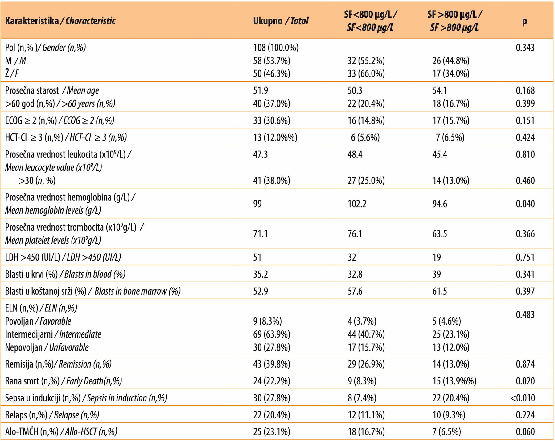
The average OS was 14.8 months (range: 11.5 – 18.8), three-year OS was 6.4%, while average DFS was 22 months (range: 12.6 – 31.2). In the group of patients with lower levels of SF, average OS was significantly longer (18.8 months, 95%CI: 11.6 – 31.2), as compared to the group with higher SF levels (12.2 months, 95%CI: 7.3 – 15.4) (p = 0.030). Our study showed that the average DFS in the group with lower SF was 29 months (95%CI: 16.9 – 41.1), while it was 12 months (95%CI: 7.2 – 16.1) in the group with higher SF, which proved to be statistically significant (p = 0.048). In addition to SF>800 µg/L, univariate analysis was used to analyze the influence of other characteristics on OS and DFS. Namely, this analysis showed that the following were significant predictors of poor OS: age >60 years (p = 0.049), unfavorable risk according to the ELN classification (p = 0.040), SF>800 µg/L (p = 0.019), and absence of allo-HSCT (p<0.010) (Appendices II, III). Also, univariate analysis showed that the following factors were significant for shorter DFS: SF >800 µg/L (p = 0.040), thrombocyte count <50x109/L (p = 0.008), and absence of allo-HSCT (p = 0.002).
Table 2. Univariate analysis showcasing the influence of some characteristics on overall survival (OS)
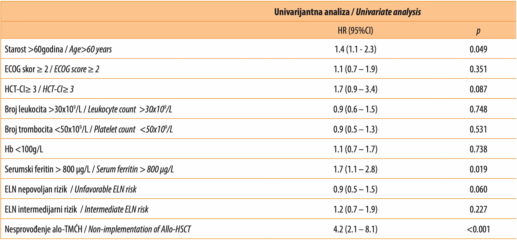

Figure 1. Kaplan-Meier curve showcasing the OS differences in patients with SF>800 µg/L vs SF<800 µg/L
DISCUSSION
Our study showed that the SF levels were elevated in almost all patients with AML in the analyzed population, which has been shown by other studies as well [5],[9],[14],[15]. SF is elevated manifold in patients who had received transfusions, due to chronic iron overload. Additionally, it is believed that hyperferritinemia is also caused by subclinical inflammation and damage to the liver [9]. In our population of patients, the average SF level was 1002.3 µg/L, which is more than was reported in a study by Ihlow et al. (769 µg/L) [15]. Namely, they used C-reactive protein (CRP) as a correction factor for SF, which is why the influence of the abovementioned inflammation on SF levels was reduced. In our study, CRP values were not available for all patients, which is why the correction could not be carried out. Analyzing the link between the SF level and the basic clinical characteristics, we found that, in the group of patients with SF levels >800 µg/L, there was a higher incidence of early death (p = 0.020) and sepsis in induction treatment (p < 0.010), while the mean hemoglobin value was significantly lower (p = 0.040), as compared to the group of patients with SF<800 µg/L. On the other hand, data on SF levels, as a predictive factor for the development of sepsis during induction therapy, is scarce [5],[9],[15]. What is known is that the higher the SF level - the more unfavorable the outcome of sepsis [16],[17]. Other studies have shown that patients with high SF levels at initial diagnosis were significantly more often men and patients aged >60 years, while such a link was not found in our study (p = 0.343, p = 0.399) [5],[9],[15]. In other studies, patients with high levels of SF significantly more often had hyperleukocytosis >30x109 /L, as compared to patients with low SF levels, while this was not registered in our study. Patients with high levels of ferritin did not show a statistically significantly higher leukocyte count in peripheral blood (p = 0.460) [5],[9]. In our study, a statistically significant difference between the group with high SF and the one with low SF was not registered in relation to the achievement of CR (p = 0.874) and the occurrence of relapse (p = 0.224). On the other hand, a study by Lebon et al. showed that patients with high levels of SF relapsed significantly more frequently, as compared to patients with lower SF levels.
In our patient population, average OS was 14.8 months, with a three-year survival of 12.0%. Our study showed that patients with SF levels <800 µg/L had statistically significantly better OS (18.8 months) and DFS (29 months), as compared to the patient group with SF> 800 µg/L, where the average OS was 12.2 months, and DFS was 12 months (p = 0.019, p = 0.040, respectively). Such significance has been confirmed by other studies as well, where patients with low levels of SF also had a statistically significantly better survival and a longer DFS [5],[9],[15].
By testing the influence of individual variables on OS with univariate analysis, we determined that the following were independent predictive factors of an unfavorable prognosis: age >60 years (p = 0.049), SF> 800 µg/L (p = 0.019), and absence of allo-HSCT (p < 0.010). Some studies have shown, in addition to these parameters, that other parameters, such as intermediate and unfavorable risk according to ELN, an ECOG score >1, and hyperleukocytosis >30x109 /L, have a statistically significant influence on OS [5],[8],[15]. Analyzing the influence of these variables on DFS, univariate analysis showed that SF>800 µg/L (p = 0.040), thrombocytopenia <50x109 (p = 0.008), and the absence of allogenic HSCT (p = 0.002) were significant independent predictive factors in our patient population Additionally, in the aforementioned studies, it has been determined that DFS is negatively influenced by ECOG>1, unfavorable risk according to ELN, and hyperleukocytosis >30x109 /L [5],[9],[15].
We must be cautious in interpreting these results, as SF levels are subject to the influence of various factors, such as the existence of comorbidities, liver failure, tumor-independent inflammation, as well as disturbance in the metabolism of iron. Standardization of certain correction factors would contribute to the decrease of the influence of these variables on SF levels, and the number of studies dealing with the role of SF in acute myeloid leukemias is insufficient.
The limitations of our study are, as follows: a relatively short follow-up period, a relatively small number of subjects, the absence of correction of SF values, as well as the inability of carrying out multivariate analyses.
CONCLUSION
Our study showed that high levels of SF at diagnosis had a negative prognostic impact on OS and DFS in patients with AML, and that they were linked to a significantly higher incidence of early death and sepsis in the induction phase of therapy. We also demonstrated that patients with high levels of SF at diagnosis significantly more often had low levels of hemoglobin, as compared to patients belonging to the group with low SF levels. The existence of larger multicentric prospective studies, which would follow patients for longer periods of time, would contribute to a better understanding of the impact of hyperferritinemia on the course and outcome of disease. The identification of SF, as an independent prognostic factor and a potential target site for the action of new drugs, may contribute to a better survival of patients with AML.
Informations

-
Keywords:
-
Received:
-
Revised:
-
Accepted:
-
Online first:
-
DOI:
-
Cite this article:

Dejan Dudok
Faculty of Medicine, University of Belgrade, Serbia
36 Salvadora Aljendea Street, 11000 Belgrade, Serbia
E-mail:
1. Janković G. Akutna mijeloblastna leukemija. U: Ćolović M, Janković G. Maligne bolesti krvi. Beograd, Zavod za udžbenike i nastavna sredstva, 1999: 81-119.
2. Short NJ, Rytting ME, Cortes JE. Acute myeloid leukaemia. Lancet. 2018 Aug 18;392(10147):593-606. doi: 10.1016/S0140-6736(18)31041-9.[CROSSREF]
3. Alkhateeb AA, Connor JR. The significance of ferritin in cancer: anti-oxidation, inflammation and tumorigenesis. Biochim Biophys Acta. 2013 Dec;1836(2):245-54. doi: 10.1016/j.bbcan.2013.07.002.[CROSSREF]
4. Pham CG, Bubici C, Zazzeroni F, Papa S, Jones J, Alvarez K, et al. Ferritin heavy chain upregulation by NF-kappaB inhibits TNFalpha-induced apoptosis by suppressing reactive oxygen species. Cell. 2004 Nov 12;119(4):529-42. doi: 10.1016/j.cell.2004.10.017.[CROSSREF]
5. Bertoli S, Paubelle E, Bérard E, Saland E, Thomas X, Tavitian S, et al. Ferritin heavy/light chain (FTH1/FTL) expression, serum ferritin levels, and their functional as well as prognostic roles in acute myeloid leukemia. Eur J Haematol. 2019 Feb;102(2):131-42. doi: 10.1111/ejh.13183.[CROSSREF]
6. Arber DA, Orazi A, Hasserjian R, Thiele J, Borowitz MJ, Le Beau MM, et al. The 2016 revision to the World Health Organization classification of myeloid neoplasms and acute leukemia. Blood. 2016 May 19;127(20):2391-405. doi: 10.1182/blood-2016-03-643544.[CROSSREF]
7. Bennett JM, Catovsky D, Daniel MT, Flandrin G, Galton DA, Gralnick HR, et al. Proposed revised criteria for the classification of acute myeloid leukemia. A report of the French-American-British Cooperative Group. Ann Intern Med. 1985 Oct;103(4):620-5. doi: 10.7326/0003-4819-103-4-620.[CROSSREF]
8. Döhner H, Estey EH, Amadori S, Appelbaum FR, Büchner T, Burnett AK, et al. Diagnosis and management of acute myeloid leukemia in adults: recommendations from an international expert panel, on behalf of the European LeukemiaNet. Blood. 2010 Jan 21;115(3):453-74. doi: 10.1182/blood-2009-07-235358.[CROSSREF]
9. Lebon D, Vergez F, Bertoli S, Harrivel V, De Botton S, Micol JB, et al. Hyperferritinemia at diagnosis predicts relapse and overall survival in younger AML patients with intermediate-risk cytogenetics. Leuk Res. 2015 Aug;39(8):818- 21. doi: 10.1016/j.leukres.2015.05.001.[CROSSREF]
10. Oken MM, Creech RH, Tormey DC, Horton J, Davis TE, McFadden ET, et al. Toxicity and response criteria of the Eastern Cooperative Oncology Group. Am J Clin Oncol. 1982 Dec;5(6):649-55.
11. Sorror ML, Storb RF, Sandmaier BM, Maziarz RT, Pulsipher MA, Maris MB, et al. Comorbidity-age index: a clinical measure of biologic age before allogeneic hematopoietic cell transplantation. J Clin Oncol. 2014 Oct 10;32(29):3249-56. doi: 10.1200/JCO.2013.53.8157.[CROSSREF]
12. Cheson BD, Bennett JM, Kopecky KJ, Büchner T, Willman CL, Estey EH, et al.; International Working Group for Diagnosis, Standardization of Response Criteria, Treatment Outcomes, and Reporting Standards for Therapeutic Trials in Acute Myeloid Leukemia. Revised recommendations of the International Working Group for Diagnosis, Standardization of Response Criteria, Treatment Outcomes, and Reporting Standards for Therapeutic Trials in Acute Myeloid Leukemia. J Clin Oncol. 2003 Dec 15;21(24):4642-9. doi: 10.1200/JCO.2003.04.036.[CROSSREF]
13. Walter RB, Othus M, Borthakur G, Ravandi F, Cortes JE, Pierce SA, et al. Prediction of early death after induction therapy for newly diagnosed acute myeloid leukemia with pretreatment risk scores: a novel paradigm for treatment assignment. J Clin Oncol. 2011 Nov 20;29(33):4417-23. doi: 10.1200/JCO.2011.35.7525.[CROSSREF]
14. Cullis JO, Fitzsimons EJ, Griffiths WJ, Tsochatzis E, Thomas DW; British Society for Haematology. Investigation and management of a raised serum ferritin. Br J Haematol. 2018 May;181(3):331-40. doi: 10.1111/bjh.15166.[CROSSREF]
15. Ihlow J, Gross S, Sick A, Schneider T, Flörcken A, Burmeister T, et al. AML: high serum ferritin at initial diagnosis has a negative impact on long-term survival. Leuk Lymphoma. 2019 Jan;60(1):69-77. doi: 10.1080/10428194.2018.1461860.[CROSSREF]
16. Connor J, Pang BM. Role of ferritin in cancer biology. J Cancer Sci Ther. 2015;7(5):155-60.
17. Lan P, Pan KH, Wang SJ, Shi QC, Yu YX, Fu Y, et al. High Serum Iron level is Associated with Increased Mortality in Patients with Sepsis. Sci Rep. 2018 Jul 23;8(1):11072. doi: 10.1038/s41598-018-29353-2.[CROSSREF]