


Examining the prevalence of thrombophilia in women with fetal loss
- University of Banja Luka, Faculty of Medicine, Banja Luka, Bosnia and Herzegovina
- University Clinical Center of the Republic of Srpska, Clinic for Internal Medicine, Department of Hematology, Banja Luka, Bosnia and Herzegovina
ABSTRACT
Introduction: In addition to antiphospholipid syndrome (APS), inherited thrombophilia is one of the most important hematologic disorders that can lead to pregnancy complications such as fetal loss, intrauterine growth restriction (IUGR), preeclampsia, and fetal death. The frequency of spontaneous abortions is up to 20% of all clinically recognized pregnancies.
Objective: The aim of this study was to determine the frequency of inherited thrombophilia in women with fetal loss, analyze the gestational period and age at which fetal loss occurred, and assess the frequency of successful pregnancies after prophylactic use of low molecular weight heparin (LMWH).
Material and methods: The study was designed as a 30-month retrospective observational study at the Clinic for Gynecology and Obstetrics of the University Clinical Center of the Republic of Srpska, involving 69 patients with one or more fetal losses in the second and third trimesters of pregnancy, or two or more repeated fetal losses in the first trimester of pregnancy. All patients underwent tests to detect the presence of thrombophilia.
Results: The average age of the patients was 30.7 years, and they had a total of 167 unsuccessful pregnancies. Thrombophilia was proven in a total of 40 patients (58%). The results showed that combined thrombophilia, including polymorphisms, occurred most frequently – 47.5% (n=19). In the group of patients with inherited thrombophilia, low molecular weight heparin (LMWH) was administered to 22 patients and it resulted in a successful pregnancy outcome in 19 patients.
Conclusion: Based on the conducted research, it can be concluded that combined thrombophilia, including combined polymorphisms MTHFR and PAI-1, carry a significantly higher risk of pregnancy loss in patients of all age groups. Age over 35 years is a risk factor for higher frequency of spontaneous abortions irrespective of the type of inherited thrombophilia. The use of LMWH significantly improves pregnancy outcomes in patients with inherited thrombophilia and previous pregnancy losses.
INTRODUCTION
Thrombophilia is a hereditary or acquired disorder of hemostasis which predisposes or increases the risk of thrombosis or recurrent thrombosis [1],[2]. There are two types of hereditary thrombophilia. The first category is characterized by a lack of coagulation inhibitors (i.e., antithrombin (AT), protein C (PC) and protein S (PS)), whereas the second category includes: activated protein C resistance (APCR), Factor V Leiden, prothrombin (F2) gene mutation, elevated levels of both FVIII and FIX, a decreased level of FXII and dysfibrinogenemia [3]. A lack of coagulation inhibitors implies rare but strong thrombophilias. Mutations in Factor Leiden and F2 gene account for up to 70% of diagnosed hereditary thrombophilias and these are considered significant thrombophilias [3]. According to the current recommendations, polymorphisms in the methylenetetrahydrofolate reductase (MTHFR) and plasminogen activator inhibitor-1 (PAI-1) are not considered hereditary thrombophilias, although these two polymorphisms used to be tested as part of laboratory tests for thrombophilia [3],[4],[5].
Pregnancy is physiologically characterized by hypercoagulability due to changes in homeostasis during pregnancy and postpartum. Hypercoagulability is a consequence of changes in coagulation and fibrinolytic system which are manifested by an increase in coagulation factors (factors II, VII, VIII, X, XII) and fibrinogen, i.e., increased fibrin formation and a reduced fibrinolytic activity [6]. Recent research has shown that thrombophilia can heighten the negative effects of these changes, and in addition to maternal complications it can also cause problems related to pregnancy itself: fetal loss, fetal growth retardation (IUGR), preeclampsia, and fetal death [7],[8].
Recurrent pregnancy loss is not only an emotional issue, but a social and economic problem as well [9],[10]. It is believed that fetal loss in early stages of pregnancy can result from various factors including anatomical abnormalities of the uterus, karyotypic abnormalities, and a defect in progesterone production. Additionally, hematological disorders such as the presence of antiphospholipid antibodies and congenital thrombophilia are considered significant contributors to fetal loss [11]. A successful pregnancy outcome depends on the development of adequate placental circulation. The placenta has dual circulation: maternal, in the intervillous space, and fetal, which consists of the chorionic vessels and chorionic villi. The success of pregnancy depends on the mechanisms that prevent coagulation in villous and fetal blood circulation [6]. If there is hypercoagulability on the part of the mother, there is a reduced flow in the intervillous space, with a high risk for the formation and deposition of fibrin deposits in the placenta that can lead to placental infarction. If more than 5% of the placental mass is affected, perinatal morbidity is increased. In pregnant women with thrombophilia, endothelial cell dysfunction, coagulation activation, fibrin deposition, and utero-placental thrombosis lead to decreased trophoblast invasion into spiral arteries and ultimately lead to implantation disorders, recurrent abortions, IUGR, preeclampsia, and fetal death [12],[13].
The placenta is formed at the end of the third month of pregnancy. Placental cells express the anticoagulant system comprising thrombomodulin, protein C, and the endothelial protein C receptor on their surface. This system is a very important inhibitory coagulation system, but also a receptor for tissue factor [14]. For adequate placentation and embryonic development, the activity of the protein C anticoagulant is of great importance as it controls the thrombin generated on the surface of the trophoblast or the intervillous space. Thrombin generation is a precisely controlled process limited to the generation of a limited amount of thrombin sufficient to form a blood clot at the site of a vascular trauma. Physiological thrombin inhibitors, with antithrombin being the most important among them, deactivate all excess thrombin. The thrombotic tendency in most congenital thrombophilic conditions arises either from heightened thrombin production or the inability to promptly and efficiently deactivate it [15]. Antithrombin deficiency results in diminished inhibition of thrombin, FXa, FXIa and FIXa, which leads to elevated levels of activated coagulation factors and blood hypercoagulability [15]. The protein C system takes an important place in the control of thrombin formation. In the presence of its co-factor protein S, activated protein C deactivates factors V and VIII. Additionally, free protein S exerts an anticoagulant effect by neutralizing prothrombinase, which consists of FXa, FVa and phospholipids. In case of protein C or protein S deficiency, thrombin generation increases due to the absence of regulatory mechanisms that would inhibit its production [15]. The generated thrombin has a negative effect on embryonic development by inducing the formation of fibrin degradation products, which, in turn, trigger cell apoptosis in the trophoblast.
Pregnancy loss is one of the leading health concerns affecting women during their fertile years. The rate of miscarriage in clinically recognized pregnancies ranges between 8% and 20%, whereas during pre-implantation and early implantation period it is higher and ranges between 13% and 26% [16],[17]. Three or more consecutive spontaneous abortions with the same partner without achieving a single pregnancy are called primary habitual abortions, while secondary habitual abortions are defined as abortions in women who previously had a successful pregnancy. The likelihood of a woman, without treatment, who has experienced a primary habitual abortion delivering a live child is approximately 25-45%.
The laboratory practice of testing for thrombophilic conditions has become increasingly common. With laboratory testing, it is possible to rule out only known causes of congenital thrombophilia, but a negative result does not mean the absence of a hereditary tendency to thrombosis. There are controversial opinions about the connection between hereditary thrombophilia and the occurrence of spontaneous abortions [18],[19],[20].
The objective of this study was to gather and analyze data regarding the prevalence of congenital thrombophilia among individuals experiencing fetal loss. Additionally, it aimed to examine the timing of pregnancy loss and the age range during which it occurred, as well as to assess the rate of successful pregnancies following the prophylactic administration of LMWH in individuals with congenital thrombophilia and a history of spontaneous fetal losses.
METHODS
The study of the prevalence of thrombophilia among participants experiencing fetal loss was carried out at the Clinic for Gynecology and Obstetrics of the University Clinical Centre of the Republic of Srpska. The research was designed as a retrospective observational study spanning 30 months. It included 69 participants treated at the Clinic for Gynecology and Obstetrics of the University Clinical Centre of the Republic of Srpska. These participants experienced either one or more losses of anatomically normal fetuses in the second and third trimesters of pregnancy, or two or more recurrent fetal losses in the first trimester. All participants were tested to prove the presence of thrombophilia, but other diagnostic analyses were also performed to rule out other causes of miscarriages, i.e., hormonal, infectious and autoimmune.
The analysis incorporated subject data extracted from medical records: demographics, information on prior pregnancies and their outcomes, details on complications during previous pregnancies such as recurrent spontaneous abortions, intrauterine growth restriction, intrauterine fetal demise, incidences of placental abruption or preeclampsia, as well as diagnosed medical conditions. Additionally, the research encompassed the following laboratory parameters: counts of leucocytes (Le) in x109 /l, platelets (Tr) in x109 /l, hemoglobin (Hb) in g/l, as well as measurements of prothrombin time (PT), activated partial thromboplastin time (aPTT), and fibrinogen levels (µg/ml). The methods employed to assess natural inhibitors incuded functional and immunological methodologies to ascertain AT concentration (with reference values of 70- 120%), functional and chromogenic assays to determine protein C concentration (with reference values of 70-120%), and functional assays for assessing protein S concentration (with reference values of 60-120%). Mutation-specific PCR tests were used to identify mutations including heterozygous/homozygous for factor V Leiden, heterozygous/homozygous for F2, heterozygous/homozygous for MTHFR C677T, as well as variants 4G/4G, 4G/5G, and 5G/5G of PAI-1.
All data were entered into an electronic database. Lists of respondents were compiled based on the obtained data and respecting the alphabetical order and then they were entered into the database. Each respondent was assigned a specific code to ensure anonymity and avoid data duplication.
Data were processed using descriptive statistical methods including calculation of the arithmetic mean, median value, range of variation (min-max), standard deviation, and frequency expressed in both absolute numbers and percentages.
RESULTS
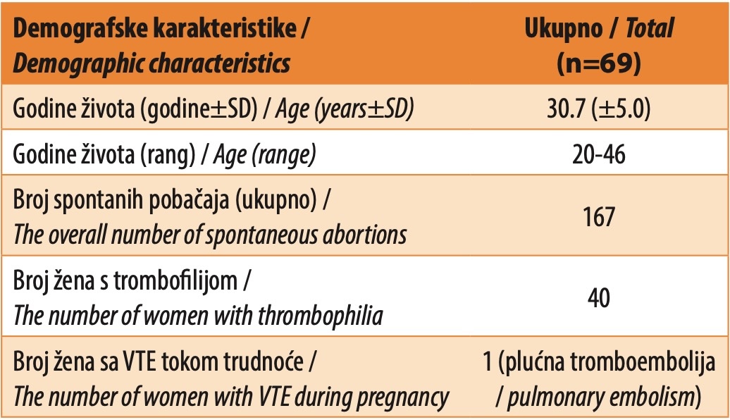
The study involved the analysis of medical histories from a total of 69 participants, aged 20 to 46 years. The average age of the participants was 30.7 years. Collectively, they experienced a total of 167 unsuccessful pregnancies, including two instances of placental abruption occurring in the 36th and 37th weeks of pregnancy, resulting in stillborn fetuses in the same participant.
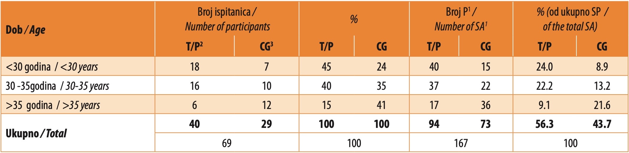
Thrombophilia was found in a total of 40 participants (58%). Table 1 displays the demographic characteristics of all subjects with spontaneous abortion, whereas Table 2 shows the age distribution among subjects with thrombophilia and MTHFR and PAI-1 polymorphisms, along with subjects in the control group (CG) lacking confirmed thrombophilia or polymorphism but experiencing spontaneous abortions. Based on the results, the highest number of unsuccessful pregnancies was found in the largest group of respondents. However, having calculated the average number of abortions per respondent within each group, it was observed that the group of older subjects with thrombophilia (>35 years) had a higher average number of abortions, 2.83 per respondent. Additionally, within the control group of respondents, the highest number of abortions occurred among those aged over 35 years (3.0 per respondent).
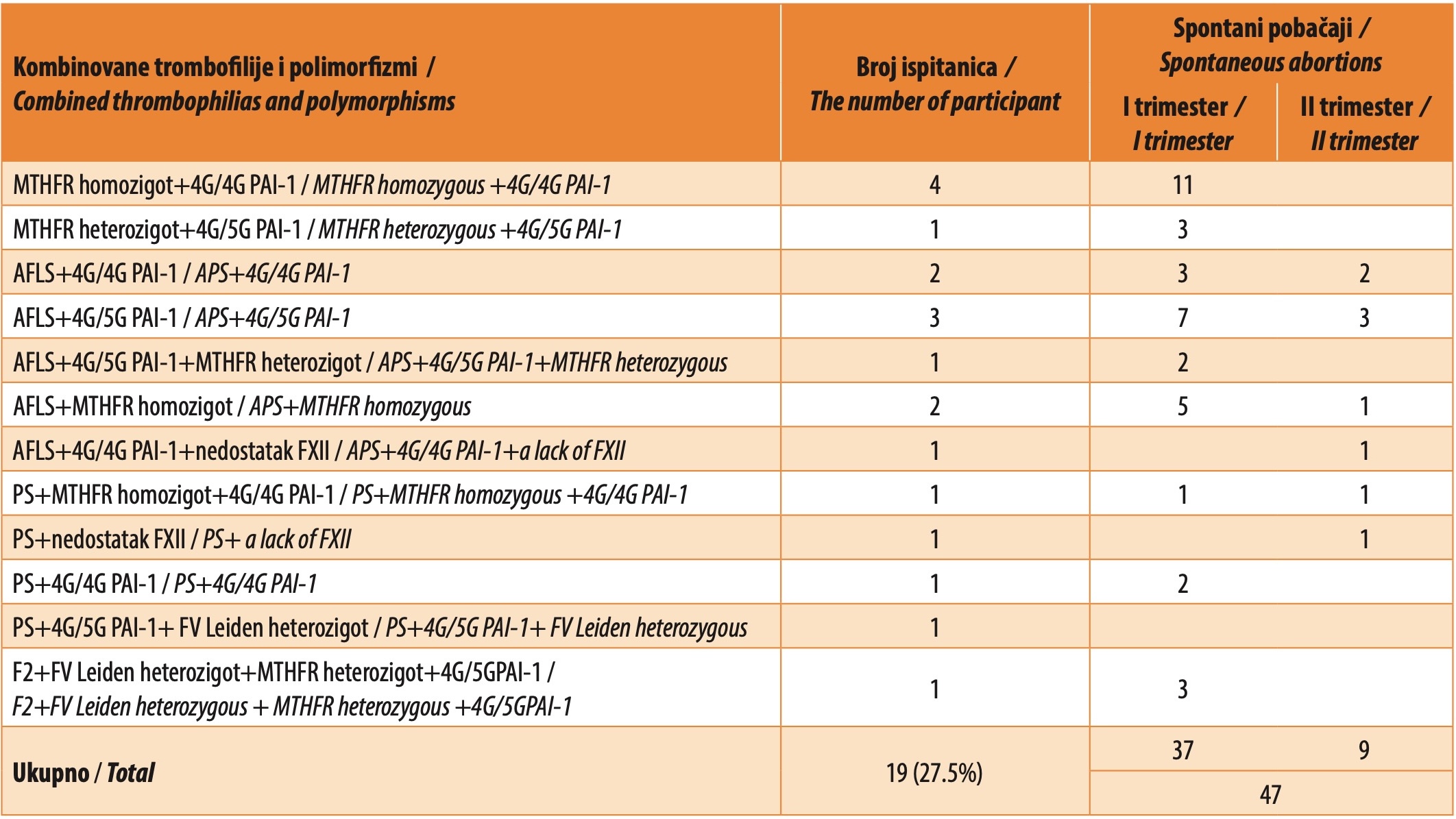
The results showed that combined thrombophilias (including combined polymorphisms and APS) occurred with the highest frequency of 47.5% (n=19). A lower frequency was associated with the isolated polymorphism for PAI-1, found in 27.5% of subjects (n=11). The mutations with the lowest prevalence were FV Leiden, detected in only 5.0% of subjects (n=2), isolated homozygous MTHFR, observed in 7,5% of subjects (n=3), and MTHFR heterozygous, present in 12.5% of subjects (n=5). Protein S deficiency in combination with other thrombophilic conditions was found in four subjects. A detailed overview of the frequency of all combined thrombophilias is shown in Table 2.
The study revealed that within the group of test subjects with detected PAI-1, the majority exhibited the 4G/4G polymorphism (n=6), whereas five test subjects showed the 4G/5G variant. In this group, there were a total of 25 spontaneous abortions, with 20 occurring in the first trimester, two in the second trimester, and three in the third trimester. Among the latter, one involved a stillborn fetus at 32 weeks of gestation, while two were placental abruptions resulting in stillborn fetuses – one at 36 weeks and the other at 37 weeks – both observed in a subject with the 4G/5G polymorphism. The number of spontaneous abortions in the 4G/4G PAI-1 group was 2.33 per test subject, and in the 4G/5G PAI-1 there were 2.4 spontaneous abortions per test subject.
Two test subjects with a heterozygous mutation for FV Leiden were identified, each experiencing one unsuccessful pregnancy that terminated in the first and second trimesters, respectively.
Isolated homozygous mutation for MTHFR was found in three subjects. Two subjects exhibited elevated homocysteine levels, with one subject’s values reaching the upper limit of the reference range. A total of 7 unsuccessful pregnancies were recorded in this group, 6 of which ended in the first trimester, and one in the second trimester.
In the group of subjects with hereditary thrombophilia, LMWH was applied to 22 subjects. In all subjects, the drug was administered as prophylactic anticoagulant therapy due to previous unsuccessful pregnancies. A successful pregnancy outcome upon the application of LMWH was observed in 19 subjects. In three subjects, fetal loss occurred irrespective of the administered drug, with two cases observed in the second trimester of pregnancy and one in the third trimester.
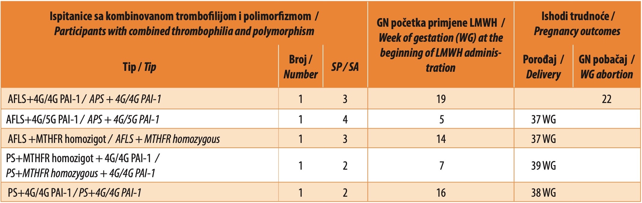
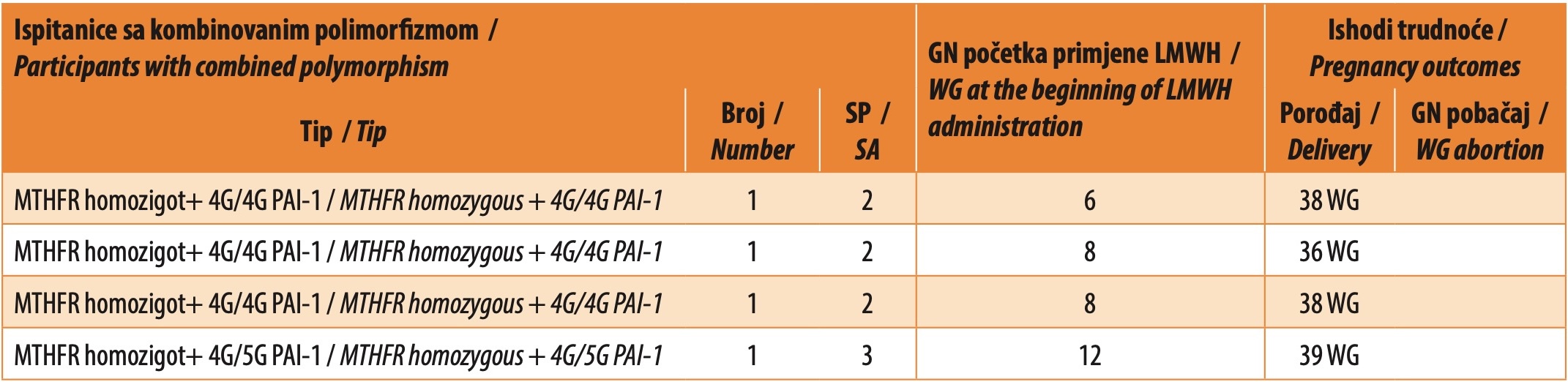
In the group of subjects with combined thrombophilia, a total of 47 unsuccessful pregnancies were recorded, of which one occurred in the third trimester of pregnancy, 9 in the second trimester, while the remaining unsuccessful pregnancies occurred in the first trimester of pregnancy. In 9 subjects, LMWH was administered in a prophylactic dose. The therapy was administered during the period spanning from the 5th to the 19th week of pregnancy. In one subject, who started therapy in the 19th week of pregnancy immediately after testing for congenital thrombophilia, fetal loss occurred in the 22nd week of pregnancy. In the remaining eight subjects, the pregnancies concluded without any complications, resulting in full-term births. Table 3 shows the pregnancy outcomes in 9 subjects with combined thrombophilia who were treated with a prophylactic dose of LMWH.
A total of 25 unsuccessful pregnancies (2.27 abortions per test subject) were observed in the group of subjects with isolated polymorphism for PAI-1. In all cases, LMWH was administered prophylactically during subsequent pregnancies. The outcomes were successful in nine subjects, while two subjects experienced spontaneous fetal loss regardless of the medication administered, as shown in Table 4. The average number of abortions per subject under LMWH therapy was 0.18, markedly lower than the average number of abortions per subject without LMWH therapy, which was 2.27.
The research showed that among the group of subjects with a homozygous MTHFR mutation (n=3), there were a total of 7 unsuccessful pregnancies prior to the study. In two subjects LMWH therapy was initiated up to the 12th week of pregnancy. In both cases pregnancies were successful. Throughout the follow-up period of this study, no pregnancies occurred in subjects with a confirmed heterozygous mutation for FV Leiden.
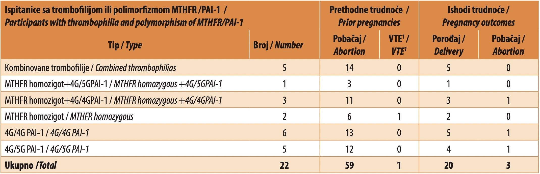
The study compared pregnancy outcomes in the group of subjects with thrombophilia before the diagnosis of hereditary thrombophilia was established with pregnancy outcomes after the therapy was administered during the follow-up period. Among the 40 subjects with thrombophilia, there were a total of 94 previous unsuccessful pregnancies and one instance of pulmonary thromboembolism (PTE) during pregnancy. During the follow-up period, pregnancy occurred in 22 test subjects. A total of 59 adverse pregnancy outcomes and one case of pulmonary thromboembolism during pregnancy were recorded in this group (n=22). LMWH was administered to all subjects. In 20 subjects, the pregnancies were carried to term and concluded with childbirth, while unfavorable pregnancy outcomes were recorded in 3 subjects, as shown in Table 5.
There were 8 successful deliveries in the group of subjects who did not have confirmed thrombophilia in the period of observation. A statistically significant difference (p < 0.05) was observed in the number of deliveries between subjects with combined thrombophilia and combined polymorphisms who received LMWH treatment and the group of subjects without thrombophilia (OR=3.179; 95% CI=1.071-9.405; p = 0.037). However, no statistically significant difference was found in the number of deliveries between subjects with combined thrombophilia (excluding MTHFR and PAI-1 polymorphisms) who received LMWH treatment and the group without thrombophilia (OR=2.607; 95% CI=0.689-9.853; p = 0.158). No statistically significant difference (p > 0.05) was observed in the number of successful deliveries between the group of all subjects with thrombophilia and the group of subjects without confirmed thrombophilia (OR=1.941; 95% CI=0.809- 4.658; p = 0.137). However, a statistically significant difference was found in the number of deliveries between the group of patients who used LMWH and the control group (OR=3.093; 95% CI=1.272-7.524; p = 0.013).
DISCUSSION
Reproductive failure has significant personal and social consequences and a major impact on public health. Apart from being an emotional problem, recurrent pregnancy losses are also a social and economic problem [20].
In this study, the presence of hereditary thrombophilia was identified in 58% of patients. In their study, Kupferminic et al. followed 100 subjects experiencing unexplained recurrent pregnancy loss alongside 43 subjects from the control group. They discovered a significantly elevated prevalence of thrombophilia, with 65% of individuals in the first group exhibiting this condition compared to only 18% in the control group [21]. In the HABENOX study conducted by Visser et al., thrombophilia was the cause of repeated spontaneous abortions in 50% of the test subjects. These individuals experienced either three or more spontaneous abortions during the first trimester of pregnancy, or two or more during the second and third trimesters, or one in each of the first and third trimesters[22]. Brenner et al. obtained similar results in their study, analyzing the origins of spontaneous abortions in 76 subjects. These individuals had experienced three or more spontaneous abortions during the first trimester of pregnancy, two or more during the second trimester, and at least one during the third trimester. This research documented that thrombophilia accounted for 49% of spontaneous abortions among the respondents [23].
Surprisingly, in this study thrombophilia was the most common cause of spontaneous abortions in 47.5% of the respondents. The majority of spontaneous abortions occurred during the first trimester of pregnancy, constituting one-third of all such cases within the group affected by thrombophilia. Combined thrombophilias, whether arising from a combination of acquired and congenital factors or from multiple congenital conditions, have been recognized as causes of both early and late spontaneous abortions in women. In the study conducted by Ivanov et al., including 52 test subjects experiencing fetal loss between the 10th and 20th week of pregnancy, the occurrence of combined thrombophilia was identified in 7.7% of the participants. The most prevalent combination involved the FV Leiden mutation along with the 4G polymorphism/5G PAI-1 [24]. The EPCOT study (European Prospective Cohort on Thrombophilia) included 843 subjects with thrombophilia, among whom 571 individuals experienced a total of 1524 pregnancies, compared to a control group comprising 541 subjects, of whom 395 had a collective total of 1019 pregnancies. The incidence of pregnancy loss was higher in the group exhibiting multiple established thrombophilias (OR 14.3) [25].
In slightly more than a quarter of the respondents (27.5%), the 4G/4G PAI-1 and 4G/5G PAI-1polymorphisms were found. Twenty-five spontaneous abortions were recorded in this group, and those were mostly spontaneous abortions in the first trimester of pregnancy (n=20, 80%). In a study conducted by Dossenbach et al., analyzing the prevalence of specific thrombophilias among a cohort of women (n=49) experiencing two or more spontaneous abortions in the first trimester of pregnancy, a relatively high prevalence of the 4G/5G PAI-1 polymorphism was observed in 57% of the subjects. Meanwhile, the 4G/4G PAI-1 variant was detected in 25% of the respondents. When comparing subjects with the 4G/4G versus 5G/5G PAI-1 polymorphism, a significantly elevated relative risk for early pregnancy loss was noted (OR 2.5). In contrast, the groups 4G/5G versus 5G/5G (OR 1.9) and 4G/4G versus 4G/5G PAI-1 (OR 1.3) exhibited comparatively lower relative risks [26]. In their research, Subrt et al. found a substantial and statistically significant correlation between repeated pregnancy losses and the 4G/4G PAI-1 polymorphism. However, they did not establish a statistically significant association between recurrent pregnancy losses and the 4G/5G PAI-1 polymorphism [27].
Heterozygous mutation for Factor V Leiden was identified in 10% of patients. In their study, Koher et al. found a statistically significant association between the presence of the FV Leiden mutation and stillbirth, with an OR of 10.9 [28]. In the NOHA study (Nimes Obstetricians and Hematologists), employing a case-control design and including over 32,000 participants, there was a significant finding: 18% of individuals experiencing unsuccessful pregnancies showed a significant correlation with heterozygous mutations for FV Leiden (OR 3.46) and F2 mutations (OR 2.6) [29]. Unsuccessful pregnancies were mostly related to fetal loss after the 10th week of pregnancy and could not be associated with early pregnancy loss.
Numerous studies have investigated the effectiveness of anticoagulant therapy during pregnancy in subjects with thrombophilia and recurrent fetal losses. One of the studies included 160 subjects who experienced pregnancy loss beyond the 10th week, each exhibiting a confirmed hypercoagulable state marked by the presence of either the FV Leiden mutation, F2 mutation, or protein S deficiency. Patients administered enoxaparin experienced an 86% success rate in pregnancies, compared to the 28% success rate observed in the aspirin group [30]. The use of thromboprophylaxis during pregnancy resulted in a lower pregnancy loss rate (0% versus 45%).
In their research, Brenner et al. discovered that employing LMWH in individuals with a history of spontaneous abortions and confirmed thrombophilia not only increased the incidence of favorable pregnancy outcomes but also led to a rise in live births. Additionally, the same author proved the enoxaparin dose of 40 mg once a day was sufficiently effective in the prophylaxis of spontaneous abortions [31]. In his research, Greer presents data indicating that 85% of pregnant women who used LMWH experienced positive pregnancy outcomes, whereas Bauersach cites an even higher figure of 94% [32],[33]. Unlike these authors, the ALIFE 2 study concludeed that the administration of LMWH did not result in increased rates of live births among individuals with a history of two or more spontaneous abortions and confirmed hereditary thrombophilia [34].
The small sample of respondents is one of the shortcomings of this research, which indicates the need for conducting a larger study including a larger sample of respondents with an extended follow-up duration. Another shortcoming is the insufficient diagnostic treatment received by certain test subjects experiencing recurrent fetal losses. This is compounded by the fact that some participants did not undergo the full range of available diagnostic analyses following repeated fetal losses, including immunological assessments, pathohistological analyses post-abortion, and investigations into potential infectious causes.
This study is a modest contribution to previous retrospective research exploring the impact of hereditary thrombophilia on pregnancy outcomes. It revealed an 82% frequency of successful pregnancies without complications among subjects with hereditary thrombophilia, without prior births, who received LMWH during pregnancy.
CONCLUSION
Based on the conducted research, it can be concluded that combined thrombophilia poses a significantly heightened risk of pregnancy loss across all age groups. Age over 35 years is a risk for a higher frequency of spontaneous abortions, irrespective of the specific type of hereditary thrombophilia. The application of LMWH significantly improves pregnancy outcomes in subjects with congenital thrombophilia and previous pregnancy losses.
Informations

-
Keywords:
-
Received:
-
Revised:
-
Accepted:
-
Online first:
-
DOI:
-
Cite this article:

Danijela Mandić
University Clinical Center of the Republic of Srpska
12 beba Street, 78000 Banja Luka, Bosnia and Herzegovina
E-mail:
1. British Committee for Standards in Haematology. Guidelines on investigation and management of thrombophilia. J Clin Pathol. 1999;43:703-10. doi: 10.1136%2Fjcp.43.9.703. [CROSSREF]
2. Campello E., Spiezia L., Adamo A., Simioni P. Thrombophilia, risk factors and prevention. Expert Rev. Hematol. 2019;12:147-58. doi: 10.1080/17474086.2019.1583555. [CROSSREF]
3. Samfireag M, Potre C, Potre O, Tudor R, Hoinoiu T, Anghel A. Approach to Thrombophilia in Pregnancy-A Narrative Review. Medicina (Kaunas). 2022;58(5):692. doi: 10.3390%2Fmedicina58050692. [CROSSREF]
4. Deloughery TG, Beverley J, Hunt BJ, Barnes GD, Connors JM. A call to action: MTHFR polymorphisms should not be a part of inherited thrombophilia testing. Res Pract Thromb Haemost. 2022 (4):e12739. doi: 10.1002/rth2.12739. [CROSSREF]
5. Middeldorp S, Nieuwlaat R, Baumann Kreuziger L, Coppens M, Houghton D, James AH et al. American Society of Hematology 2023 guidelines for management of venous thromboembolism: thrombophilia testing. Blood Adv. 2023;7(22):7101-38. doi: 10.1182/bloodadvances.2023010177. [CROSSREF]
6. Hellgren M. Hemostasis during normal pregnancy and puerperium. Semin. Thromb. Hemost. 2003;29:125-30. doi: 10.1055/s-2003-38897. [CROSSREF]
7. Simcox LE, Ormesher L, Tower C, Greer IA. Thrombophilia and Pregnancy Complications. Int. J. Mol. Sci. 2015;16:28418-28. doi: 10.3390%2Fijms161226104. [CROSSREF]
8. Ahangari N, Doosti M, Mousavifar N, Attaran M, Shahrokhzadeh S, Memarpour S, et al. Hereditary thrombophilia genetic variants in recurrent pregnancy loss. Arch. Gynecol. Obstet. 2019;300:777-82. doi: 10.1007/s00404- 019-05224-7. [CROSSREF]
9. Sattar N, Greer IA, Rumley A, Stewart G, Shepherd J, Packard CJ, et al. A longitudinal study of the relationships between haemostatic, lipid and oestradiol changes during normal human pregnancy. Thromb Haemost 1999;81:71-5. [CROSSREF]
10. Bremme K. Haemostasis in normal pregnancy. In Brenner B, Mardar V, Conard J, editors. Woomens issues in thrombosis and haemostasis. Martin Dunitz Ltd, a member of the Taylor and Francis Group; 2002: 151-65. [HTTP]
11. Elezović I. Osnovni principi laboratorijskog pristupa u dijagnostici hemoragijskih sindroma i tromboze. U: Marisavljević (urednik) Klinička hematologija. Zavod za udžbenike, Beograd 2012: 729-39.
12. Bhave AA. Coagulopathies in Pregnancy: What an Obstetrician Ought to Know! J Obstet Gynaecol India. 2019;69:479-82. doi: 10.1007%2Fs13224- 019-01290-8 [CROSSREF]
13. Vodnik T, Ignjatovic S, Majkic N, Singh. Parametri hemostaze kao pokazatelji hiperkoagulabilnosti u trudnoći; Jugoslovenska medicinska biohemija 2003; 22.
14. Castillo MM, Yang Q, Sigala AS, McKinney DT, Zhan M, Chen KL, et al. The endothelial protein C receptor plays an essential role in the maintenance of pregnancy. Sci Adv. 2020;6(45):eabb6196. doi: 10.1126/sciadv.abb6196. [CROSSREF]
15. Miljić P. Urođena trombofilna stanja. U: Marisavljević (urednik) Klinička hematologija. Zavod za udžbenike, Beograd 2012:795-803.
16. Wang X, Chen C, Wang L, Chen D, Guang W, French J. Conception, early pregnancy loss, and time to clinical pregnancy: a population-based prospective study. Fertil Steril 2003;79(3):577-84. doi: 10.1016/s0015-0282(02)04694-0. [CROSSREF]
17. Lohstroh PN, Overstreet JW, Stewart DR, Nakajima ST, Cragun JR, Boyers SP, et al. Secretion and excretion of human chorionic gonadotropin during early pregnancy. Fertil Steril 2005;83(4):1000-11. doi: 10.1016/j.fertnstert.2004.10.038. [CROSSREF]
18. Dizon-Townson D, Miller C, Sibai B, Spong CY, Thom E, Wendel G, et al. The relationship of the factor V Leiden mutation and pregnancy outcomes for mother and fetus. Obstet Gynecol 2005;106(3):517-24. doi: 10.1097/01. aog.0000173986.32528.ca. [CROSSREF]
19. Said JM, Higgins JR, Moses EK, Walker SP, Borg AJ, Monagle PT, et al. Inherited thrombophilia polymorphisms and pregnancy outcomes in nulliparous women. Obstet Gynecol 2010;115(1):5-13. doi: 10.1097/aog.0b013e3181c68907. [CROSSREF]
20. Kagami M, Maruyama T, Koizumi T, Miyazaki K, Nishikawa-Uchida S, Oda H, et al. Psychological adjustment and psychosocial stress among Japanese couples with a history or recurrent pregnancy loss. Human reprod 2012;27:787- 94. doi: 10.1093/humrep/der441. [CROSSREF]
21. Kupferminic MJ, Eldor A, Steinman N, Many A, Bar-Am A, Jaffa A, et al. Icreased frequency of genetic thrombophilia in women with complications of pregnancy. N Engl J Med 1999;340:9-13. doi: 10.1056/nejm199901073400102. [CROSSREF]
22. Visser J, Ulander VM, Helmerhorst FM, Lampinen K, Morin-Papunen L, Bloemenkamp KWM, et al. Thromboprophylaxis for recurrent miscarriage in women with or without thrombophilia. HABENOX: a randomized multicenter trial. Thromb Hemost 2011;105:295-301. doi: 10.1160/th10-05-0334. [CROSSREF]
23. Brenner B, Sarig G, Weiner Z, Younis J, Blumenfeld Z, Lanir N. Thrombophilic polymorphisms are common in women with fetal loss without apparent cause. Thromb Hemost 1999;82:6-9. [CROSSREF]
24. Ivanov P, Komsa-Penkova R, Konova E, Gecheva S, Ivanov I, Kovacheva K, et al. Combined thrombophilic factors among women with late recurrent spontaneous abortions. Akush Ginekol 2011;50:8-12.
25. Preston FE, Rosendaal FR, Walker ID, Briet E, Berntorp E, Conard J, et al. Increased fetal loss in women with heritable thrombophilia. Lancet 1996;348(9032):913-6. doi: 10.1016/s0140-6736(96)04125-6. [CROSSREF]
26. Dossenbach M, Van Trotsenburg M, Dossenbach-Glaninger A. Plasminogen activator inhibitor I 4G/5G polymorphism and coagulation factor XIII Val34Leu polymorphism: impaired fibrinolysis and early pregnancy loss. Clinical Chemistry 2003;49:1081-6. doi: 10.1373/49.7.1081. [CROSSREF]
27. Subrt I, Ulcova-Gallova Z, Bibkova K, Micanova Z, Hejnalova M, Cerna M, et al. Recurrent pregnancy loss and frequency of eight antiphospholipid antibodies and genetic thrombophilic factors in Czech women. American J of Reprod Immunolo. 2008;59:193-200. doi: 10.1111/j.1600-0897.2007.00554.x. [CROSSREF]
28. Kocher O, Cirovic C, Malynn E, Rowland CM, Bare LA, Young BA, et al. Obstetric complications in patients with hereditary thrombophilia identified using the 8 Thrombosis LCx microparticle enzyme immunoassay: a controlled study of 5,000 patients. Am J Clin Pathol. 2007;127:68-75. doi: 10.1309/JWL27GRGU71VP5QL. [CROSSREF]
29. Lissalde-Lavigne G, Fabbro-Peray G, Cochery-Nouvellon E, Mercier E, Ripart-Neveu S, Balducchi JP, et al. Factor V Leiden and prothrombin G20210A polymorphisms as risk factors for miscarriage during a first intended pregnancy: the matched case-control “NOHA First” study”. J Thromb Haemost. 2005;3:2178-84. doi: 10.1111/j.1538-7836.2005.01581.x. [CROSSREF]
30. Gris JC, Mercier E, Qu´er´e I, Lavigne-Lissalde G, Cochery-Nouvellon E, Hoffet M, et al. Low-molecular-weight heparin versus low-dose aspirin in women with one fetal loss and a constitutional thrombophilic disorder. Blood. 2004;103:3695-9. doi: 10.1182/blood-2003-12-4250. [CROSSREF]
31. Brenner B, Bar J, Ellis M, Yarom I, Yohai D, Samueloff A, et al. Effects of enoxaparin on late pregnancy complications and neonatal outcome in women with recurrent pregnancy loss and thrombophilia: results from the Live-Enox study. Feril Steril. 2005;84:770-3. doi: 10.1016/j.fertnstert.2005.03.048. [CROSSREF]
32. Bauersach RM; Dudenhausen J, Feridi A, Fischer T, Fung, Geisen U, et al. Risk stratification and heparin profilaxys to preven venous thromboembolism in pregnant women. Thromb Hemost. 2007;98:1237-45. doi: 10.1160/th07-05- 0329. [CROSSREF]
33. Greer IA, Nelson-Piercy C. Low-molecular-weight heparins for thromboprophylaxis and treatment of venous thromboembolism in pregnancy: a systemic review of safety and efficacy. Blod. 2005;6:401-7. doi: 10.1182/ blood-2005-02-0626. [CROSSREF]
34. Quenby S, Booth K, Hiller L, Coomarasamy A, de Jong PG, Hamulyák EN, et al. ALIFE2 Block Writing Committee; ALIFE2 Investigators. Heparin for women with recurrent miscarriage and inherited thrombophilia (ALIFE2): an international open-label, randomised controlled trial. Lancet. 2023 Jul 1;402(10395):54-61. doi: 10.1016/S0140-6736(23)00693-1. [CROSSREF]
- Table 1. Demographic characteristics of all participants experiencing fetal loss
- Table 2. Distribution of all participants categorized by age group
- Table 3. Combined thrombophilias and polymorphisms of MTHFR and PAI-1 in participants with spontaneous abortions
- Table 4. Applied LMWH and pregnancy outcomes in combined thrombophilia and polymorphism of MTHFR and PAI-1
- Table 5. Administered LMWH and pregnancy outcomes in combined polymorphisms MTHFR and PAI-1
- Table 6. Comparison of pregnancy outcomes before and after LMWH administration in participants with thrombophilia and polymorphism of MTHFR and PAI-1