


Lessons learnt from inappropriate d-dimer testing – case report
- University Clinical Center of Serbia, Clinic of Hematology, Belgrade, Serbia
- University of Belgrade, Faculty of Medicine, Belgrade, Serbia
ABSTRACT
Introduction: Since the beginning of the COVID-19 pandemic, D-dimer testing has been widely used in not-yet-approved indications. This has led to misdiagnosis, overuse of diagnostic procedures, and unnecessary costs.
Case report: Herein, we report the case of a 50-year-old female patient who came to the emergency department with chest pain, shortness of breath, fatigue, and pain in her right leg. Because of an elevated level of D-dimer (4.73 mg/l (N:< 0.5)), deep vein thrombosis and pulmonary embolism, as well as COVID-19 were excluded. Therapy with rivaroxaban, 10 mg/day, was initiated. Seven days later, the D-dimer level continued to rise (17.52 mg/l), which was why rivaroxaban was replaced with low molecular weight heparin (LMWH). After another five days, the level of D-dimer continued to rise (27.26 mg/l). A complete blood count revealed significant thrombocytopenia (54 x 109 /l). As the 4Ts score for heparin-induced thrombocytopenia was 5, the anti-heparin/PF4 antibody test was performed, and it came back strongly positive (4+). LMWH was replaced with fondaparinux sodium, and the patient was referred to a hematologist. On examination, cutaneous bleeding and gingivorrhagia were present. Laboratory analyses registered pancytopenia (hemoglobin = 101 g/l; white blood cell count = 1.7 x 10⁹ /l; platelet count = 29 x 109 /l) and consumption coagulopathy (INR = 1.7; aPTT = 27.1 s; fibrinogen = 0.8 g/l; D-dimer = 30.9 mg/l). Bone marrow aspirate was analyzed and the diagnosis of acute monoblastic leukemia, 47, XX, +12 [4] / 46, XX [16], wild type FLT3-ITD and NPM1, was established.
Conclusion: D-dimer is a powerful diagnostic tool when used properly. It should never be treated as a single, isolated result and the decision to introduce anticoagulant therapy should never be made based on its value alone.
INTRODUCTION
D-dimer molecules are formed as the result of the degradation of fibrin polymers. In everyday practice, D-dimer serves as a marker of the activation of coagulation and fibrinolytic systems [1],[2]. Indications for D-dimer testing, such as exclusion of venous thromboembolism, assessment of risk of recurrent thrombosis, and the diagnosis and management of disseminated intravascular coagulation (DIC), are clearly defined [3]. However, since the beginning of the COVID-19 pandemic, D-dimer testing has been widely used in notyet-approved indications. This may lead to the overuse of imaging diagnostics and unnecessary costs [4].
We present the case of a patient wherein inappropriate D-dimer testing led to inadequate anticoagulant therapy complicated with heparin-induced thrombocytopenia, bleeding, and delayed diagnosis of acute leukemia.
CASE REPORT
A 50-year-old female patient came to the emergency department (ED) with chest pain, lasting two days prior and occurring at 15-minute intervals. Moreover, she complained of shortness of breath, fatigue, and pain in the right leg, which had been present for three days. In her personal history, she reported arterial hypertension, mammary dysplasia, and COVID-19, eight months prior to the exam. She was a non-smoker and reported no thrombotic events in her family history.
The attending physician ordered laboratory tests, although her physical exam and electrocardiogram were normal. A complete blood count revealed leukopenia (3.2 x 10⁹ /l) with a normal differential and a normal hemoglobin level and platelet count. At the time, a blood smear was not performed. The level of lactate dehydrogenase (LDH) was elevated (534 U/l (N:< 460)) as well as D-dimer (4.73 mg/l (N:< 0.5)). Cardiac enzymes (troponin T and creatine kinase MB) were normal. COVID-19 was excluded with antigen, and later, with polymerase chain reaction (PCR) testing. Wells criteria for deep venous thrombosis (DVT) [5] and pulmonary embolism (PE) [6] were not calculated at that point, but retrospective calculation showed values of 1 (moderate risk group) for DVT and 3 (moderate risk group) for PE. Computed tomography angiography (CTA) of the pulmonary artery was performed and revealed no signs of thromboembolism. Therapy with rivaroxaban, 10 mg/day, was introduced and the patient was discharged.
Two days later, duplex ultrasonography of the lower extremities was performed with a normal finding. Seven days later, D-dimer levels continued to rise (17.52 mg/l). Rivaroxaban was replaced with a prophylactic dose of low molecular weight heparin (LMWH). The patient was referred for a mammography and to a gynecologist. The mammography showed no signs of malignancy. The abdominal CT finding was normal.
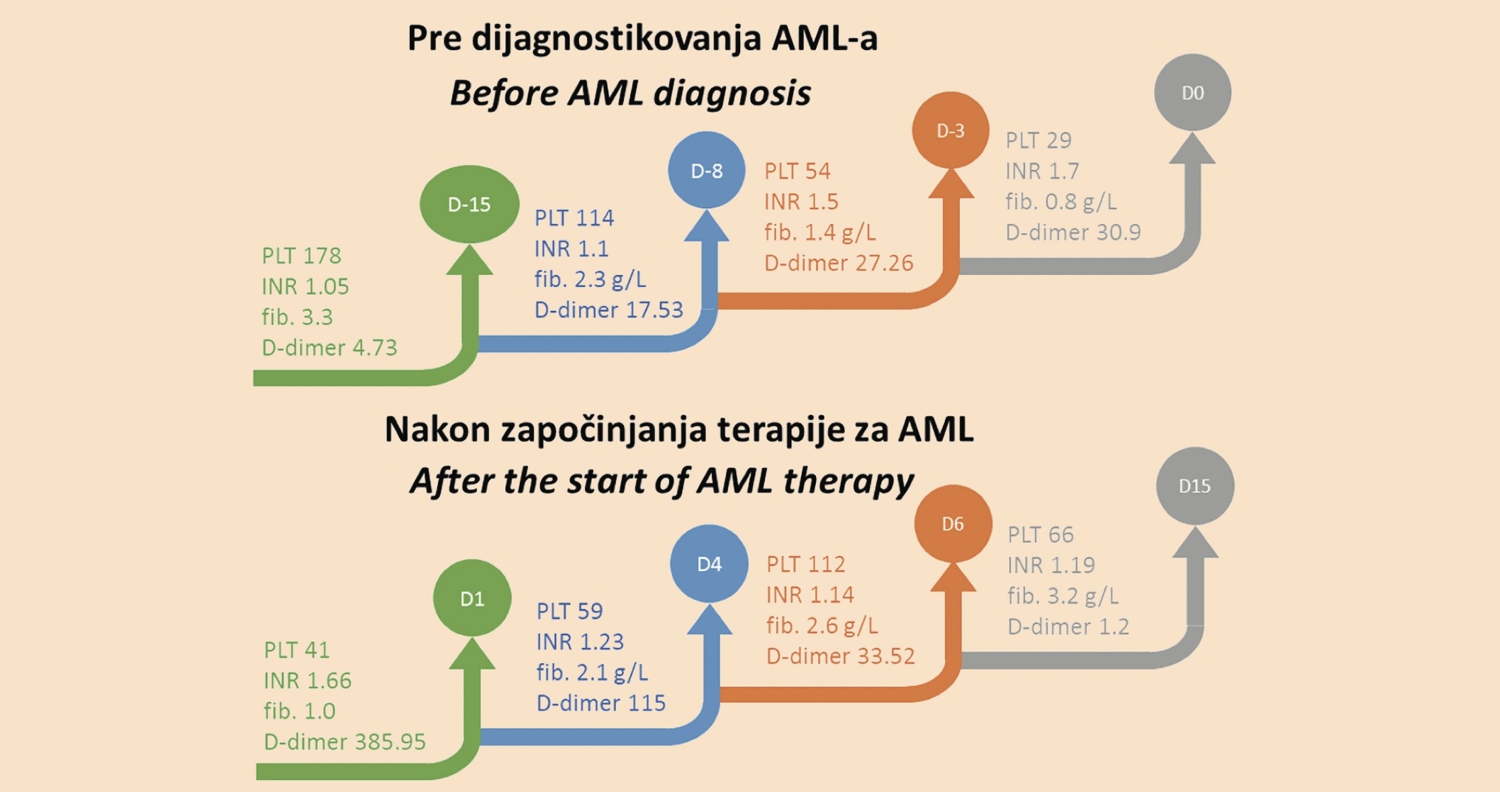
After another five days, D-dimer and LDH continued to rise (27.26 mg/l and 1,730 U/l, respectively). CBC revealed significant thrombocytopenia (54 x 109 /l), along with leukopenia (WBC = 2.4 x 10⁹ /l; neutrophils = 1.3 x 10⁹ /l; lymphocytes = 0.8 x 10⁹ /l; monocytes = 0.2 x 10⁹ /l), while the hemoglobin level was normal (124 g/l). As the 4Ts score for heparin-induced thrombocytopenia (HIT) was 5 [7], the anti-heparin-PF4 antibody test was performed, and it came back strongly positive (4+). LMWH was replaced with fondaparinux sodium, and the patient was referred to a hematologist.
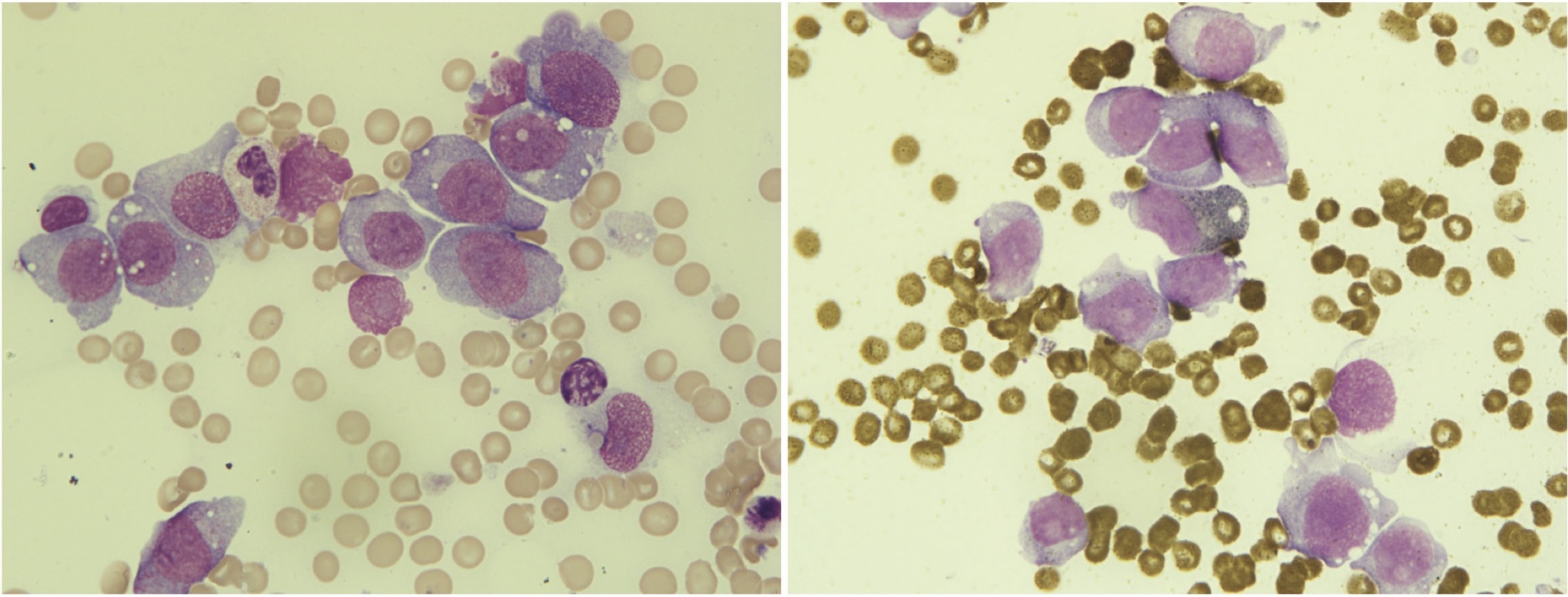
On examination at our center, cutaneous bleeding was noted at the venepuncture and LMWH application sites, along with diffuse limb petechiae and gingivorrhagia. Laboratory analyses registered pancytopenia (hemoglobin = 101 g/l; MCV = 84.8 fl, WBC = 1.7 x 10⁹ /l; PC = 29 x 109 /l), consumption coagulopathy (INR = 1.7; aPTT = 27.1 s; fibrinogen = 0.8 g/l; D-dimer = 30.9 mg/l; ISTH DIC score = 7) [8]. The peripheral blood smear revealed the following elements: neutrophils 38%, lymphocytes 46%, monocytes 4%, basophils 1%, metamyelocytes 1%, myelocytes 3%, and lymphoid elements 7%. The bone marrow aspirate was analyzed on the same day, and the diagnosis of acute monoblastic leukemia (AML), 47, XX, +12 [4] / 46, XX [16], wild-type FLT3-ITD and NPM1, was established (Figure 1).
Induction chemotherapy treatment comprising idarubicin and cytarabine was initiated along with supportive measures for DIC management [9]. After the fourth day of treatment, no signs of consumption coagulopathy were present (Graph 1).
Fondaparinux was administered for a period of four weeks. Bone marrow aspirate assessed on the 27th day of treatment showed that the patient had achieved complete remission.
DISCUSSION
D-dimer is a useful and inexpensive test, readily available in most hospitals. It provides relevant diagnostic and prognostic information in patient workup when used appropriately [1]. However, studies looking at real-world D-dimer testing have found that 40% – 60% of the tests performed are unnecessary [10],[11]. Once measured, high D-dimer values cannot be ignored, bearing in mind potentially fatal complications such as PE [12]. This can lead to a phenomenon known as the diagnostic cascade resulting in more diagnostic tests as well as patient discomfort, harm to the patient, and increased healthcare costs [10],[11].
In our case, the main reason for unnecessary testing is speeding up the patient flow in the ED. It has been reported that in some EDs, blood tests are performed by nurses and healthcare assistants before medical assessment so as to reduce the time that the patient spends in the ER. This practice has led to unnecessary diagnostics, even in patients with a clear diagnosis [12].
Furthermore, the inappropriate use of D-dimer may be due to the lack of a clear understanding of the limitations of this test. There is a general perception that a D-dimer test is the investigative tool of choice for the diagnosis of venous thromboembolism in any patient, without enough consideration having been given to their clinical status and comorbidities [13]. D-dimer can be elevated in numerous conditions. A study by Schutte et al. showed that among patients with extremely elevated D-dimer levels (> 10 x cutoff), the following conditions were present: pulmonary embolism (32%), cancer (29%), sepsis (24%), trauma/ surgery (24%), and deep vein thrombosis (13%). This is why these authors propose that an extremely elevated D-dimer level should raise suspicion of cancer even in the presence of thrombosis [14]. Knowing which patients will benefit from a testing the D-dimer level and selecting them appropriately in the ED will reduce unnecessary imaging tests [1]. It has been shown that a D-dimer test should be requested only in patients with a low clinical probability of venous thromboembolism after the assessment of the clinical probability scores. In those with high clinical probability, this test is unlikely to add any useful diagnostic value [13].
The case of our patient shows that starting anticoagulant therapy without a clearly determined reason for D-dimer elevation can lead to different complications and delayed diagnosis. One of the first complications in our patient was HIT. In patients with HIT, the platelet count often returns to basal levels after the cessation of treatment with heparin derivates. However, in our patient, PC continued to decline even after LMWH was discontinued. Further tests and a physical examination revealed DIC (ISTH DIC score = 7). The prevalence of DIC in acute leukemia varies in different studies between 8.5% and 25% among non-acute promyelocytic AML and acute lymphoblastic leukemia patients [15]. In our patient, DIC was probably caused by the underlying AML, however, we should also take into consideration HIT-associated DIC, which could be another complication of the inappropriate use of LMWH [16].
CONCLUSION
D-dimer is a powerful diagnostic tool when used properly. It should never be treated as a single, isolated result and the decision to introduce anticoagulant therapy should never be made based on its value alone. In this way, serious complications like bleeding or HIT may be avoided. On the other hand, thorough diagnostic workup in patients with elevated D-dimer should be performed and nothing ought to be overlooked.
Informations

-
Keywords:
-
Received:
-
Revised:
-
Accepted:
-
Online first:
-
DOI:
-
Cite this article:

Mirjana Mitrović
Clinic of Hematology, University Clinical Center of Serbia
Address: 2 Dr Koste Todorovića Street, 11000 Belgrade, Serbia
E-mail:
1. Innocenti F, Lazzari C, Ricci F, Paolucci E, Agishev I, Pini R. D-dimer tests in the emergency department: current insights. Open Access Emerg Med. 2021 Nov 11;13:465-79. doi: 10.2147/OAEM.S238696. [CROSSREF]
2. Johnson ED, Schell JC, Rodgers GM. The D‐dimer assay. Am J Hematol. 2019 Jul;94(7):833-9. doi: 10.1002/ajh.25482. [CROSSREF]
3. Thachil J, Lippi G, Favaloro EJ. D-dimer testing: laboratory aspects and current issues, Methods Mol Biol. 2017;1646:91-104. doi: 10.1007/978-1-4939- 7196-1_7. [CROSSREF]
4. Auditeau C, Khider L, Planquette B, Sanchez O, Smadja DM, Gendron N. D‐ dimer testing in clinical practice in the era of COVID‐19. Res Pract Thromb Haemost. 2022 May 25;6(4):e12730. doi: 10.1002/rth2.12730. [CROSSREF]
5. Wells PS, Anderson DR, Rodger M, Forgie M, Kearon C, Dreyer J, et al. Evaluation of D-dimer in the diagnosis of suspected deep-vein thrombosis. N Engl J Med. 2003 Sep 25;349(13):1227-35. doi: 10.1056/NEJMoa023153. [CROSSREF]
6. Wells PS, Anderson DR, Rodger M, Ginsberg JS, Kearon C, Gent M, et al. Derivation of a simple clinical model to categorize patients probability of pulmonary embolism: increasing the models utility with the SimpliRED D-dimer. Thromb Haemost. 2000 Mar;83(3):416-20. [CROSSREF]
7. Lo GK, Juhl D, Warkentin TE, Sigouin CS, Eichler P, Greinacher A. Evaluation of pretest clinical score (4 T’s) for the diagnosis of heparin‐induced thrombocytopenia in two clinical settings. J Thromb Haemost. 2006 Apr;4(4):759-65. doi: 10.1111/j.1538-7836.2006.01787.x. [CROSSREF]
8. Taylor FB, Toh CH, Hoots WK, Wada H, Levi M; Scientific Subcommittee on Disseminated Intravascular Coagulation (DIC) of the International Society on Thrombosis and Haemostasis (ISTH). Towards definition, clinical and laboratory criteria, and a scoring system for disseminated intravascular coagulation. Thromb Haemost. 2001 Nov;86(5):1327-30. [CROSSREF]
9. Levi M, Toh CH, Thachil J, Watson HG. Guidelines for the diagnosis and management of disseminated intravascular coagulation. Br J Haematol. 2009 Apr;145(1):24-33. doi: 10.1111/j.1365-2141.2009.07600.x. [CROSSREF]
10. Kamolratanapiboon K, Tantanate C. Inappropriate use of D-dimer and impact on the test characteristics for deep vein thrombosis exclusion. Scand J Clin Lab Invest. 2019 Oct;79(6):431-6. doi: 10.1080/00365513.2019.1658214. [CROSSREF]
11. Oliver M, Karkhaneh M, Karathra J, Goubran M, Wu CM. A Review of inappropriate D-dimer ordering at a Canadian tertiary care centre. Blood 2019;134(Suppl 1):5778. doi: 10.1182/blood-2019-122617. [CROSSREF]
12. Jones P, Elangbam B, Williams NR. Inappropriate use and interpretation of D-dimer testing in the emergency department: an unexpected adverse effect of meeting the “4-h target.” Emerg Med J. 2010 Jan;27(1):43-7. doi: 10.1136/emj.2009.075838. [CROSSREF]
13. Thachil J, Fitzmaurice DA, Toh CH. Appropriate use of D-dimer in hospital patients. Am J Med. 2010 Jan;123(1):17-9. doi: 10.1016/j.amjmed.2009.09.011. [CROSSREF]
14. Schutte T, Thijs A, Smulders YM. Never ignore extremely elevated D-dimer levels: they are specific for serious illness. Neth J Med. 2016 Dec;74(10):443- 8.
15. Ten Cate H, Leader A. Management of disseminated intravascular coagulation in acute leukemias. Hamostaseologie. 2021 Apr;41(2):120-6. doi: 10.1055/ a-1393-8302. [CROSSREF]
16. Greinacher A, Selleng K, Warkentin TE. Autoimmune heparin‐induced thrombocytopenia. J Thromb Haemost. 2017 Nov;15(11):2099-114. doi: 10.1111/jth.13813. [CROSSREF]
- Figure 1. The bone marrow aspirate showed infiltration of leukemic blasts; 100 x; May-Grünwald Giemsa staining
- Graph 1. Coagulopathy-related parameters before and after AML treatment: AML – acute monoblastic leukemia; PLT – platelet count; INR – international normalized ratio; fib. – fibrinogen; D0 – day of diagnosis; D1 – day when the treatment started