


Clinical characteristics and survival outcomes in acute myeloid leukemia patients: single center experience
- University Clinical Center of Serbia, Clinic for Hematology, Belgrade, Serbia
- University of Belgrade, Faculty of Medicine, Belgrade, Serbia
ABSTRACT
Introduction/Aim:Acute myeloid leukemia (AML) is a heterogeneous disease, in terms of its biological characteristics and response to therapy. Numerous prognostic factors at diagnosis related to the disease itself, the treatment, and the response to treatment influence the outcome of AML. Despite the high rate of complete remission, overall survival (OS) is still poor. This study aims to determine the clinical and biological profile of patients with AML and its prognostic impact on the OS rate and disease-free survival (DFS).
Materials and methods:The retrospective analysis included 271 patients diagnosed with acute myeloid leukemia at the University Clinical Center of Serbia (UCCS) Hematology Clinic, between January 2018 and January 2023. Demographic parameters, the Eastern Cooperative Oncology Group performance status (ECOG PS), comorbidities, and the parameters of laboratory analysis and hematological diagnosis of AML were analyzed as potential risk factors for OS and DFS, using the univariate Cox regression model.
Results: Out of a total of 271 patients, 206 (76%) were treated with intensive chemotherapy, of whom 108 were men and 98 were women. The average age of these respondents was 50.3 years. According to the European Leukemia Network (ELN) risk classification, the patients were mostly in the group with intermediate risk, i.e. 128 (62.1%) patients. The most common subtype of AML was AML NOS (AML not otherwise specified), present in 123 (59.7%) patients. Univariate analysis showed that age ≥ 60 years (p = 0.009) and WBC ≥ 30 x 109 /l (p = 0.031) were unfavorable prognostic parameters for OS. AML subtype, MRC (myelodysplasia-related changes) was the most significant risk factor for shorter disease-free survival (p = 0.027).
Conclusion: In the analyzed group of AML patients treated with intensive therapy, we demonstrated a low OS rate and showed that age and the MRC subtype of AML represent unfavorable risk factors for shorter OS and DFS, respectively.
INTRODUCTION
Acute myeloid leukemia (AML) is a heterogeneous malignant disease of the hematopoietic system characterized by clonal proliferation of undifferentiated myeloid precursors, i.e. blasts [1]. As a result of the accumulation of immature cells, normal hematopoietic cells are suppressed and damaged, which consequently manifests clinically as infection, anemia, and bleeding [2]. Occasionally, AML can have an extramedullary presentation, including central nervous system (CNS) involvement [3].
AML is a disease of older age – the average age of patients at the time of diagnosis is 69 years. It belongs to rare diseases, with an annual incidence of 4.2/100,000 inhabitants, and is more common in men (M : F = 5.1/100,000 : 3.5/100,000) [4].
Several risk factors are known to be associated with the development of AML: exposure to mutagenic environmental factors, previous chemotherapy, and other hematological diseases [2]. More recent studies have shown an association between AML and congenital inheritance within the family [5],[6].
Technological advances in molecular diagnostics have led to new insights into the biology of AML. The previous World Health Organization (WHO) classification of AML, from 2016, required ≥ 20% blasts as a diagnostic criterion (except in AML with recurrent cytogenetic abnormalities – AML with RCA), and included four main groups: AML with RCA, AML with myelodysplasia-related changes (AML with MRC), therapy-related AML (t-AML), and AML not otherwise specified (AML NOS) [5]. The International Consensus Classification (ICC) and the WHO classification, from 2022, improved the previous classification of AML by introducing new genetically defined entities for which the threshold value of the percentage of blasts was lowered [6],[7]. Both 2022 classifications are based on the assumption that abnormal morphology stems from disrupted cell biology, which is caused by somatic mutations or altered gene expression, and therefore focus more on extensive genetic profiling in defining disease entities.
According to the WHO, the final diagnosis of AML is established based on an integrated diagnostic algorithm that includes the results of several complementary methods: cytomorphological, immunophenotypic, cytogenetic, and molecular genetic analysis in the form of a unified report [6],[7].
The outcome of AML is very heterogeneous and depends on several factors related to the patient (age, Eastern Cooperative Oncology Group performance status – ECOG PS, comorbidities), as well as to the disease itself (white blood cell count (WBC) at diagnosis, previous myelodysplastic syndrome (MDS), pathological karyotype, molecular markers, previous cytotoxic therapy) [2]. Genetic abnormalities are a strong risk factor for unfavorable prognosis [1],[8]. According to the recommendations of the European Leukemia Network (ELN), the risk stratification of AML, which is based on cytogenetic and molecular aberrations, includes three risk groups: favorable, intermediate, and unfavorable [7]. In addition, assessment of measurable residual disease (MRD) by flow cytometry and the polymerase chain reaction (PCR) method [7] showed an association with the occurrence of disease relapse and shorter overall survival (OS) [9].
In addition to ECOG PS and comorbidities, the choice of therapeutic approach is influenced by the prognostic molecular profile of AML [7]. The standard of intensive treatment for patients with AML consists of initial “7+3” induction therapy (7 days of cytarabine and 3 days of anthracycline) and post-remission consolidation therapy (cytarabine-based protocols), which is followed by hematopoietic stem cell transplantation (HSCT), if indicated [7],[10],[11]. Depending on age, the rate of achieving complete remission (CR) varies. For patients aged ≤ 60 years, the average CR is 60-85%, with a five-year OS of about 30% [4],[10], while for patients aged > 60 years, the CR rate is 40-60% and the OS is less than 10% [10],[12].
Current trends are directed towards a personalized therapeutic approach based on the specific molecular profile of the disease [13]. The genomic heterogeneity of AML has led to the development of therapies that directly target mutations, such as FMS-related receptor tyrosine kinase 3 (FLT3) and isocitrate dehydrogenase 1/2 (IDH1/IDH2) inhibitors. These treatments have become part of standard treatment. In addition, other innovative drugs such as gemtuzumab ozogamicin (GO), (anti CD33 monoclonal antibody), and CPX-351 (a liposomal formulation of daunorubicin and cytarabine) have been introduced [7]. With the introduction of lower-intensity therapies, the therapeutic possibilities for all patients have increased, especially for older patients or those who are not eligible for intensive treatment. Thus, the application of a protocol with the B cell lymphoma 2 (BCL-2) inhibitor in combination with low doses of cytarabine or with hypomethylating agents has shown an improvement in the treatment of this group of patients [14],[15].
Despite recent promising breakthroughs and the introduction of new therapeutic approaches, the outcome of patients with AML is still not completely satisfactory [16]. Consequently, numerous studies of innovative drugs are underway aimed at increasing the cure rate of AML. Their preliminary results seem encouraging.
This study aims to determine the clinical and biological profile of patients with AML and its prognostic impact on OS rate and disease-free survival (DFS).
MATERIALS AND METHODS
Patients
The retrospective analysis included 271 patients with AML, diagnosed and treated at the Clinic for Hematology of the University Clinical Center of Serbia (UCCS), between January 2018 and January 2023. Patients with acute promyelocytic leukemia were not included in this study. The diagnosis of AML was established based on an integrated diagnostic approach, as per the 2016 WHO recommendations [5]. Risk stratification of patients with AML (favorable, intermediate, adverse) was carried out in keeping with ELN recommendations [11]. The standard G-banding technique was used in the cytogenetic analysis of bone marrow samples [17]. Immunophenotyping was performed by flow cytometry on bone marrow samples [18]. The presence of internal tandem duplication (FLT3-ITD) and tyrosine kinase domain (TKD) mutations, as well as of nucleophosmin (NPM1) mutations was analyzed by PCR methods [19],[20]. Organomegaly, lymphadenopathy, gingival hypertrophy, and CNS infiltration were defined as extramedullary disease (EMD). Examination of the presence of leukemic cells in the cerebrospinal fluid was carried out by flow cytometry in symptomatic and asymptomatic patients with characteristics associated with CNS infiltration (lactate dehydrogenase (LDH) ≥ 450, WBC ≥ 30x109 /l, myelomonocytic and/or monocytic phenotype, CD56 expression, FLT3-ITD/TDK mutations and core binding factor (CBF)) in AML [21]. The general functioning status was recorded based on the ECOG PS scale [22]. The significance of existing comorbidities was also determined based on the HCT-CI (hematopoietic cell transplantation-specific comorbidity index) score [23]. The following laboratory test were performed in all patients: white blood cell count - WBC (x109 /l), platelet count - PLT (x109 /l), hemoglobin – Hb (g/l), LDH (U/l), percentage of blasts in peripheral blood and bone marrow.
Treatment
Most of the patients (206/271) were treated with intensive therapy (daunorubicin and cytarabine 3+7 regimen), followed by consolidation with cytarabine and HSCT. Patients who were not eligible for intensive treatment (65/271) received non-intensive chemotherapy (daunorubicin and cytarabine regimens 2+5 and 1+5, low-dose cytarabine) or supportive therapy (6-mercaptopurine, hydroxyurea), depending on their general functioning status and comorbidity index. Patients who relapsed and those who were primarily refractory were treated with salvage protocols (fludarabine, cytarabine, idarubicin, according to the FLAGIda protocol, or mitoxantrone, etoposide, cytarabine, according to the MEC protocol).
Assessment of therapeutic response
The assessment of therapeutic response was defined according to ELN guidelines [11]: CR < 5% blasts in the bone marrow, absence of circulating blasts, absence of EMD, absolute neutrophil count ≥ 1.0×10⁹ /l, and PLT ≥ 100 ×109 /l. Relapse was defined as the presence of ≥ 5% blasts in the bone marrow or the appearance of blasts in peripheral blood, as well as the presence of EMD. Early death was defined as death within 28 days of starting induction chemotherapy. Refractory disease (RD), OS, and DFS were defined according to the proposed guidelines [11].
Statistical analysis
Descriptive analysis methods (arithmetic mean, median, standard deviation, frequency). were used to describe the data. The length of survival was analyzed using the Kaplan-Meier method, while the log-rank test was used to compare survival among the studied groups. The univariate Cox proportional regression model was used to identify risk factors. Statistical significance was defined at p < 0.05. Statistical tests were performed with the statistical analysis software IBM® SPSS® Statistics V25, IBM, USA.
RESULTS
Clinical characteristics
Out of a total of 271 patients with AML, 206 (76%) were treated with intensive chemotherapy, while 65 (24%) were treated with non-intensive therapy. The clinical characteristics of patients treated with intensive therapy are shown in Table 1. The study included 108 men and 98 women (M : F 52.4% : 47.6%) The average age of the patients was 50.3 ± 13.9 years.
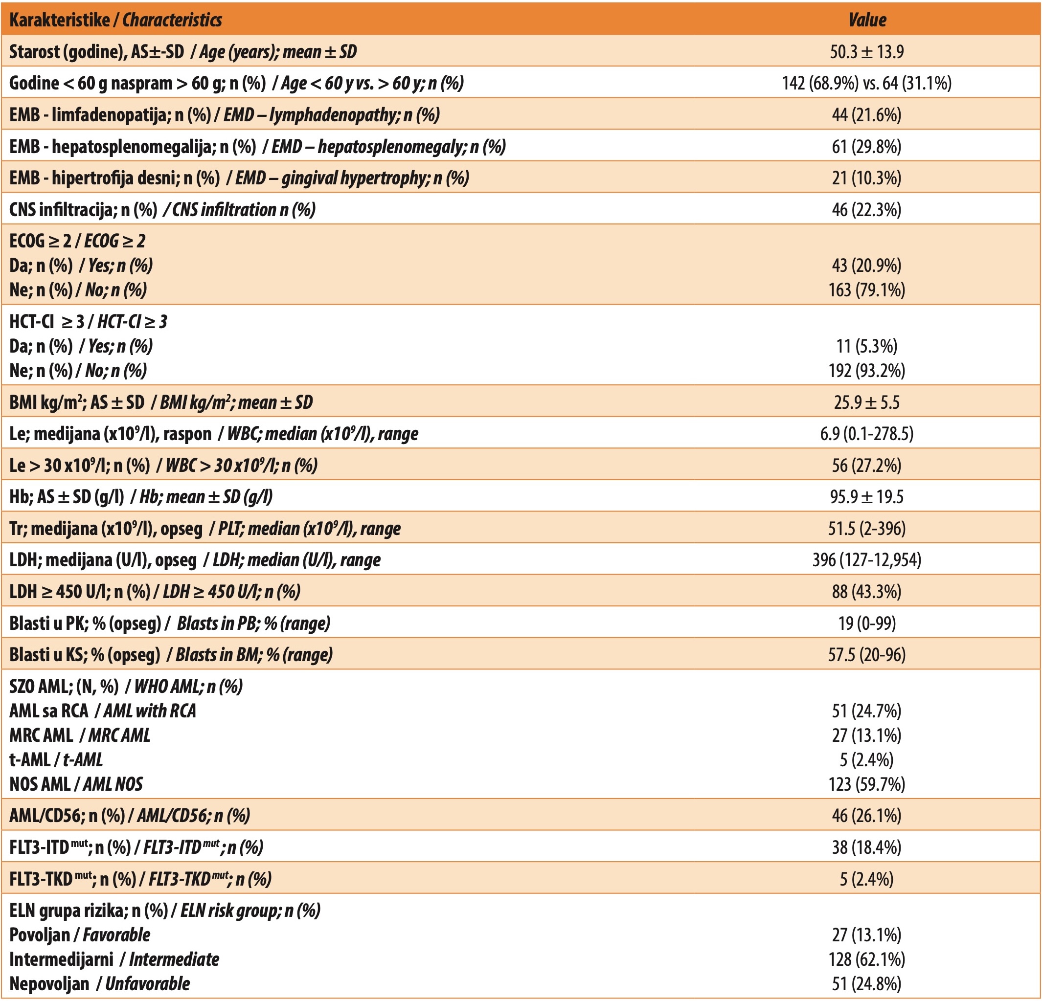
The distribution of patients according to AML subtype, and per the WHO classification from 2016, was as follows: 51 (24.76%) patients had AML with RCA, of whom there were 32 (15.5%) patients with NPM1, 12 (5.6%) patients with inv(16) and 7 (3.4%) patients with t(8;21); 27 (13.1%) patients had MRC AML; while t-AML was present in 5 (2.4%) patients. The majority were patients with AML NOS, 123 (59.7%) patients; there were 7 (7%) cases of minimally differentiated AML, 21 (20%) cases of non-maturing AML, 26 (25%) cases of maturing AML, 33 (32%) cases of acute myelomonocytic leukemia, 15 (15%) of cases of acute monoblastic and monocytic leukemia. FLT3-ITD positivity was observed in 38 (18.4%) cases, while FLT3-TKD mutation was registered in 5 (2.4%) cases. In our group of patients, the following immunophenotypic antigens on leukemic cells showed the highest prevalence: CD13 (90.7%), CD33 (93.9%), CD34 (74.4%), and CD117 (86%). A total of 26.1% of patients had CD56-positive AML.
Treatment outcome
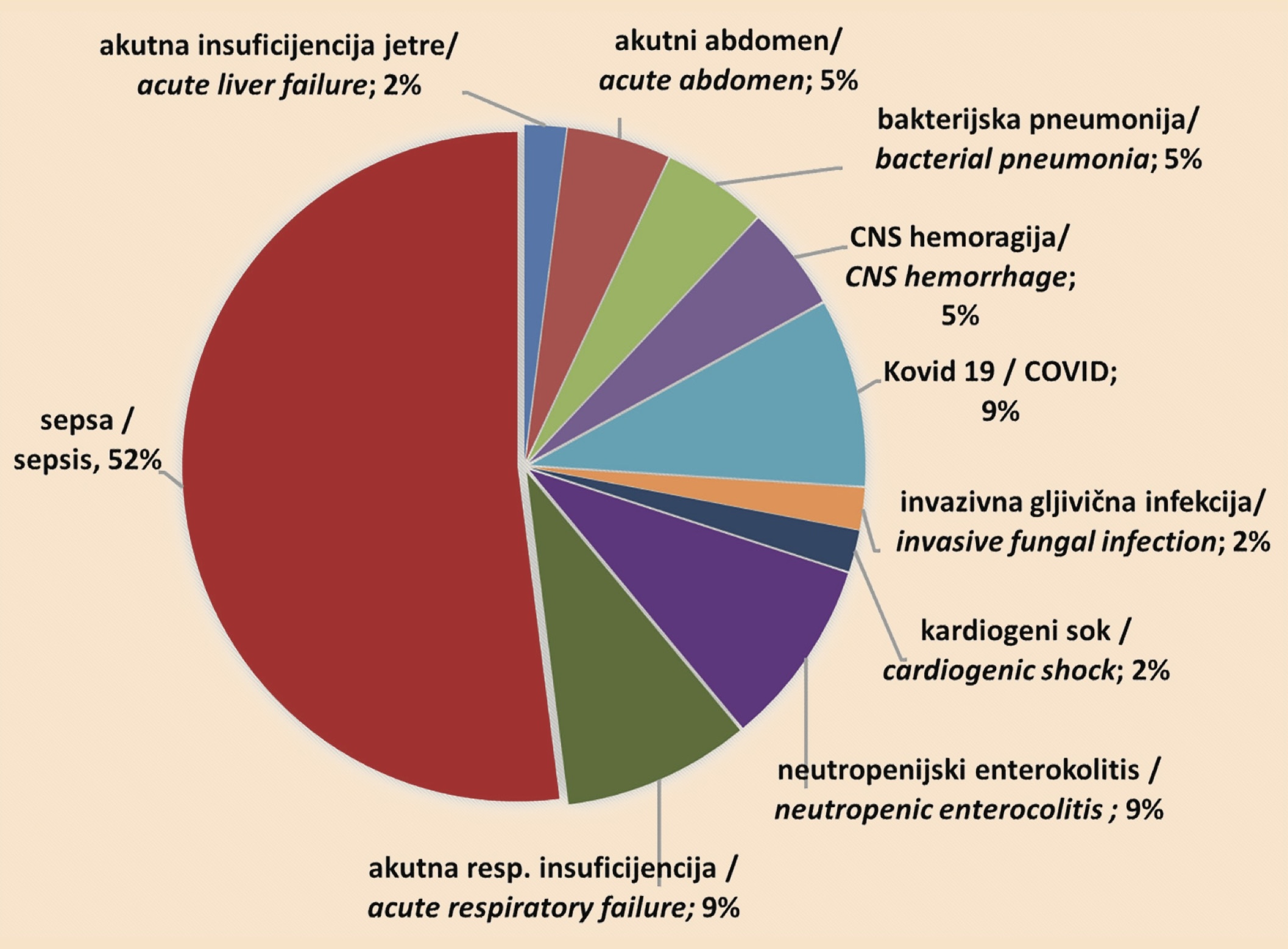
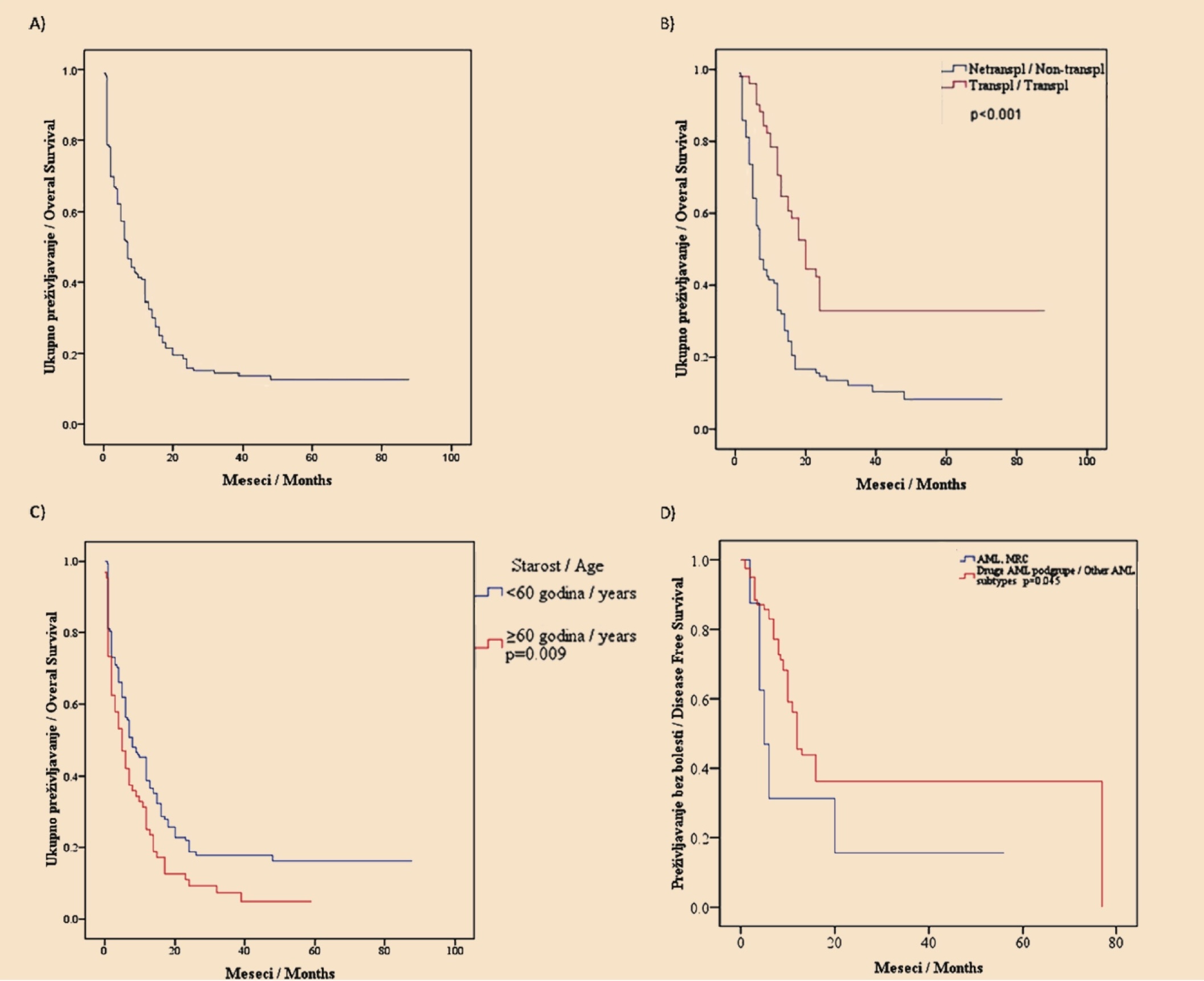
Complete remission was achieved in 92 out of 158 (79.7%) cases. Sixty-two (67.4%) patients had disease relapse, while 32 (20.3%) were primarily refractory patients. Early death was recorded in 48 (23.3%) cases. The main causes of early death are presented in Graph 1. Median OS was 12 months (10-13.9 months). Overall five-year survival was 19% (Figure 1. A). The median DFS was 13 months. Fifty-one out of 157 (32.5%) patients were treated with hematopoietic stem cell transplantation. Patients treated with HSCT had statistically significantly longer OS (median 20 months vs. 7 months; p < 0.001), as compared to patients not treated with this therapy (Figure 1. B).
Prognostic factors of OS and DFS
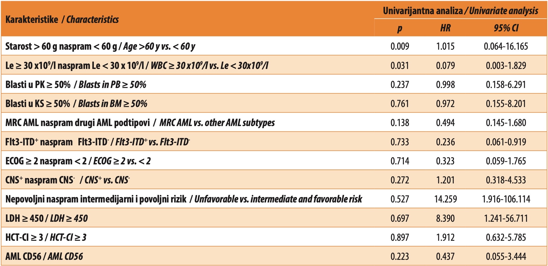
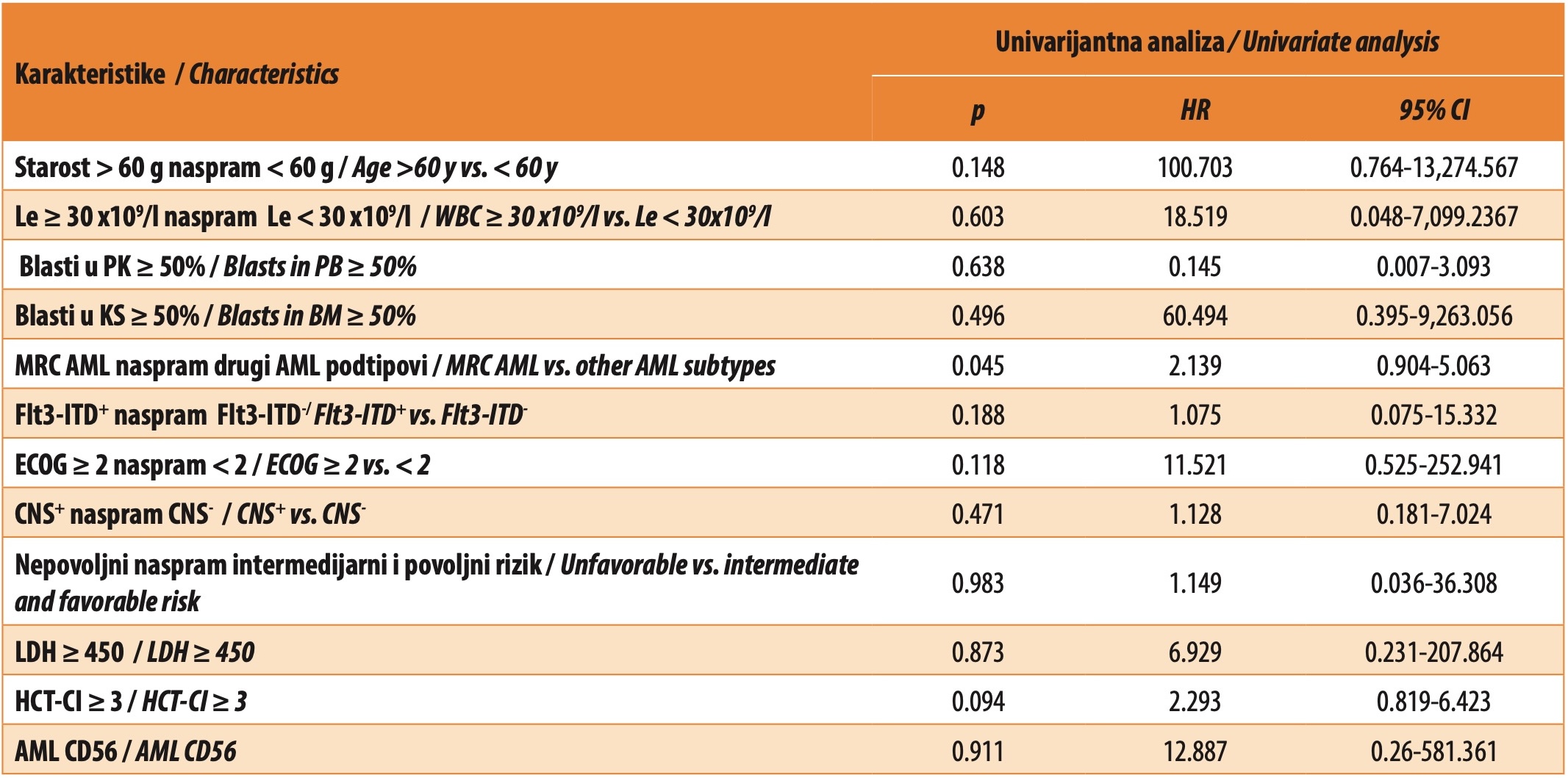
Univariate analysis showed that the following analyzed characteristics were significant predictors of shorter OS: age ≥ 60 (p = 0.009), (Figure 1. C), WBC ≥ 30 x109 /l (p = 0.031). Other examined prognostic factors did not show statistical significance (Table 2). Univariate analysis showed that poor DFS was significantly associated with the MRC AML subtype (p = 0.045) (Figure 1 D).
DISCUSSION
AML is a heterogeneous disease, in terms of its cytological, immunophenotypical, cytogenetic, and molecular characteristics, but also with regards to patient response to standard treatment and patient survival [1],[2]. Despite the high CR rate (60-85%), the average five-year OS rate is low, around 30%, and varies significantly among different age groups, reaching 50% in younger patients, while being less than 10% in patients older than 60 years [4],[10],[24]. Numerous studies have shown that prognostic factors at diagnosis (cytogenetic-molecular status, age, initial leukocytosis, percentage of blasts, AML subtype, ECOG PS, HCT-CI) have an impact on the outcome of the disease [1],[8],[25],[26],[27],[28],[29],[30]. Also, other significant predictors of outcome include the response to applied therapy and drug resistance [8],[31].
Our group comprising 206 patients treated with intensive therapy was younger, with an average age of 50.3 years, compared to literature data [4],[26], with a low ECOG PS and HCT-CI score. The majority of patients had AML NOS, 123 (59.7%), according to the WHO classification from 2016, and in this group, there was a predominance of patients with granulocytic differentiation (80/123, i.e., 65%), and the most common subtype was AML with maturation, which corresponds to literature data [5]. In our group of patients, EMD at diagnosis presented as: hepatosplenomegaly (29.8%), lymphadenopathy (21.6%), and gingival hypertrophy (10.3%), while CNS infiltration was confirmed in 46 (22.3%) patients, which is in keeping with recently published results [3].
Our patients achieved a high rate of CR (79.7%), while 32 (20.3%) patients had primary refractory disease, which corresponds to literature data [10],[16]. Early or induced death occurred in 48 (23.3%) cases. In clinical studies, carried out over the last two decades, mortality associated with induction therapy has been reduced from 15-20% to less than 5% [32]. The most common cause of early death was sepsis 52.6%. In fact, infection is described as one of the most common causes of early death [33]. In our group, relapse occurred in 62 (67.4%) patients, which is more as compared to literature data [34],[16]. Treatment with HSCT is the only method by which patients can be cured of AML, as shown by our results – there was a statically significant difference in survival between transplanted and non-transplanted patients [2],[7],[10],[11].
In keeping with numerous other studies [25],[26],[31], univariate analysis highlighted the patient’s age at diagnosis as a predictor of clinical outcome. Namely, the results of our study show that patients older than 60 years have a 50% higher risk of shorter OS, compared to younger patients. The fact that the median age of AML diagnosis is 69 years indicates that the greatest number of patients with AML belong to the group of patients with an unfavorable risk, regardless of clinical and genetic parameters, which is in accordance with earlier studies [25],[26]. This premise is supported by the fact that for every five years of age, the hazard ratio for a less favorable treatment outcome increases by 22% [29]. Numerous factors could explain the poorer prognosis in elderly patients with AML, primarily poorer ECOG PS due to numerous comorbidities related to age [25]. On the other hand, previous studies have shown a higher frequency of unfavorable cytogenetic markers in the older population, as compared to the younger. Namely, in a study in which patients younger than 56 years were compared to those older than 75 years, unfavorable cytogenetic aberrations were present in 51% of older and 35% of younger patients with AML [29]. Accordingly, favorable cytogenetic mutations in the group of elderly patients accounted for only 4%, while in the group of younger patients, 17% were carriers of favorable cytogenetic aberrations [29]. The higher presence of favorable aberrations such as CBF AML or NPM1 in younger patients somewhat reflects the different nature of AML in different age populations. With age, the percentage of favorable cytogenetic aberrations decreases, and the frequency of unfavorable ones increases, primarily complex and monosomal karyotypes, which carry a less favorable risk for CR and OS [30]. Also, in the older population, the so-called multidrug resistance phenotype is found more frequently, 57% versus 33% [30].
In our group of AML patients, ELN risk classification did not show its predictive impact. The reason for this may be the size of our sample or a possible cause may be the very small proportion of patients with favorable cytogenetic aberrations in our study group, i.e. 27 (13.1%). New drugs such as FTL3 inhibitors, GO and CPV-351, have shown their beneficial effect on the outcome of patients with AML [35],[36],[37]. The aforementioned target therapy has been slowly changing the prognostic categories of patients. Namely, the introduction of FLT3 inhibitors in the treatment of AML led to the fact that AML with FLT3 mutation, which was previously mostly chemoresistant [34],[25] and belonged to the group of unfavorable molecular aberrations, was classified as intermediate risk in the latest ELN classification [7]. It should be noted that, in our study, patients were treated with standard chemotherapy protocols, without targeted therapy.
In addition to the prior presence of MDS as well as MDS-associated cytogenetic abnormalities [5], MRC AML is associated with a complex and monosomal karyotype, as well as MDS-associated gene mutations such as the following: TP53, ASXL1, BCOR, EZH2, RUNX1, SF3B1, SRSF2, STAG2, U2AF1, ZRSR2 [7]. In our group of patients with MRC AML, univariate analysis showed an unfavorable influence on DFS. In a study comparing MCR AML patients with patients belonging to the AML NOS subtype, it was shown that MRC AML patients had a significantly lower CR rate [38]. Although therapeutic options that would significantly prolong OS in this AML subtype have not yet been investigated, therapy with CPX-351 has shown a higher five-year OS rate, compared to standard chemotherapy [37].
CONCLUSION
In our study, we confirmed that age is an independent prognostic factor of overall survival in patients with AML. Also, the results of our study indicate that MRC AML represents an unfavorable prognostic parameter for DFS. Overall five-year survival in the analyzed group was poor. In this regard, the introduction of new therapeutic approaches as part of standard therapy for the treatment of AML, as well as the inclusion of patients in studies with innovative drugs, can contribute to improving the outcome of patients with acute myeloid leukemia.
ABBREVIATIONS AND ACRONYMS
AML – acute myeloid leukemia
OS – overall survival
DFS – disease-free survival
ECOG PS – Eastern Cooperative Oncology Group performance status
ELN – European LeukemiaNet
NOS – not otherwise specified
MRC – myelodysplasia-related changes
CNS – central nervous system
WHO – World Health Organization
RCA – AML with recurrent cytogenetic abnormalities
t-AML – therapy-related AML
ICC – International Consensus Classification
MDS – myelodysplastic syndrome
MRD – measurable residual disease
PCR – polymerase chain reaction
HSCT – hematopoietic stem cell transplantation
CR – complete remission
FLT3 – fms-related receptor tyrosine kinase 3
FLT3-ITD – mutation an internal tandem duplication
FLT3-TKD – tyrosine kinase domain
NPM1 – nucleophosmin
IDH1/IDH2 – isocitrate dehydrogenase ½
GO – gemtuzumab ozogamicin
CPX-351 – liposomal daunorubicin and cytarabine
BCL-2 – B cell lymphoma 2
EMD – extramedullary disease
LDH – lactate dehydrogenase
CBF – core binding factor
HCT-CI – hematopoietic cell transplantation-specific comorbidity index
RD – refractory disease
Informations

-
Keywords:
-
Received:
-
Revised:
-
Accepted:
-
Online first:
-
DOI:
-
Cite this article:

Marijana Virijević
Clinic for Hematology, University Clinical Center of Serbia
2 Dr Koste Todorovića Street, 11000 Belgrade, Serbia
E-mail:
1. Papaemmanuil E, Gerstung M, Bullinger L, Gaidzik VI, Paschka P, Roberts ND, et al. Genomic classification and prognosis in acute myeloid leukemia. N Engl J Med. 2016 Jun 9;374(23):2209-21. doi: 10.1056/NEJMoa1516192. [CROSSREF]
2. Short NJ, Rytting ME, Cortes JE. Acute myeloid leukaemia. Lancet. 2018 Aug 18;392(10147):593-606. doi: 10.1016/S0140-6736(18)31041-9. [CROSSREF]
3. Virijevic M, Kraguljac-Kurtovic N, Mitrovic M, Jakovic L, Bukumuric Z, Pantic N, et al. Incidence, risk factors, and outcome of asymptomatic central nervous system involvement in adult patients with acute myeloid leukemia. Hematol Oncol. 2024 Mar;42(2):e3253. doi: 10.1002/hon.3253. [CROSSREF]
4. SEER Cancer Stat Facts: Leukemia – Acute Myeloid Leukemia (AML) [Internet]. Bethesda (MD): National Cancer Institute (US). [datum nepoznat] – [citirano 10. januar 2024]. Dostupno: https://seer.cancer.gov/statfacts/html/amyl.html [HTTP]
5. Arber DA, Orazi A, Hasserjian R, Thiele J, Borowitz MJ, Le Beau MM, et al. The 2016 revision to the World Health Organization classification of myeloid neoplasms and acute leukemia. Blood. 2016 May 19;12 (20):2391-405. doi: 10.1182/blood-2016-03-643544. [CROSSREF]
6. Khoury JD, Solary E, Abla O, Akkari Y, Alaggio R, Apperley JF, et al. The 5th edition of the World Health Organization classification of haematolymphoid tumours: myeloid and histiocytic/dendritic neoplasms. Leukemia. 2022 Jul;36(7):1703-19. doi: 10.1038/s41375-022-01613-1. [CROSSREF]
7. Döhner H, Wei AH, Appelbaum FR, Craddock C, DiNardo CD, Dombret H, et al. Diagnosis and management of AML in adults: 2022 recommendations from an international expert panel on behalf of the ELN. Blood. 2022 Sep 22;140(12):1345-77. doi: 10.1182/blood.2022016867. [CROSSREF]
8. Bullinger L, Döhner K, Döhner H. Genomics of acute myeloid leukemia diagnosis and pathways. J Clin Oncol. 2017 Mar 20;35(9):934-46. doi: 10.1200/ JCO.2016.71.2208. [CROSSREF]
9. Short NJ, Zhou S, Fu C, Berry DA, Walter RB, Freeman SD, et al. Association of measurable residual disease with survival outcomes in patients with acute myeloid leukemia: a systematic review and meta-analysis. JAMA Oncol. 2020 Dec 1;6(12):1890-9. doi: 10.1001/jamaoncol.2020.4600. [CROSSREF]
10. Döhner H, Weisdorf DJ, Bloomfield CD. Acute myeloid leukemia. N Engl J Med. 2015 Sep 17;373(12):1136-52. doi: 10.1056/NEJMra1406184. [CROSSREF]
11. Döhner H, Estey E, Grimwade D, Amadori S, Appelbaum FR, Büchner T, et al. Diagnosis and management of AML in adults: 2017 ELN recommendations from an international expert panel. Blood. 2017 Jan 26;129(4):424-47. doi: 10.1182/blood-2016-08-733196. [CROSSREF]
12. Récher C, Röllig C, Bérard E, Bertoli S, Dumas PY, Tavitian S, et al. Long-term survival after intensive chemotherapy or hypomethylating agents in AML patients aged 70 years and older: a large patient data set study from European registries. Leukemia. 2022 Apr;36(4):913-22. doi: 10.1038/s41375-021- 01425-9. [CROSSREF]
13. Kantarjian H, Kadia T, DiNardo C, Daver N, Borthakur G, Jabbour E, et al. Acute myeloid leukemia: current progress and future directions. Blood Cancer J. 2021 Feb 22;11(2):41. doi: 10.1038/s41408-021-00425-3. [CROSSREF]
14. Wei AH, Strickland SA Jr, Hou JZ, Fiedler W, Lin TL, Walter RB, et al. Venetoclax combined with low-dose cytarabine for previously untreated patients with acute myeloid leukemia: results from a phase ib/ii study. J Clin Oncol. 2019 May 20;37(15):1277-84. doi: 10.1200/JCO.18.01600. [CROSSREF]
15. DiNardo CD, Jonas BA, Pullarkat V, Thirman MJ, Garcia JS, Wei AH, et al. Azacitidine and venetoclax in previously untreated acute myeloid leukemia. N Engl J Med. 2020 Aug 13;383(7):617-29. doi: 10.1056/NEJMoa2012971. [CROSSREF]
16. Thol F, Döhner H, Ganser A. How I treat refractory and relapsed acute myeloid leukemia. Blood. 2024 Jan 4;143(1):11-20. doi: 10.1182/blood.2023022481. [CROSSREF]
17. McGowan-Jordan J, Hastings RJ, Moore S, editors. ISCN 2020: An International System for Human Cytogenomic Nomenclature. Basel: Karger; 2020.
18. Béné MC, Nebe T, Bettelheim P, Buldini B, Bumbea H, Kern W, et al. Immunophenotyping of acute leukemia and lymphoproliferative disorders: a consensus proposal of the European LeukemiaNet Work Package 10. Leukemia. 2011 Apr;25(4):567-74. doi: 10.1038/leu.2010.312. [CROSSREF]
19. Kiyoi H, Naoe T, Yokota S, Nakao M, Minami S, Kuriyama K, et al. Internal tandem duplication of FLT3 associated with leukocytosis in acute promyelocytic leukemia. Leukemia Study Group of the Ministry of Health and Welfare (Kohseisho). Leukemia. 1997 Sep;11(9):1447-52. doi: 10.1038/sj.leu.2400756. [CROSSREF]
20. Falini B, Mecucci C, Tiacci E, Alcalay M, Rosati R, Pasqualucci L, et al; GIMEMA Acute Leukemia Working Party. Cytolasmatic nucleophosmin in acute myelogenous leukemia with normal karyotype. N Engl J Med. 2005 Jan 20;352(3):254-66. doi: 10.1056/NEJMoa041974. [CROSSREF]
21. Kraan J, Gratama JW, Haioun C, Orfao A, Plonquet A, Porwit A, et al. Flow cytometric immunophenotyping of cerebrospinal fluid. Curr Protoc Cytom. 2008 Jul; Chapter 6: Unit 6.25. doi: 10.1002/0471142956.cy0625s45. [CROSSREF]
22. Oken MM, Creech RH, Tormey DC, Horton J, Davis TE, McFadden ET, et al. Toxicity and response criteria of the Eastern Cooperative Oncology Group. Am J Clin Oncol. 1982 Dec;5(6):649-55. [CROSSREF]
23. Sorror ML, Marris MB, Storb R, Baron F, Sandmaier BM, Maloney DG, et al. Hematopoetic cell transplantation (HCT)-specific comorbidity index: a new tool for risk assessment before allogenic HCT. Blood. 2005 Oct 15;106(8): 2912-9. doi: 10.1182/blood-2005-05-2004. [CROSSREF]
24. Sasaki K, Ravandi F, Kadia TM, DiNardo CD, Short NJ, Borthakur G, et al. De novo acute myeloid leukemia: A population-based study of outcome in the United States based on the Surveillance, Epidemiology, and End Results (SEER) database, 1980 to 2017. Cancer. 2021 Jun 15;127(12):2049-61. doi: 10.1002/cncr.33458. [CROSSREF]
25. Gbadamosi B, Ezekwudo D, Bastola S, Jaiyesimi I. Predictive and prognostic markers in adults with acute myeloid leukemia: a single-institution experience. Clin Lymphoma Myeloma Leuk. 2018 Jul;18(7):e287-94. doi: 10.1016/j. clml.2018.05.005. [CROSSREF]
26. Appelbaum FR, Gundacker H, Head DR, Slovak ML, Willman CL, Godwin JE, et al. Age and acute myeloid leukemia. Blood. 2006 May 1;107(9):3481-5. doi: 10.1182/blood-2005-09-3724. [CROSSREF]
27. Etienne A, Esterni B, Charbonnier A, Mozziconacci MJ, Arnoulet C, Coso D, et al. Comorbidity is an independent predictor of complete remission in elderly patients receiving induction chemotherapy for acute myeloid leukemia. Cancer. 2007 Apr 1;109(7):1376-83. doi: 10.1002/cncr.22537. [CROSSREF]
28. Slovak ML, Kopecky KJ, Cassileth PA, Harrington DH, Theil KS, Mohamed A, et al. Karyotypic analysis predicts outcome of preremission and postremission therapy in adult acute myeloid leukemia: a Southwest Oncology Group/ Eastern Cooperative Oncology Group Study. Blood. 2000 Dec 15;96(13):4075- 83. [CROSSREF]
29. Karami K, Akbari M, Moradi MT, Soleymani B, Fallahi H. Survival prognostic factors in patients with acute myeloid leukemia using machine learning techniques. PLoS One. 2021 Jul 21;16(7):e0254976. doi: 10.1371/journal. pone.0254976. [CROSSREF]
30. Breems DA, Van Putten WL, De Greef GE, Van Zelderen-Bhola SL, Gerssen-Schoorl KB, Mellink CH, et al. Monosomal karyotype in acute myeloid leukemia: a better indicator of poor prognosis than a complex karyotype. J Clin Oncol. 2008 Oct 10; 26(29):4791-7. doi: 10.1200/JCO.2008.16.0259. [CROSSREF]
31. Pravdić Z, Suvajdžić Vuković N, Virijević M, Mitrović M, Pantić N, Sabljić N, et al. Can pharmacogenetics impact the therapeutic effect of cytarabine and anthracyclines in adult acute myeloid leukaemia patients?: A Serbian experience. J Med Biochem. 2024;43(4):545-55. doi: 10.5937/jomb0-47459. [CROSSREF]
32. Othus M, Kantarjian H, Petersdorf S, Ravandi F, Godwin J, Cortes J, et al. Declining rates of treatment-related mortality in patients with newly diagnosed AML given ‘intense’ induction regimens: a report from SWOG and MD Anderson. Leukemia. 2014 Feb;28(2):289-92. doi: 10.1038/leu.2013.176. [CROSSREF]
33. Kayal S, Sengar M, Jain H, Bonda A, George B, Kulkarni OP. et al. Induction related mortality in acute myeloid leukemia: multivariate model of predictive score from the Indian Acute Leukemia Research Database (INwARD) of the Hematology Cancer Consortium (HCC). Blood. 2019 Nov 13;134(Suppl 1):2615. doi: 10.1182/blood-2019-127623. [CROSSREF]
34. Ganzel C, Sun Z, Cripe LD, Fernandez HF, Douer D, Rowe JM, et al. Very poor long-term survival in past and more recent studies for relapsed AML patients: The ECOG-ACRIN experience. Am J Hematol. 2018 Aug;93(8):1074-81. doi: 10.1002/ajh.25162. [CROSSREF]
35. Stone RM, Mandrekar SJ, Sanford BL, Laumann K, Geyer S, Bloomfield CD, et al. Midostaurin plus chemotherapy for acute myeloid leukemia with a FLT3 mutation. N Engl J Med. 2017 Aug 3;377(5):454-64. doi: 10.1056/NEJMoa1614359. [CROSSREF]
36. Lambert J, Pautas C, Terré C, Raffoux E, Turlure P, Caillot D, et al. Gemtuzumab ozogamicin for de novo acute myeloid leukemia: final efficacy and safety updates from the open-label, phase III ALFA-0701 trial. Haematologica. 2019 Jan;104(1):113-9. doi: 10.3324/haematol.2018.188888. [CROSSREF]
37. Lancet JE, Uy GL, Newell LF, Lin TL, Ritchie EK, Stuart RK, et al. CPX-351 versus 7+ 3 cytarabine and daunorubicin chemotherapy in older adults with newly diagnosed high-risk or secondary acute myeloid leukaemia: 5-year results of a randomised, open-label, multicentre, phase 3 trial. Lancet Haematol. 2021 Jul;8(7):e481-91. doi: 10.1016/S2352-3026(21)00134-4. [CROSSREF]
38. Wang L, Chu X, Wang J, An L, Liu Y, Li L, et al. Clinical characteristics and optimal therapy of acute myeloid leukemia with myelodysplasia-related-changes: a retrospective analysis in a cohort of Chinese patients. Turk J Haematol. 2021 Aug 25;38(3):188-94. doi: 10.4274/tjh.galenos.2021.2021.0009. [CROSSREF]
- Table 1. Clinical and biological characteristics of patients treated with intensive chemotherapy
- Graph 1. Causes of early death in patients with AML treated with intensive therapy
- Figure 1. Survival curves of patients with AML
- Table 2. Univariate analysis of risk factors for OS in AML patients treated with intensive therapy
- Table 3. Univariate analysis of risk factors for DFS in AML patients treated with intensive therapy