


Individualized treatment for posthysterectomy vaginal eversion – Case report and literature review
- University Clinical Center of Serbia, Clinic for Gynecology and Obstetrics, Belgrade, Serbia
- University of Belgrade, Faculty of Medicine, Belgrade, Serbia
ABSTRACT
Introduction: Currently, there are different recommendations for obtaining optimal treatment of vaginal eversion. The presented case highlights current options for individualized treatment of complete vaginal eversion.
Case report: A 61-year-old woman presented with complete vaginal eversion stage IV seventeen years after classic abdominal hysterectomy with bilateral adnexectomy. For anterior compartment prolapse, it was decided to perform anterior colpoplasty and bladder elevation using sutures, plication by Kelly. Nichols bilateral sacrospinal fixation was the chosen technique for correction of the rectocele/enterocele and prolapse of the posterior vaginal wall. The postoperative course was uneventful, and the resolution of vaginal eversion was confirmed on a six-month check-up.
Conclusions: The surgical repair incorporating anterior colpoplasty combined with bilateral sacrospinous ligament fixation is advised for the advanced stage of complex vaginal eversions. This treatment option is an effective and safe technique.
INTRODUCTION
Complete vaginal eversion (cuff/vault/dome prolapse) represents a descent of the apical, i.e., upper, midportion, and lower third of the vagina beyond the introitus. Vaginal cuff eversion can occur as a consequence of prior hysterectomy, both vaginal and abdominal. Additional risk factors are congenital or age-dependent insufficiency of soft tissue, pelvic floor defect, damage of the pelvic floor after birth, heavy physical work, neurological illnesses, and obesity with abdominopelvic imbalance [1],[2].
With the aging of the female population, more patients require treatment for both pelvic organ prolapse. Vaginal vault prolapse following hysterectomy is a common complication (0.2% to even 36%) and can cause significant discomfort for patients as pain, itching, vaginal erosion, and issues with urinary and stool retention. Moreover, it seems that rates of posthysterectomy vaginal eversion show a relationship with indications for hysterectomy (1.6% following hysterectomy for prolapse and 1.8% for other pathologies) [1],[3].
Various treatments are available (conservative and surgical), and there are no clear guidelines to recommend which is the best. Conservative or mechanical treatments are considered for women with a mild degree of prolapse or those who are unwilling or unable to undergo surgery due to different contraindications. Surgical intervention is often necessary when conservative treatment does not succeed. The surgical technique should be adapted to each patient based on anatomy, age, comorbidity, and other factors. Still, all surgical methods, regardless of their approach (vaginal, abdominal open, or laparoscopic), aim at resuspending the vaginal apex (with native tissue or by synthetic material) to firm pelvic structures that can enable its further support (sacrum or sacrospinous ligaments) [4],[5].
This case shows the available options for the treatment of vaginal eversion in the individual patient.
CASE REPORT
A 61-year-old woman was referred to our institution because of complete vaginal eversion. The patient had undergone a classic abdominal hysterectomy with bilateral adnexectomy 17 years ago due to a sizeable uterine myoma. The patient had no gynecological complaints for 12 years, after which she started noticing that during physical activity, a small bulge was slightly protruding from the vaginal introitus. The protuberance increased over time, and the condition progressed with significant discomfort and difficulties in voiding commencement but without urinary incontinence. The patient declined any vaginal bleeding, abdominal and/or pelvic pain but had vaginal erosion, defecation difficulties, and urinary retention. The patient had chronic hypertension, diabetes mellitus type II, and obesity (Body Mass Index 34.53 kg/m2 ). She was interested in maintaining sexual function.
During the physical examination upon admission to our institution, a stage IV vaginal vault prolapse was identified using the Pelvic Organ Prolapse Quantification System, with decubital changes on the mucosa. Although stress urinary incontinence wasn’t present, postvoid residual urine was measured at 57 ml during catheterization. Thus, operative treatment was indicated. It was decided to correct the anterior compartment prolapse with anterior colpoplasty and bladder elevation, while Nichols’s bilateral sacrospinous fixation was the chosen technique for correction of the rectocele/ enterocele and prolapse of the posterior vaginal wall. Before surgery, detailed medical and gynecological history and standard laboratory and microbiological analyses were taken. An ultrasonographic scan of the pelvis and abdomen showed no pathological findings.
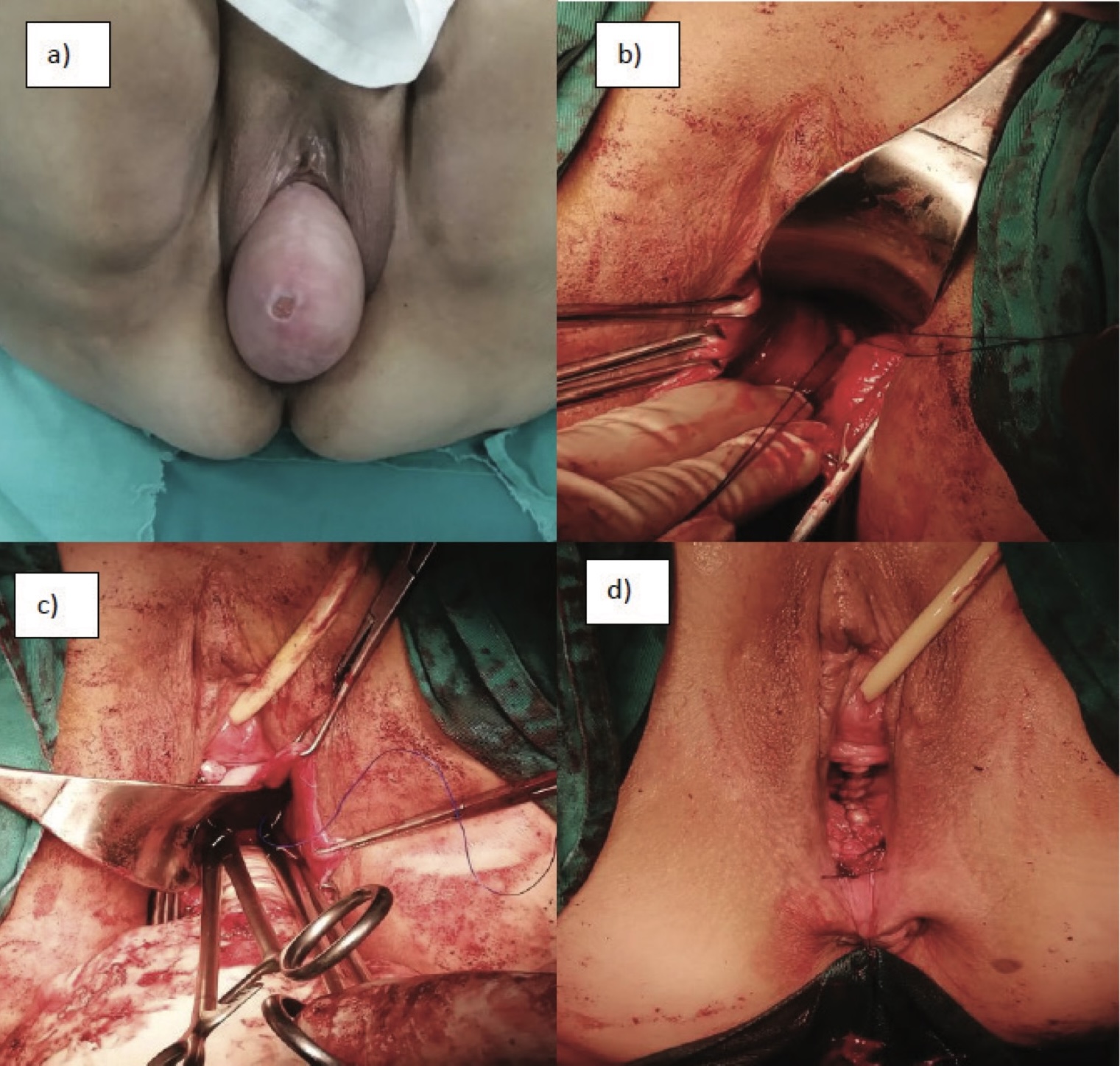
Following the administration of general anesthesia, the patient was positioned in lithotomy and prepared aseptically. A urethral catheter was placed. The vaginal apex was identified and pulled downwards. Anteriorly, the vertical midline incision was made approximately one centimeter from the urethral meatus to the vaginal apex. Using sharp and blunt dissection, the bladder and intestines were detached from the vaginal wall. Transverse sutures were placed on the vesicovaginal fascia to elevate and support the bladder. Excess vaginal mucosa was cut off, and the anterior vaginal wall was sutured. Afterward, the vertical midline incision was made from the vaginal apex to the perineum posteriorly. Pararectal, blunt dissection was made toward the ischial spine, primarily on the right and then on the left side. The sacrospinous ligament was digitally and visually identified. With Breisky-Navratil, the retractor rectum was displaced medially, and the visualization of the ligament was enabled. Using a long straight needle, one resorptive and one nonresorptive suture was placed through the sacrospinous ligament on the right and left side, approximately 2 to 3 cm medial to the ischial spine, and fixed to the vaginal apex. The vaginal prolapse was repositioned into the pelvis by securing the four sutures with a Pulley stitch. Excess vaginal mucosa was cut off, and the posterior vaginal wall was sutured.
After the surgery, the vagina maintained a length of 7 cm. On the first day after surgery, the catheter was removed, and the patient was able to empty her bladder spontaneously. She was discharged on the third postoperative day in a stable condition. The postoperative recovery lasted without any complications, and the resolution of vaginal eversion was confirmed on a six-month check-up. Patient follow-up continues.
DISCUSSION
The genital organs are maintained in the pelvic cavity mainly by the support of the endopelvic fascia (including the vesicovaginal fascia, rectovaginal septum, sacrouterine ligament, and cardinal ligament) and the pelvic diaphragm (which comprises the levator ani muscles) [1],[6]. The apical prolapse accompanied by the prolapse of both walls, i.e., compartments, occurs in 67% of cases and is referred to as complex vaginal eversion. Complex eversions consist of apical prolapse with cystocele in 7%, rectocele in 30%, and both cystocele and rectocele in 30% of cases. Complete vaginal eversion or complex eversion to the point lower than 2cm of the total vaginal length is classified as stage IV in the Pelvic Organ Prolapse Quantification system [1],[6]. This was the case for our patient.
Vaginal eversion, i.e., vault prolapse after abdominal or vaginal hysterectomy, is one of the most challenging conditions in pelvic reconstruction, and it usually necessitates combined surgical techniques as well as the use of synthetic materials. Other treatment methods, medication, and exercises for strengthening pelvic floor muscles have been shown to be inadequate and do not entirely resolve the problem. Although the abdominal (sometimes open, but more often laparoscopic) approach is frequently used, the vaginal operative approach is generally considered optimal for the correction of all three compartmental defects of the pelvic floor [3],[4],[5].
The literature describes the advantages of using synthetic materials for correcting anterior vaginal wall prolapse. Synthetic materials provide durable longterm support for the vaginal walls and apex. Literature data indicate that commonly used mesh kits have pretty high (up to 95%) success rates for vaginal eversion correction on one-year follow-up. Moreover, the application of polypropylene mesh is minimally invasive with fast postoperative recovery and significantly reduced morbidity rate compared to the open or laparoscopic approach [7],[8],[9].
However, an increasing number of warnings about the safety of synthetic vaginal material use has been released in recent years (infrequent but serious complications such as infections, deep fascial necrosis, fistulae, and dyspareunia). The most prominent complication is mesh extrusion (4 to 20%), especially in aging women whose vaginal tissue and ligamentous support reduce over time even more. Mesh extrusion may cause damage to adjacent organs and, therefore, necessitates reintervention [10]. Due to these severe complications that were reported, mesh kits in some countries have been reclassified as high-risk medical devices, and their use has even been abolished. Therefore, the transvaginal application of any mesh type should be performed only in complex cases accompanied by severe urinary incontinence, where the benefit justifies the recognized risks. Consequently, native tissue repair surgeries have regained attention in pelvic reconstruction [11],[12].
The vagina can be successfully restored to its anatomic posterior inclination position by sacrospinous ligament fixation without implanting any artificial materials. Another advantage is the preservation of vaginal length and, consequently, coital function [4],[13]. This procedure is performed vaginally in a short time and with minimal blood loss, which makes it a safe and effective technique. Sacrospinous fixation was proven to give good long-term objective outcomes as well as improved quality of life with fewer complications than mesh application. Literature data indicate significant improvement of all symptoms in more than 90% of patients. Moreover, this type of surgery was found to have a lower risk of recurrent prolapse, repeat surgery for prolapse, postoperative urinary stress incontinence, and dyspareunia than other vaginal operations [5],[14]. Sacrospinous ligament fixation is performed unilaterally or bilaterally, traditionally using the posterior and occasionally anterior dissection techniques. The bilateral technique was found to enable a better quality of life than unilateral fixation as it seems to reduce dyspareunia and bowel symptoms caused by deviation of the vaginal axis to one side that might occur in the case of unilateral fixation [15],[16]. As for the presented patient, bilateral sacrospinous ligament fixation was performed, and it proved to have no complications and a favorable outcome six months after the operation.
The most significant complications associated with sacrospinous ligament fixation include vascular injury and hemorrhage (approximately 5%), rectal damage (0.5%), pudendal or sciatic nerve injury (up to 2%), and chronic pain (2%). Additionally, recurrent vaginal vault prolapse may occur in up to 19% of cases after the procedure. After sacrospinous ligament fixation, recurrent defects most commonly arise in the anterior compartment due to the postoperative vaginal retroversion making the anterior vaginal more exposed to abdominal pressure, but recurrent rectocele and urethral syndrome have also been reported [13]. Therefore, in order to prevent recurrence, simultaneous treatment of the anterior wall is suggested with every colpopexy, as we presented in this case report. Still, experience and good operative technique, along with appropriate indications and selection of patients, mainly reduce complications and incidence of recurrences [17].
The transperitoneal approach with uterosacral suspension, usually by laparoscopy today, was described by Shull et al., with 88.2% successful suspensions, but it was associated with a risk of ureteral occlusion of up to 11% [18].
Alternative minor transvaginal operations are obliterative procedures (colpocleisis). However, following these procedures, there is a higher risk of stress urinary incontinence, difficulties in diagnosing uterine malignancy, and loss of sexual function. Abdominal sacral colpopexy, either open or laparoscopic, can also be performed in some cases. Nevertheless, the vaginal approach is always better, as it also enables the repair of the pelvic floor while potential complications associated with general anesthesia, laparotomy, or laparoscopy can be avoided, which is particularly important for elderly patients who are often medically compromised [4],[5],[13].
Finally, it can be concluded that surgical repair of posthysterectomy vaginal eversion using bilateral sacrospinous ligament fixation is both effective and safe, even for the most advanced stages of vaginal eversion.
Informations

-
Keywords:
-
Received:
-
Revised:
-
Accepted:
-
Online first:
-
DOI:
-
Cite this article:

Ivana Likić Lađević
Clinic for Gynecology and Obstetrics, University Clinical Center of Serbia
26 Dr Koste Todorovića Street, 11000 Belgrade, Serbia
E-mail:
1. Kayondo M, Geissbüehler V, Migisha R, Kajabwangu R, Njagi J, Kato PK, et al. Risk factors for recurrence of pelvic organ prolapse after vaginal surgery among Ugandan women: a prospective cohort study. Int Urogynecol J. 2022 Jul;33(7):1933-9. doi: 10.1007/ s00192-021-04930-8. [CROSSREF]
2. Brunes M, Ek M, Drca A, Söderberg M, Bergman I, Warnqvist A, et al. Vaginal vault prolapse and recurrent surgery: A nationwide observational cohort study. Acta Obstet Gynecol Scand. 2022 May;101(5):542-9. doi: 10.1111/aogs.14341. [CROSSREF]
3. Robinson D, Thiagamoorthy G, Cardozo L. Post-hysterectomy vaginal vault prolapse. Maturitas. 2018 Jan; 107:39-43. doi: 10.1016/j.maturitas.2017.07.011. [CROSSREF]
4. Lua-Mailland LL, Wallace SL, Khan FA, Kannikal JJ, Israeli JM, Syan R. Review of vaginal approaches to apical prolapse repair. Curr Urol Rep. 2022 Dec;23(12):335-44. doi: 10.1007/s11934-022- 01124-7. [CROSSREF]
5. Lallemant M, Grob ATM, Puyraveau M, Perik MAG, Alhafidh AHH, Cosson M, et al. Comparison of first versus second line sacrocolpopexies in terms of morbidity and mid-term efficacy. Sci Rep. 2022 Sep 29;12(1):16283. doi: 10.1038/s41598-022-20127-5. [CROSSREF]
6. Weintraub AY, Glinter H, Marcus-Braun N. Narrative review of the epidemiology, diagnosis and pathophysiology of pelvic organ prolapse. Int Braz J Urol. 2020 Jan-Feb;46(1):5-14. doi: 10.1590/ S1677-5538.IBJU.2018.0581. [CROSSREF]
7. Wang X, Chen Y, Hu C, Hua K. Long-term outcomes of transvaginal mesh surgery for pelvic organ prolapse: a retrospective cohort study. BMC Womens Health. 2021 Oct 11;21(1):362. doi: 10.1186/s12905-021-01505-z. [CROSSREF]
8. Sun MJ, Chuang YL, Lau HH, Lo TS, Su TH. The efficacy and complications of using transvaginal mesh to treat pelvic organ prolapse in Taiwan: A 10-year review. Taiwan J Obstet Gynecol. 2021 Mar;60(2):187-92. doi: 10.1016/j.tjog.2021.01.031. [CROSSREF]
9. Marcu D, Diaconu C, Iorga L, Bratu O, Mischianu D. Mesh colposacropexy in the management of anterior vaginal compartment prolapse. J Med Life. 2019 Jan-Mar;12(1):65-70. doi: 10.25122/ jml-2019-0018. [CROSSREF]
10. Deblaere S, Hauspy J, Hansen K. Mesh exposure following minimally invasive sacrocolpopexy: a narrative review. Int Urogynecol J. 2022 Oct;33(10):2713-25. doi: 10.1007/s00192-021-04998- 2. [CROSSREF]
11. Wyndaele M, Madhu C, Hashim H. Female stress urinary incontinence surgery: ‘Resurgence of the Titans’. Turk J Urol. 2021 May;47(3):210-5. doi: 10.5152/tud.2021.21073. [CROSSREF]
12. Ugianskiene A, Davila GW, Su TH; FIGO Urogynecology and Pelvic Floor Committee. FIGO review of statements on use of synthetic mesh for pelvic organ prolapse and stress urinary incontinence. Int J Gynaecol Obstet. 2019 Nov;147(2):147-55. doi: 10.1002/ ijgo.12932. [CROSSREF]
13. Leron E, Erez O, Shwarzmam P, Baessler K. Sacrospinous ligament fixation (SSLF): an old method with new horizons. Arch Gynecol Obstet. 2022 Jun;305(6):1379-82. doi: 10.1007/s00404-022-06508- 1. [CROSSREF]
14. Palmerola R, Rosenblum N. prolapse repair using non-synthetic material: what is the current standard? Curr Urol Rep. 2019 Oct 14;20(11):70. doi: 10.1007/s11934-019-0939-8. [CROSSREF]
15. Campbell J, Pedroletti C, Ekhed L, Nüssler E, Strandell A. Patient-reported outcomes after sacrospinous fixation of vault prolapse with a suturing device: a retrospective national cohort study. Int Urogynecol J. 2018 Jun;29(6):821-9. doi: 10.1007/s00192-017-3491-4. [CROSSREF]
16. Vitale SG, Laganà AS, Noventa M, Giampaolino P, Zizolfi B, Butticè S, et al. Transvaginal bilateral sacrospinous fixation after second recurrence of vaginal vault prolapse: efficacy and impact on quality of life and sexuality. Biomed Res Int. 2018 Feb 28;2018:5727165. doi: 10.1155/2018/5727165. [CROSSREF]
17. Lallemant M, Giraudet G, Delporte V, Behal H, Rubod C, Delplanque S, et al. Long-term assessment of pelvic organ prolapse reoperation risk in obese women: vaginal and laparoscopic approaches. J Clin Med. 2022 Nov 21;11(22):6867. doi: 10.3390/jcm11226867. [CROSSREF]
18. Ow LL, Walsh CE, Rajamaheswari N, Dwyer PL. Technique of extraperitoneal uterosacral ligament suspension for apical suspension. Int Urogynecol J. 2016 Apr;27(4):637-9. doi: 10.1007/ s00192-015-2873-8. [CROSSREF]